Effects of Immobilization on the Cardiovascular System and ...
54
Diplomarbeit Effects of Immobilization on the Cardiovascular System and Orthostatic Tolerance eingereicht von Bujar Berisha zur Erlangung des akademischen Grades Doktor der gesamten Heilkunde (Dr. med. univ.) an der Medizinischen Universität Graz ausgeführt am Institut für Physiologie unter der Anleitung von Ass.-Prof. Priv.- Doz. Dr.med. Nandu Goswami PhD. Univ.-Prof. Dr.med.univ. Helmut Hinghofer-Szalkay Graz, 21.11.2014
Transcript of Effects of Immobilization on the Cardiovascular System and ...
Diplomarbeit
Effects of Immobilization on the Cardiovascular System and
Orthostatic Tolerance
eingereicht von
Bujar Berisha
zur Erlangung des akademischen Grades
Doktor der gesamten Heilkunde
(Dr. med. univ.)
an der
Medizinischen Universität Graz
ausgeführt am
Institut für Physiologie
unter der Anleitung von
Ass.-Prof. Priv.- Doz. Dr.med. Nandu Goswami PhD. Univ.-Prof. Dr.med.univ. Helmut Hinghofer-Szalkay
Graz, 21.11.2014
i
Eidesstattliche Erklärung
Ich erkläre ehrenwörtlich, dass ich die vorliegende Arbeit selbstständig und ohne fremde
Hilfe verfasst habe, andere als die angegebenen Quellen nicht verwendet habe und die den
benutzten Quellen wörtlich oder inhaltlich entnommenen Stellen als solche kenntlich gemacht
habe.
Graz, 21.11.2014 Bujar Berisha eh
ii
AKNOWLEDGEMENTS
I would like to express my deepest gratitude to my supervisors Ass. Prof. Priv.-Doc. Dr. med.
Nandu GOSWAMI and Univ.Prof.Dr.med.univ.Helmut HINGHOFER-SZALKAY for their
support and guidance of my thesis.
My sincerer thanks goes to the Medcal University of Graz for giving me the opportunity to use
online Databasis, scientific journals and Books relevant to my research.
I would never have been able to finish my work without the continued support of my girlfriend
Dr.med.univ Adelina TMAVA and my family.
iii
TABLE OF CONTENTS
Heading Page
AKNOWLEDGEMENTS ................................................................................................... ii
TABLE OF CONTENTS ................................................................................................... iii
ABREVIATIONS ................................................................................................................ v
LIST OF FIGURES ........................................................................................................... vii
LIST OF TABLES.............................................................................................................viii
ABSTRACT ....................................................................................................................... ix
ZUSAMMENFASSUNG .................................................................................................... x
I. INTRODUCTION ............................................................................................................1
1.1. Physiology of cardiovascular system........................................................................1
1.1.1 Heart and its function 1
1.1.2 Blood vessels 2
1.2 Regulation of blood pressure ....................................................................................4
1.2.1 Cardiac output and mean arterial prssure 4
1.2.2 Mechanisms of blood pressure regulation 6
1.2.3 Hormones that influence blood pressure 14
1.3 Bed rest and Immobilization...................................................................................17
1.4 Orthostatic Tolerance..............................................................................................19
1.4.1 Compensatory mechanisms of orthostatic tolerance 19
1.4.2 Test techniques used to induce orthostatic stress 21
II. AIMS AND OBJECTIVES ..........................................................................................23
III. METHODOLOGY ......................................................................................................24
IV. UPDATE OF THE LITERATURE .............................................................................26
iv
4.1 Studies dealing primarily with short term bed rest period......................................26
4.2 Studies dealing primarily with medium term bed rest period .................................30
4.3 Studies dealing primarily with prolonged term bed rest period .............................33
V. SUMMARY ..................................................................................................................38
VI. REFERENCES………………………………………………………………………39
v
ABREVIATIONS
ACE Angiotensin-converting enzyme
ANP Atrial Natriuretic Peptide
ANS Autonomic nervous system
AV atrioventricular
BFV Blood flow velocity
CBF Cerebral blood flow
CNS Central nervous system
CAR Cardiac adrenergic receptors
CC Cardiac contractility
CM Chinese medicine
CMA Cerebral arterial flow velocity
CO Cardiac output
CPFR Cardiopulmonary functional reserve
ESV End-systolic volume
EDV End-diastolic volume
EF Ejection fraction
EX Exercises
GFR Glomerular filtration rate
GIT Gastro-intestinal tract
HDBR head down bed rest
HDT head down tilt
HR heart rate
HUT Head upright tilt
L2 second lumbar vertebra
LBNP lower body negative pressure
LV left ventricle
M men
MAP Mean Arterial Pressure
NOEX No exercises
vi
MRI Magnetic resonance imaging
N number of subjects
Nut Nutrition supplement group
PEPi pre-ejection period
PVR peripheral vascular resistance
RAS Renin-angiotensin system
RAAS Renin-angiotensin-aldosterone system
RR Riva Rocci
RVE resistive vibration exercise
SV stroke volume
T1 first thoracic vertebra
TPR Total Peripheral Resistance
TYP Taikong Zangxin Prescription
VO2max maximal oxygen consumption;
w women
↑ increase
↓ decrease
vii
LIST OF FIGURES
Figure Page
Figure 1: Characteristis of arterial and venous wall .......................................................... 2
Figure 2: Impact of vasoactivity in blood pressure ........................................................... 3
Figure 3: Cardiac output and mean arterial pressure. ........................................................ 5
Figure 4: Role of autonomic nervous system in cardiovascular system............................ 8
Figure 5: Role of Baroreceptors in Homeostasis ............................................................. 10
Figure 6: Renin-angiotensin system ................................................................................ 12
Figure 7: Central regulation and autoregulation of cardiovascular system ..................... 14
Figure 8: Bed rest effects by people lying in bed and astronauts .................................... 17
Figure 9: Autoregulation maintains a constant blood flow by MAP 50-150mmHg ....... 20
Figure 10: Low body negative pressure box.................................................................... 22
viii
LIST OF TABLES
Table Page
Table 1: Effects of different receptors in cardiovascular system ........................................9
Table 2: Short term bed rest studies ..................................................................................26
Table 3: Medium term bed rest studies..............................................................................30
Table 4: Long term bed rest studies...................................................................................33
ix
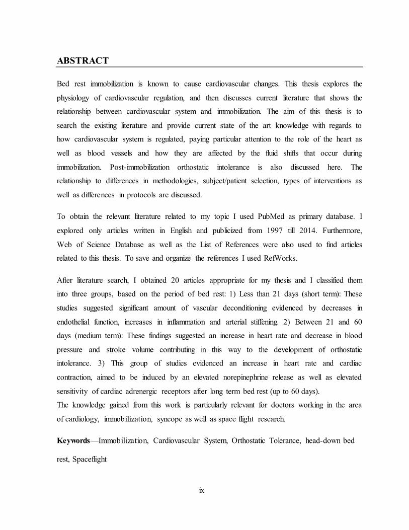
ABSTRACT
Bed rest immobilization is known to cause cardiovascular changes. This thesis explores the
physiology of cardiovascular regulation, and then discusses current literature that shows the
relationship between cardiovascular system and immobilization. The aim of this thesis is to
search the existing literature and provide current state of the art knowledge with regards to
how cardiovascular system is regulated, paying particular attention to the role of the heart as
well as blood vessels and how they are affected by the fluid shifts that occur during
immobilization. Post-immobilization orthostatic intolerance is also discussed here. The
relationship to differences in methodologies, subject/patient selection, types of interventions as
well as differences in protocols are discussed.
To obtain the relevant literature related to my topic I used PubMed as primary database. I
explored only articles written in English and publicized from 1997 till 2014. Furthermore,
Web of Science Database as well as the List of References were also used to find articles
related to this thesis. To save and organize the references I used RefWorks.
After literature search, I obtained 20 articles appropriate for my thesis and I classified them
into three groups, based on the period of bed rest: 1) Less than 21 days (short term): These
studies suggested significant amount of vascular deconditioning evidenced by decreases in
endothelial function, increases in inflammation and arterial stiffening. 2) Between 21 and 60
days (medium term): These findings suggested an increase in heart rate and decrease in blood
pressure and stroke volume contributing in this way to the development of orthostatic
intolerance. 3) This group of studies evidenced an increase in heart rate and cardiac
contraction, aimed to be induced by an elevated norepinephrine release as well as elevated
sensitivity of cardiac adrenergic receptors after long term bed rest (up to 60 days).
The knowledge gained from this work is particularly relevant for doctors working in the area
of cardiology, immobilization, syncope as well as space flight research.
Keywords—Immobilization, Cardiovascular System, Orthostatic Tolerance, head-down bed
rest, Spaceflight
x
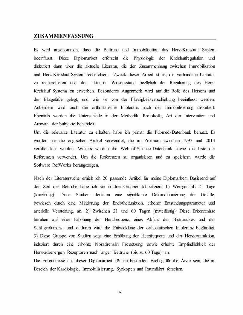
ZUSAMMENFASSUNG
Es wird angenommen, dass die Bettruhe und Immobilisation das Herz-Kreislauf System
beeinflusst. Diese Diplomarbeit erforscht die Physiologie der Kreislaufregulation und
diskutiert dann über die aktuelle Literatur, die den Zusammenhang zwischen Immobilisation
und Herz-Kreislauf-System recherchiert. Zweck dieser Arbeit ist es, die vorhandene Literatur
zu recherchieren und den aktuellen Wissensstand bezüglich der Regulierung des Herz-
Kreislauf Systems zu erwerben. Besonderes Augenmerk wird auf die Rolle des Herzens und
der Blutgefäße gelegt, und wie sie von der Flüssigkeitsverschiebung beeinflusst werden.
Außerdem wird auch die orthostatische Intoleranz nach der Immobilisierung diskutiert.
Ebenfalls werden die Unterschiede in der Methodik, Protokolle, Art der Intervention und
Auswahl der Subjekte behandelt.
Um die relevante Literatur zu erhalten, habe ich primär die Pubmed-Datenbank benutzt. Es
wurden nur die englischen Artikel verwendet, die im Zeitraum zwischen 1997 und 2014
veröffentlicht wurden. Weiters wurden die Web-of-Science-Datenbank sowie die Liste der
Referenzen verwendet. Um die Referenzen zu organisieren und zu speichern, wurde die
Software RefWorks herangezogen.
Nach der Literatursuche erhielt ich 20 passende Artikel für meine Diplomarbeit. Basierend auf
der Zeit der Bettruhe habe ich sie in drei Gruppen klassifiziert: 1) Weniger als 21 Tage
(kurzfristig): Diese Studien deuteten eine signifikante Dekonditionierung der Gefäße,
bewiesen durch eine Minderung der Endothelfunktion, erhöhte Entzündungsparameter und
arterielle Versteifung, an. 2) Zwischen 21 und 60 Tagen (mittelfristig): Diese Erkenntnisse
beruhen auf einer Erhöhung der Herzfrequenz, eines Abfalls des Blutdruckes und des
Schlagvolumens, und dadurch wird die Entwicklung der orthostatischen Intoleranz begünstigt.
3) Diese Gruppe von Studien zeigt eine Erhöhung der Herzfrequenz und der Herzkontraktion,
induziert durch eine erhöhte Noradrenalin Freisetzung, sowie erhöhte Empfindlichkeit der
Herz-adrenergen Rezeptoren nach langer Bettruhe (bis zu 60 Tage), an.
Die Erkenntnisse aus dieser Diplomarbeit können besonders wichtig für die Ärzte sein, die im
Bereich der Kardiologie, Immobilisierung, Synkopen und Raumfahrt forschen.
1
I. INTRODUCTION
1.1. Physiology of cardiovascular system
Organs are part of the systems and only together and coordinated they can fulfill their
functions. Cardiovascular system is a closed elastic system with an important function that can
be categorized in three main groups: transportation, regulation and protection. Cardiovascular
system supplies our body with essential nutrients and removes waste (such as urea) from the
cells. It also transports hormones between the tissues and regulates the temperature of the
body. Furthermore, this system protects against diverse pathogens due to immune function of
the leukocytes. Cardiovascular system persists of: heart, blood vessels and blood.
1.1.1 Heart and its function
Heart is a muscular organ of vital importance that through its rhythmical contractions
furnishes the body with blood. The heart wall persists of three layers. Epicardium is the outer
layer of the wall of the heart (visceral layer of pericardium); the muscular middle layer is
known as Myocardium. This is the thickest part of the wall. Endocardium is the inner layer of
the heart (one layer epithelium). Heart is organized in two separated pump systems. Each of
them contains one atrium and one ventricle.
Blood flow- Right atrium becomes the systemic blood continually from vena cava superior,
vena cava inferior and the coronary sinus. From the right atrium flows blood in the right
ventricle, this pumps the blood in the pulmonary trunk. Then goes the blood in to the small
circulation, get reached with oxygen in the alveolar capillary level and reaches the left atrium
through pulmonary veins. Left atrium than pumps it into the left ventricle. The last one pumps
the oxygenated blood through Aorta in the whole body. The one direction blood flow in heart
is possible thanks to heart valves. The tricuspid and bicuspid valve let the blood flow in
ventricles whereas the pulmonary and aortic valve let the blood flow out of the ventricles. This
blood flow through the heart chambers makes possible the coordinated contractions of heart
muscle.
2
Heart contraction- Heart muscle is an involuntary striated muscle made of cells known as
cardiac myocytes or myocardiocytes. Myocytes contain one nucleus in the center, myofibrils
and many mitochondria. The Myofibrils are made of sarcomeres. The arrangement of actin
and myosin filaments in sarcomeres is the same as in skeletal muscle. Myocardial cells are
connected parallel with each other through gap junctions, which allow the free diffusion of
Ions in intracellular fluid of neighbor cells and with it also the spreading of action potential.
This feature makes cardiac cells act together as a unit and is known as functional syncytium.
The heart insists of two syncytium: the atrial syncytium and the ventricular syncytium. They
are connected by a conductive system called the A-V bundle (1).
Like all tissues in the body, cardiac muscle cells need to be supplied with oxygen and nutrients
and released from the waste products. This makes possible the coronary arteries.
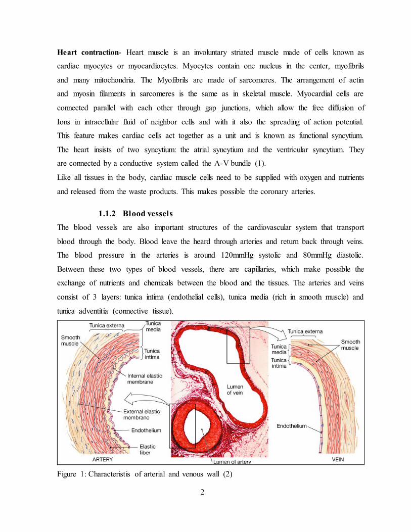
1.1.2 Blood vessels
The blood vessels are also important structures of the cardiovascular system that transport
blood through the body. Blood leave the heard through arteries and return back through veins.
The blood pressure in the arteries is around 120mmHg systolic and 80mmHg diastolic.
Between these two types of blood vessels, there are capillaries, which make possible the
exchange of nutrients and chemicals between the blood and the tissues. The arteries and veins
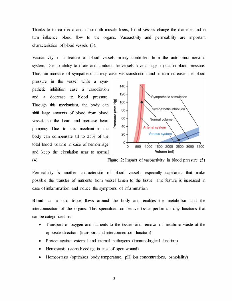
consist of 3 layers: tunica intima (endothelial cells), tunica media (rich in smooth muscle) and
tunica adventitia (connective tissue).
Figure 1: Characteristis of arterial and venous wall (2)
3
Thanks to tunica media and its smooth muscle fibers, blood vessels change the diameter and in
turn influence blood flow to the organs. Vasoactivity and permeability are important
characteristics of blood vessels (3).
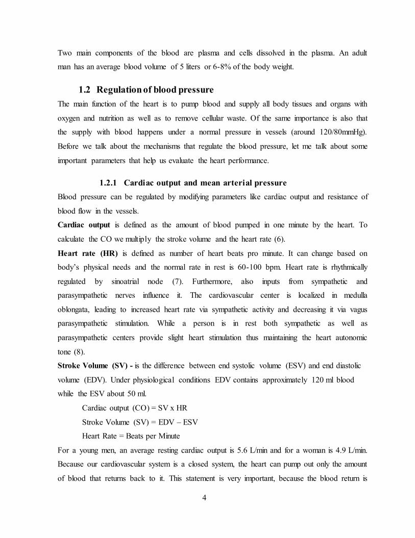
Vasoactivity is a feature of blood vessels mainly controlled from the autonomic nervous
system. Due to ability to dilate and contract the vessels have a huge impact in blood pressure.
Thus, an increase of sympathetic activity case vasoconstriction and in turn increases the blood
pressure in the vessel while a sym-
pathetic inhibition case a vasodilation
and a decrease in blood pressure.
Through this mechanism, the body can
shift large amounts of blood from blood
vessels to the heart and increase heart
pumping. Due to this mechanism, the
body can compensate till to 25% of the
total blood volume in case of hemorrhage
and keep the circulation near to normal
(4). Figure 2: Impact of vasoactivity in blood pressure (5)
Permeability is another characteristic of blood vessels, especially capillaries that make
possible the transfer of nutrients from vessel lumen to the tissue. This feature is increased in
case of inflammation and induce the symptoms of inflammation.
Blood- as a fluid tissue flows around the body and enables the metabolism and the
interconnection of the organs. This specialized connective tissue performs many functions that
can be categorized in:
Transport of oxygen and nutrients to the tissues and removal of metabolic waste at the
opposite direction (transport and interconnection function)
Protect against external and internal pathogens (immunological function)
Hemostasis (stops bleeding in case of open wound)
Homeostasis (optimizes body temperature, pH, ion concentrations, osmolality)
4
Two main components of the blood are plasma and cells dissolved in the plasma. An adult
man has an average blood volume of 5 liters or 6-8% of the body weight.
1.2 Regulation of blood pressure
The main function of the heart is to pump blood and supply all body tissues and organs with
oxygen and nutrition as well as to remove cellular waste. Of the same importance is also that
the supply with blood happens under a normal pressure in vessels (around 120/80mmHg).
Before we talk about the mechanisms that regulate the blood pressure, let me talk about some
important parameters that help us evaluate the heart performance.
1.2.1 Cardiac output and mean arterial pressure
Blood pressure can be regulated by modifying parameters like cardiac output and resistance of
blood flow in the vessels.
Cardiac output is defined as the amount of blood pumped in one minute by the heart. To
calculate the CO we multiply the stroke volume and the heart rate (6).
Heart rate (HR) is defined as number of heart beats pro minute. It can change based on
body’s physical needs and the normal rate in rest is 60-100 bpm. Heart rate is rhythmically
regulated by sinoatrial node (7). Furthermore, also inputs from sympathetic and
parasympathetic nerves influence it. The cardiovascular center is localized in medulla
oblongata, leading to increased heart rate via sympathetic activity and decreasing it via vagus
parasympathetic stimulation. While a person is in rest both sympathetic as well as
parasympathetic centers provide slight heart stimulation thus maintaining the heart autonomic
tone (8).
Stroke Volume (SV) - is the difference between end systolic volume (ESV) and end diastolic
volume (EDV). Under physiological conditions EDV contains approximately 120 ml blood
while the ESV about 50 ml.
Cardiac output (CO) = SV x HR
Stroke Volume (SV) = EDV – ESV
Heart Rate = Beats per Minute
For a young men, an average resting cardiac output is 5.6 L/min and for a woman is 4.9 L/min.
Because our cardiovascular system is a closed system, the heart can pump out only the amount
of blood that returns back to it. This statement is very important, because the blood return is
5
from different body conditions influenced (bed rest immobilization, space flight), and in turn
also the cardiac output.
To make this blood amount flow into the systemic circulation, the heart must pump against the
resistance of the vascular bed, known as Total Peripheral Resistance (TPR). It is affected
primarily by changes in the diameter of the vessels.
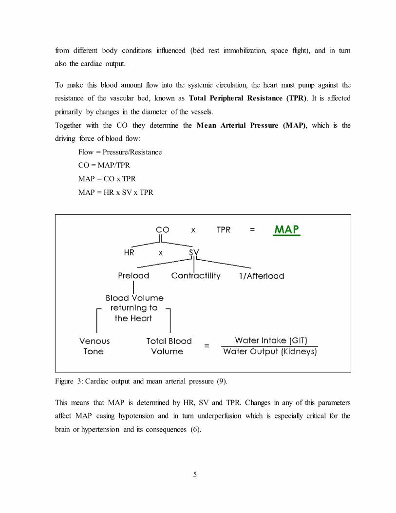
Together with the CO they determine the Mean Arterial Pressure (MAP), which is the
driving force of blood flow:
Flow = Pressure/Resistance
CO = MAP/TPR
MAP = CO x TPR
MAP = HR x SV x TPR
Figure 3: Cardiac output and mean arterial pressure (9).
This means that MAP is determined by HR, SV and TPR. Changes in any of this parameters
affect MAP casing hypotension and in turn underperfusion which is especially critical for the
brain or hypertension and its consequences (6).
6
1.2.2 Mechanisms of blood pressure regulation
Tissues have various needs for Blood amounts in different loading conditions. It is essential
that the circulatory regulation mechanisms supply the organs always optimally with Blood.
This is possible due to:
Short term mechanisms in regulation of blood pressure
Long term mechanisms in regulation of blood pressure
Local mechanisms (1)
1.2.2.1 Short term regulation of blood pressure
These mechanisms of blood pressure control are able to regulate the arterial pressure in
seconds.(10) If there is needed an increase of blood pressure than the nervous system affects
vasoconstriction and cardioacceleration together and at the same time inhibits parasympathetic
signals to the heart. Neuronal control mechanisms consist of:
a) Cardiovascular control centers in Medulla
b) Effector pathways (Autonomic nervous system) and
c) Afferent signals to the centers in central nervous system (CNS).
a) Cardiovascular control centers in CNS
Vasomotor center in CNS- is placed in retinacular substance of medulla and sends its fibers
between T1 and L2. The parasimpathetical fibers go to the heart through vagus nerves,
otherwise the peripheral sympathetic nerves and spinal cord innervate all arteries and veins.
Important areas of this center are: a vasoconstrictor area, vasodilator area and a sensory area.
Cardio Acceleratory Center- sends sympathetic fibers down the spine to T1-T5 where they
exit to periphery. When activated, it increases the heart rate and myocardial contractility.
The third center is Cardio Inhibitory Center, which originate in Medulla and it sends
parasympathetic fibers through vagus nerve (1).
b) The autonomic nervous system (ANS)
Autonomic neuronal system through its sympathetic and parasympathetic activity affects
arteries, veins and heart and makes a great job in rapid control of blood pressure. The main
regulating functions of the neuronal mechanisms in cardiovascular system are: increase of the
heart rate, rapid control of arterial pressure, redistribution of blood amounts to different parts
7
of the body. Principally, sympathetic nerve fibers increase the heart rate and contraction and
parasympathetic nerves case the opposite. Sympathetic nerves contain mainly vasoconstrictor
fibers. At the endings of this nerves is secreted Norepinephrine, which acts on α-adrenergic
receptors and affects smooth muscle contraction.
These centers send their signals to the organs and tissues through effector pathways of
autonom nervous system. An important part of the neuronal mechanisms are also the
feedbacks from periphery to the center. This is possible primarily due to baroreceptors and
chemoreceptors.
This part of nervous system is responsible for the main visceral functions of the body. The
autonomic nervous system plays a huge role in control of cardiovascular system. For example
it can double the heart rate in 3-5 seconds, in 10-15 seconds it can increase or decrease the
blood pressure twice normal. ANS centers are located in the hypothalamus, brain steam and
spinal cord. They come in contact with organs they innervate through sympathetic nervous
system and parasympathetic nervous system.
Sympathetic nervous system- The fibers of this system originate in the spinal cord (between
cord segments T1-L2) and then they get into the sympathetic chain. From here they travel to
the target tissues. Principally, the sympathetic nervous fibers are composed of 2 neurons. The
first neuron is called preganglionic neuron and connects ANS centers in the CNS and
peripheral ganglia. (1) The axon of the preganglionic neuron through rr. communicantes albi
reach the postganglionic neuron in the ganglion area and make a synapse between. The body
of the postganglionic neuron lies on the ganglia of the sympathetic chain and from here sends
postganglionic axons to the effector organs.
Parasympathetic nervous system- run away from the CNS through cranial nerves (III, VII,
IX and X) and sacral spinal roots. The parasympathetic nervous system reaches the
cardiovascular system by means of vagus around four fifths of parasympathetic fibers reach
thoracic and abdominal organs by means of vagus. This system has also its pre- and
postganglionic neurons. The ganglions of parasympathetic system are mostly located in the
wall of the organ.(11)
8
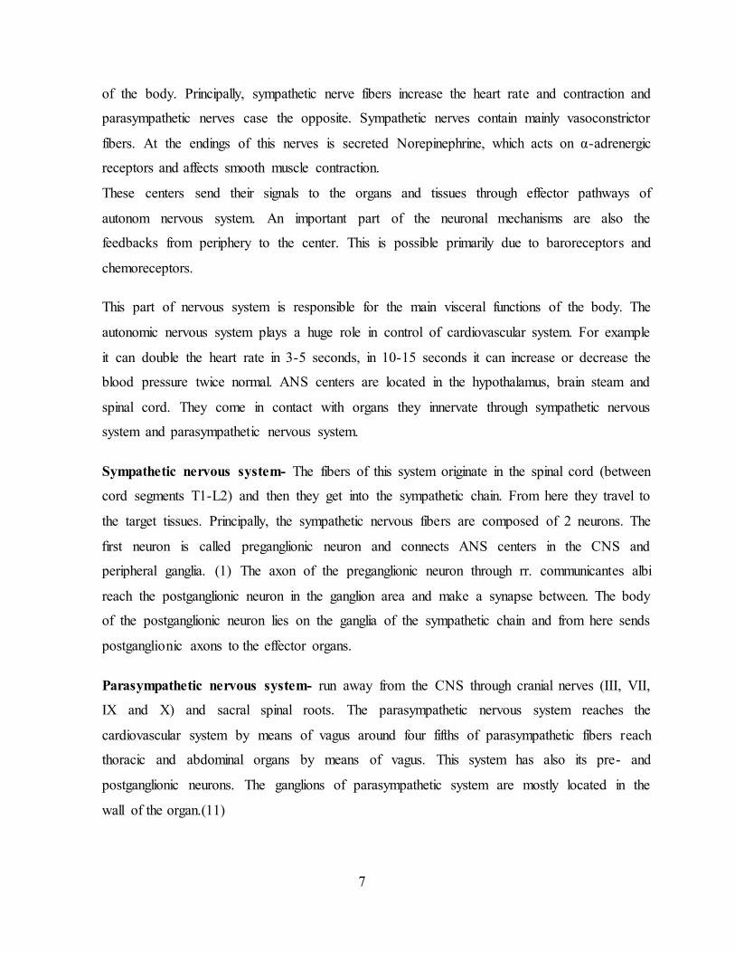
The sympathetic and parasympathetic preganglionic fibers secrete acetylcholine, which is also
a transmitter in the parasympathetic postganglionic fibers. All these fibers that secrete
acetylcholine are called cholinergic fibers. Conversely, almost all of the sympathetic
postganglionic fibers secrete norepinephrine and are said to be adrenergic.
Usually, have postganglionic nerves at their ends an enlargement (varicosities) where they
store the transmitter vesicles filled with acetylcholine or norepinephrine. (12) Under the effect
of action potential release the nerve endings their transmitter in to the effector organs.
Figure 4: Role of autonomic nervous system in cardiovascular system (13).
The transmitters act on specific receptors on the effector organs. There are two types of
receptors for acetylcholine: muscarinic and nicotinic receptors. Nicotinic receptors are located
in the autonomic ganglia (between first and second neuron) and the muscarinic receptors are
found in the effector organs and cells. There are also two types of adrenergic receptors: alpha
receptors and beta receptors and their subtypes alpha1,2 and beta 1,2 receptors. In alpha
receptors binds mainly norepinephrine and one of the actions is the contraction of blood
vessels. In beta receptors binds norepinephrine and epinephrine. Beta1 receptors are
9
responsible for cardioacceleration and increased myocardial strength, while beta2 receptors
case vasodilation (14). Detailed information to receptors are listed in Table 2.
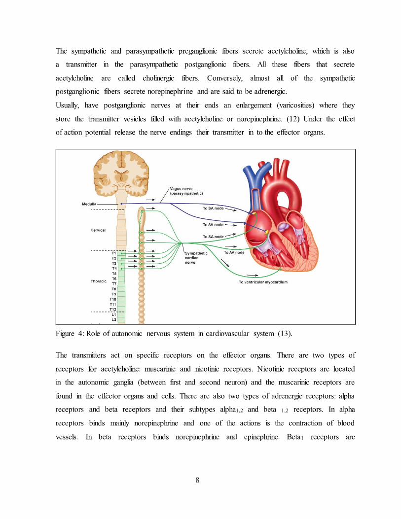
Table 2: Effects of different receptors in cardiovascular system (1).
Target Sympathetic (adrenergic) Parasympathetic (muscarinic)
cardiac output β1, (β2): increases M2: decreases
SA node: heart rate (chronotropic)
β1, (β2): increases(15) M2: decreases
Atrial cardiac muscle: contractility (inotropic)
β1, (β2): increases(15) M2: decreases
AV node
β1: increases conduction increases cardiac muscle activity
M2: decreases conduction Atrioventricular block(15)
Ventricular cardiac muscle β1, (β2): inotropic, increases cardiac muscle contractility(15)
c) Afferent signals
Baroreceptor reflex- Baroreceptors are located in the aortic sinuses, carotid sinuses and in the
right atrial walls. The baroreceptor reflexes adjust the cardiac output and peripheral resistance
being this way the best known nervous mechanisms in control of blood pressure. The
baroreceptors respond in fraction of second in change of arterial pressure. They are most
sensible in blood pressures around 100mmHg, where it is most needed. Additionally, they
respond better to acute changes of blood pressure than to stationary pressure.
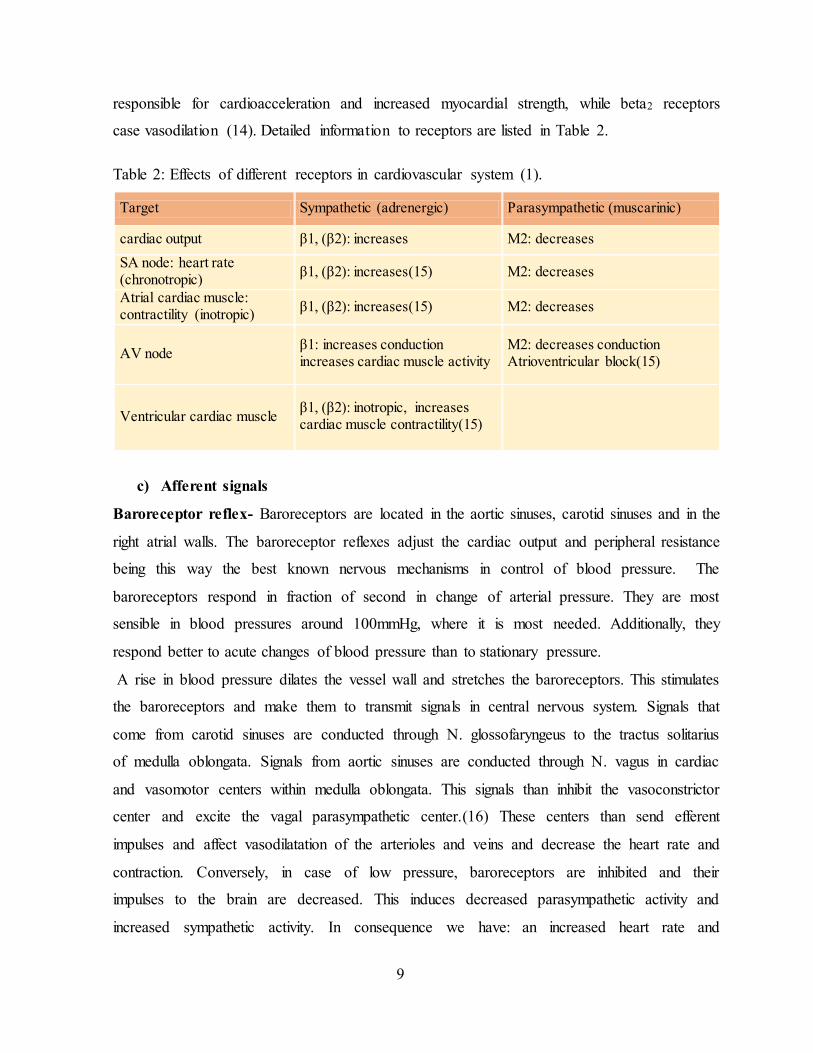
A rise in blood pressure dilates the vessel wall and stretches the baroreceptors. This stimulates
the baroreceptors and make them to transmit signals in central nervous system. Signals that
come from carotid sinuses are conducted through N. glossofaryngeus to the tractus solitarius
of medulla oblongata. Signals from aortic sinuses are conducted through N. vagus in cardiac
and vasomotor centers within medulla oblongata. This signals than inhibit the vasoconstrictor
center and excite the vagal parasympathetic center.(16) These centers than send efferent
impulses and affect vasodilatation of the arterioles and veins and decrease the heart rate and
contraction. Conversely, in case of low pressure, baroreceptors are inhibited and their
impulses to the brain are decreased. This induces decreased parasympathetic activity and
increased sympathetic activity. In consequence we have: an increased heart rate and
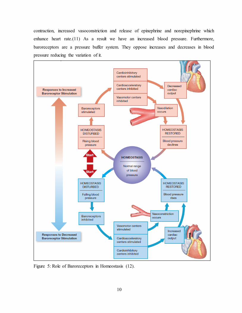
10
contraction, increased vasoconstriction and release of epinephrine and norepinephrine which
enhance heart rate.(11) As a result we have an increased blood pressure. Furthermore,
baroreceptors are a pressure buffer system. They oppose increases and decreases in blood
pressure reducing the variation of it.
Figure 5: Role of Baroreceptors in Homeostasis (12).
11
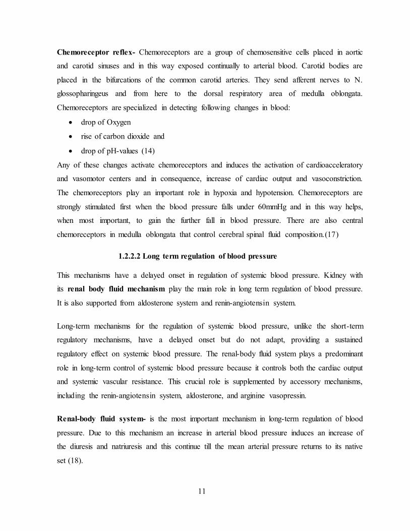
Chemoreceptor reflex- Chemoreceptors are a group of chemosensitive cells placed in aortic
and carotid sinuses and in this way exposed continually to arterial blood. Carotid bodies are
placed in the bifurcations of the common carotid arteries. They send afferent nerves to N.
glossopharingeus and from here to the dorsal respiratory area of medulla oblongata.
Chemoreceptors are specialized in detecting following changes in blood:
drop of Oxygen
rise of carbon dioxide and
drop of pH-values (14)
Any of these changes activate chemoreceptors and induces the activation of cardioacceleratory
and vasomotor centers and in consequence, increase of cardiac output and vasoconstriction.
The chemoreceptors play an important role in hypoxia and hypotension. Chemoreceptors are
strongly stimulated first when the blood pressure falls under 60mmHg and in this way helps,
when most important, to gain the further fall in blood pressure. There are also central
chemoreceptors in medulla oblongata that control cerebral spinal fluid composition.(17)
1.2.2.2 Long term regulation of blood pressure
This mechanisms have a delayed onset in regulation of systemic blood pressure. Kidney with
its renal body fluid mechanism play the main role in long term regulation of blood pressure.
It is also supported from aldosterone system and renin-angiotensin system.
Long-term mechanisms for the regulation of systemic blood pressure, unlike the short-term
regulatory mechanisms, have a delayed onset but do not adapt, providing a sustained
regulatory effect on systemic blood pressure. The renal-body fluid system plays a predominant
role in long-term control of systemic blood pressure because it controls both the cardiac output
and systemic vascular resistance. This crucial role is supplemented by accessory mechanisms,
including the renin-angiotensin system, aldosterone, and arginine vasopressin.
Renal-body fluid system- is the most important mechanism in long-term regulation of blood
pressure. Due to this mechanism an increase in arterial blood pressure induces an increase of
the diuresis and natriuresis and this continue till the mean arterial pressure returns to its native
set (18).
12
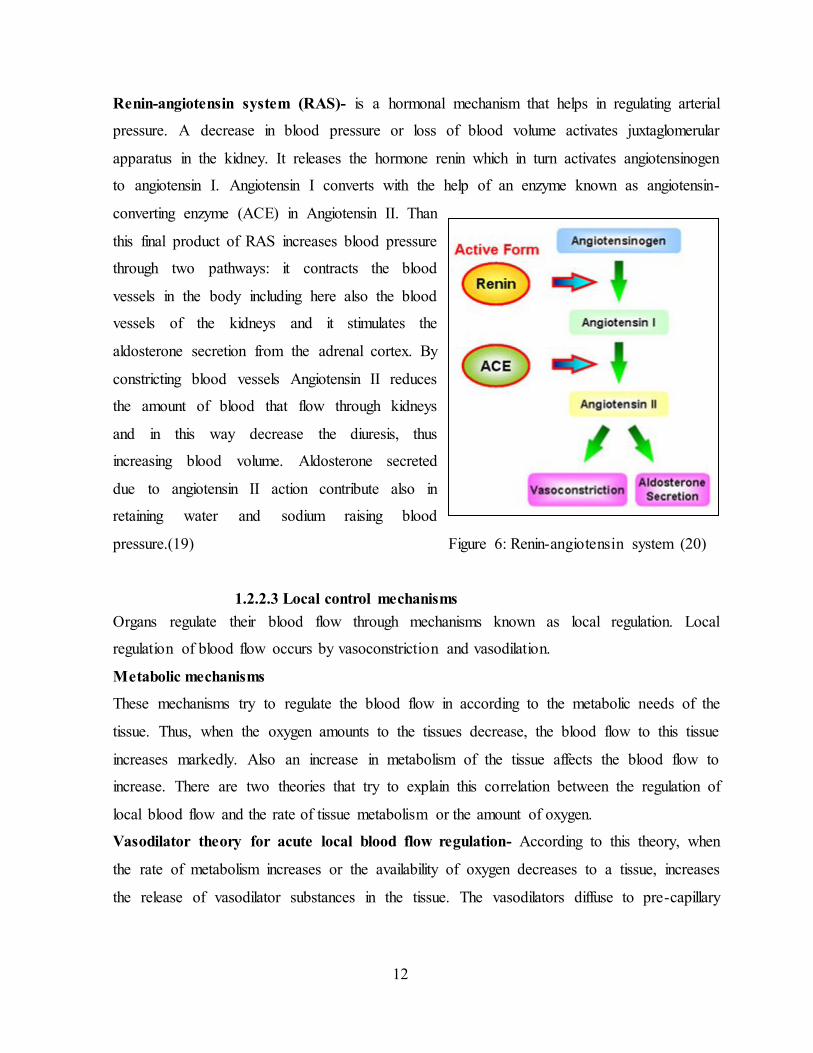
Renin-angiotensin system (RAS)- is a hormonal mechanism that helps in regulating arterial
pressure. A decrease in blood pressure or loss of blood volume activates juxtaglomerular
apparatus in the kidney. It releases the hormone renin which in turn activates angiotensinogen
to angiotensin I. Angiotensin I converts with the help of an enzyme known as angiotensin-
converting enzyme (ACE) in Angiotensin II. Than
this final product of RAS increases blood pressure
through two pathways: it contracts the blood
vessels in the body including here also the blood
vessels of the kidneys and it stimulates the
aldosterone secretion from the adrenal cortex. By
constricting blood vessels Angiotensin II reduces
the amount of blood that flow through kidneys
and in this way decrease the diuresis, thus
increasing blood volume. Aldosterone secreted
due to angiotensin II action contribute also in
retaining water and sodium raising blood
pressure.(19) Figure 6: Renin-angiotensin system (20)
1.2.2.3 Local control mechanisms
Organs regulate their blood flow through mechanisms known as local regulation. Local
regulation of blood flow occurs by vasoconstriction and vasodilation.
Metabolic mechanisms
These mechanisms try to regulate the blood flow in according to the metabolic needs of the
tissue. Thus, when the oxygen amounts to the tissues decrease, the blood flow to this tissue
increases markedly. Also an increase in metabolism of the tissue affects the blood flow to
increase. There are two theories that try to explain this correlation between the regulation of
local blood flow and the rate of tissue metabolism or the amount of oxygen.
Vasodilator theory for acute local blood flow regulation- According to this theory, when
the rate of metabolism increases or the availability of oxygen decreases to a tissue, increases
the release of vasodilator substances in the tissue. The vasodilators diffuse to pre-capillary
13
sphincters and case vasodilation. Important vasodilator substances are: adenosine, histamine,
K+ Ions and H+ Ions.
Oxygen lack theory for local blood flow control- While oxygen is required in muscle
contraction, according to this theory, the blood vessels will dilate in lack of oxygen. Oxygen
lack theory is explained in relation to the pre-capillary sphincters, which regulate the amount
of the blood flow to the tissue or organ. The higher the oxygen concentration in a tissue, the
stronger is the contraction of the pre-capillary sphincters and in this way the less is the amount
of blood flow respectively oxygen to the tissue. (1) An example of metabolic control of blood
flow is hyperemia.
Myogenic mechanisms
Increase of intraluminal pressure stretches the smooth muscle of the vessel. This in turn affects
a reactive contraction mediated by stretch-activated Calcium channels in the vascular smooth
muscle cells. This myogenic contraction reduces blood flow nearly normal. An example of
metabolic and myogenic mechanisms is the autoregulation.
Autoregulation of blood flow- is the intrinsic mechanism that keeps relatively stable the
blood flow despite changes of arterial pressure. Thanks to this mechanism, the blood flow can
be hold relatively constant even though the arterial pressure is elevated. Autoregulation is for a
long time argumented by metabolic and myogenic mechanisms.
14
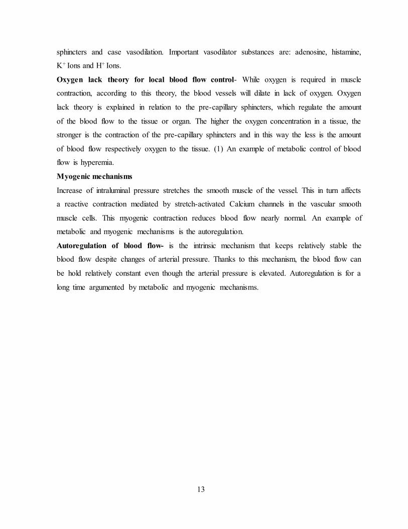
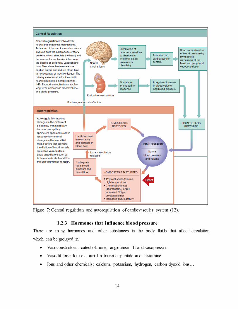
Figure 7: Central regulation and autoregulation of cardiovascular system (12).
1.2.3 Hormones that influence blood pressure
There are many hormones and other substances in the body fluids that affect circulation,
which can be grouped in:
Vasoconstrictors: catecholamine, angiotensin II and vasopressin.
Vasodilators: kinines, atrial natriuretic peptide and histamine
Ions and other chemicals: calcium, potassium, hydrogen, carbon dyoxid ions…
15
a) Vasoconstrictors
Epinephrine- is a hormone released immediately in stress situations from adrenal medulla. It
stimulates alpha- and beta-receptors, increases heart rate and blood pressure. Adrenalin
stimulates alpha-adrenoreceptors in small blood vessels that affects a vasoconstriction. Parallel
stimulates adrenalin also beta2-adrenoreceptors in central vessels effecting a vasodilatation.
Due to its effect in beta1-adrenozeptors increases the epinephrine the heart rate. It’s important
to note that chronically increased adrenalin levels in blood case hypertrophy of heard.
Norepinephrine- has a greater impact in alpha- than in beta-receptors. For this reason it acts a
generalized vasoconstriction.
Angiotensin-II- is an effector hormone of the renin-angiotensin system. It is produced by
kidneys in response to low blood pressure. Angiotensin-II is a strong vasoconstrictive peptide
and through its effect in central neural system the release of Aldosterone from adrenal cortex.
Aldosterone increases the reabsorption of water and sodium in kidneys thus increasing the
volume of fluids in the body and also blood pressure (4).
Antidiuretic Hormone (Vasopressin) - is a peptide hormone with a very short half-life time
(16-24min.) produced in Hypothalamus and stored at the posterior pituitary. It increase water
absorption in the collecting ducts and distal convoluted tubule, retaining in this way the water
in body. In high concentrations affects also vasoconstriction, which in turn increases blood
pressure. It is released in response of increased plasma solute concentration, decreased blood
pressure or blood volume and affects also the homeostasis, by regulating the amount of water,
salts and glucose in the body.(21)
b) Vasodilators
Atrial Natriuretic Peptide (ANP)- is a peptide hormone secreted from the right atria cells in
response to high blood pressure and plays a role in homeostatic. It is released in response to
stretch of atria walls, sympathetic stimulation of beta-receptors, hypernatremia, Angiotensin II
and Endothelin. ANP reduces blood pressure through different mechanisms: loss of water and
sodium, inhibition of sympathetic stimulation of adrenal medulla and vasodilation. This
hormone bins to its specific receptors and aim its goal in blood control affecting many organs
or systems of organs. When ANP released induce water and sodium waste by increasing blood
flow to the kidneys. This in turn increases the GFR; decreases the reuptake of sodium and
16
inhibits RAAS system. In vessels, relaxes the smooth muscle cells by elevation of GMPc and
in heart inhibits cardiac hypertrophy.(12)
Kinins are polypeptides that in specific concentrations act to dilate the blood vessels. There
are two forms of Kinins: Bradykinin, found in the plasma and Lysylbradikinin, found in body
tissues. This vasoactive substances are expected to play a role in regulating blood flow
especially to skin, GIT glands and salivary glands. They are also formed during active
secretion of this glands.(1)
Bradykinin is a peptidhormone, synthetized from the kinin-kallikrein system by protheolythic
cleavage of circulating kininogen. It connects to its bradykininrecceptors (B2 receptors-
responsible for the vasoactive effects) in vessel walls, which in turn cases dilation and
increased permeability of the vessel. As a result decreases the blood pressure. Based on its
effect in lowering blood pressure there are made also medicaments against high blood pressure
(ACE-inhibitors) that increase the bradykinin levels and thus in turn decrease blood pressure.
Histamine- is a biogenic amine, involved in many physiological and pathological processes
and especially in the inflammatory response. It is produced and stored in mast cells and
basophils. Histamine is released when complement components C3a and C5a interact with
specific membrane receptors or when antigens bind to mast cell fixed-IgE.
This amine is classified to the tissue hormones and exerts its actions due to specific receptors
in target cells. Histamine cases through H1 receptors a dilation and increase of the permeability
of blood vessels. This leads to an edema of the skin and mucosal membrane. Stimulation of
the same receptors in the bronchial walls case smooth muscle contraction. H2 receptors
primarily case smooth muscle dilation, but also stimulate the parietal cells of stomach to secret
gastric acid (14).
17
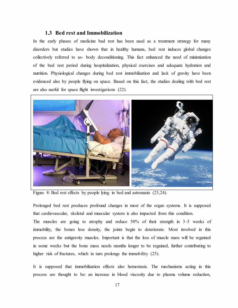
1.3 Bed rest and Immobilization
In the early phases of medicine bad rest has been used as a treatment strategy for many
disorders but studies have shown that in healthy humans, bed rest induces global changes
collectively referred to as- body deconditioning. This fact enhanced the need of minimization
of the bed rest period during hospitalization, physical exercises and adequate hydration and
nutrition. Physiological changes during bed rest immobilization and lack of gravity have been
evidenced also by people flying on space. Based on this fact, the studies dealing with bed rest
are also useful for space flight investigations (22).
Figure 8: Bed rest effects by people lying in bed and astronauts (23,24).
Prolonged bed rest produces profound changes in most of the organ systems. It is supposed
that cardiovascular, skeletal and muscular system is also impacted from this condition.
The muscles are going to atrophy and reduce 50% of their strength in 3-5 weeks of
immobility, the bones loss density, the joints begin to deteriorate. Most involved in this
process are the antigravity muscles. Important is that the loss of muscle mass will be regained
in some weeks but the bone mass needs months longer to be regained, further contributing to
higher risk of fractures, which in turn prolongs the immobility (25).
It is supposed that immobilization effects also hemostasis. The mechanisms acting in this
process are thought to be: an increase in blood viscosity due to plasma volume reduction,
18
decrease in oxygen demand because of muscle atrophy (26) as well as a hemostatic dysbalance
between pro and anti-coagulator factors which can lead to pathological formation of thrombus.
Due to bed rest immobilization the lungs reduce their forced vital capacity as well as
bronchociliar activity. These increase the risk of respiratory infections (26). Constipation, skin
ulcers, body fat increase, mental disorders, loss of minerals and electrolytes some other
consequences of immobility.
During bed rest, the body fluids shift from extremities to the thorax. This stimulates in atriums
the release of ANP hormone, in response to high blood pressure. ANP reduces blood pressure
through different mechanisms: loss of water and sodium, inhibition of sympathetic stimulation
of adrenal medulla and vasodilation leading to decreased stroke volume. To counteract this
effect, the body increase the heart rate (26). Decreased vagal activity and noradrenalin release
are thought to be the mechanisms that cause this heart rate increase (27). Furthermore the heart
muscle undergo atrophy. The cardiovascular changes due to bed rest immobilization will
impact the body also when a person starts to move. During this remobilization period the
blood pool to the lower limbs and because the cardiovascular system is already deconditioned,
it cannot be quickly corrected from it. As a result it comes to orthostatic hypotension, which
decreases cerebral perfusion and thus in turn leads to dizziness and a tendency to fall (26).
Furthermore bed rest decreases also the maximal oxygen consumption (VO2max).
The correlation between cardiovascular system and bed rest immobilization is the main point
of my thesis and is explored below.
19
1.4 Orthostatic Tolerance
Orthostatic tolerance is defined as ability of the body to maintain consciousness during
postural changes (29). Postural changes (standing up) induce orthostatic stress, which causes
shift of blood volume from chest to dependent body parts (lower extremities). This blood
translocation reduces venous return as well as induces pooling of blood and extravasal fluid to
lower extremities. During upright standing 750 ml of chest blood is going to shift downward
(30). Vein blood reservoirs below the heart are going to be filled, heart venous return impedes
and because of the hydrostatic blood pressure changes, cerebral perfusion is going to be
reduced. Upright standing is associated with approximately 6-10 % reduce of cerebral
perfusion due to hydrostatic change between brain and heart. Furthermore also the cardiac
output decreases approximately 25% while heart rate increases (30). The splanchnic
circulation is the largest venous reservoir, followed by low extremities. Similarly to orthostasis
also hemorrhage induces akin loss of central blood volume. In these conditions, intact
compensatory mechanisms are required to maintain cerebral perfusion, consciousness and
blood pressure. An important mechanism for the brain is the cerebral blood flow
autoregulation (30).
1.4.1 Compensatory mechanisms of orthostatic tolerance
Our body use this mechanisms to keep in normal range the cerebral perfusion, consciousness
and blood pressure. Important compensatory mechanisms are going to be discussed below.
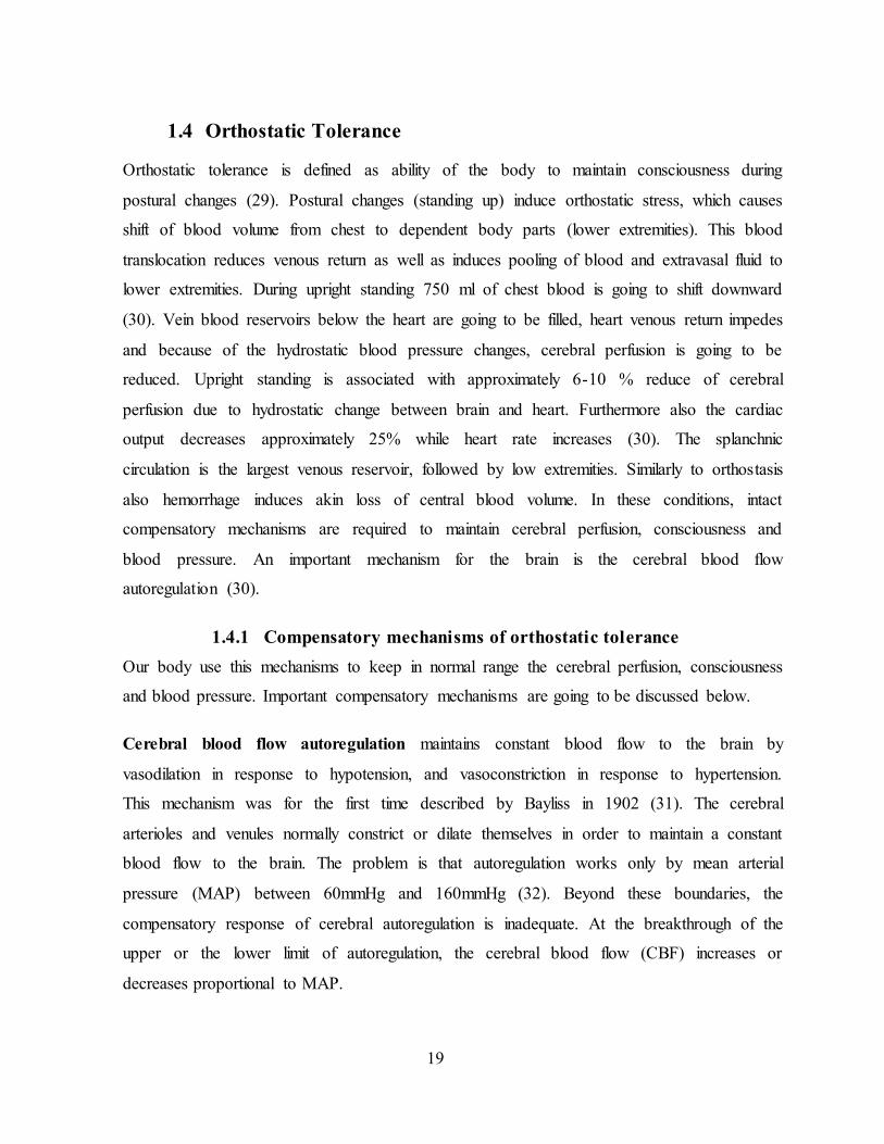
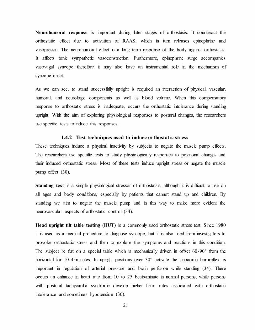
Cerebral blood flow autoregulation maintains constant blood flow to the brain by
vasodilation in response to hypotension, and vasoconstriction in response to hypertension.
This mechanism was for the first time described by Bayliss in 1902 (31). The cerebral
arterioles and venules normally constrict or dilate themselves in order to maintain a constant
blood flow to the brain. The problem is that autoregulation works only by mean arterial
pressure (MAP) between 60mmHg and 160mmHg (32). Beyond these boundaries, the
compensatory response of cerebral autoregulation is inadequate. At the breakthrough of the
upper or the lower limit of autoregulation, the cerebral blood flow (CBF) increases or
decreases proportional to MAP.
20
Figure 9: Autoregulation maintains a constant blood flow by MAP 50-150mmHg (33).
Muscle Pump effect is the first compensatory mechanism that counteract blood pooling. The
muscles of leg and gluteal region contract and this propel the pooled blood to the heart.
Muscle pump impacts also the lymphatic drainage of the low extremities and through
chemoreceptors can be also involved in neurogenic compensatory system. Exactly this
mechanism is crucial by people lying in bed for a long time and by astronauts. Because the
muscle pump undergo atrophy during physical inactivity and/or less of gravity, this persons
are vulnerable toward orthostatic stress.
The second line that counteract orthostatic intolerance is the neurovascular respond.
Vasoconstriction of the arteries promote passive empting of the splanchnic and extremity
circulation. This effect is associated with norepinephrine spillover from the nerve endings. The
reflective compensatory mechanism that occurs during orthostatic stress are primarily
controlled from baroreceptors in the carotid arch, sinus carotids and proximal coronary
arteries. Through these high pressure baroreceptors occurs vasoconstriction and changes the
heart rate. Some other mechanisms included on the compensation are: metabolic, local
myogenic and arteriovenous responses. Also the inflammatory molecules, neuropeptides,
prostacyclin, nitric oxide are thought to play an important role on this defense response.
21
Neurohumoral response is important during later stages of orthostasis. It counteract the
orthostatic effect due to activation of RAAS, which in turn releases epinephrine and
vasopressin. The neurohumoral effect is a long term response of the body against orthostasis.
It affects tonic sympathetic vasoconstriction. Furthermore, epinephrine surge accompanies
vasovagal syncope therefore it may also have an instrumental role in the mechanism of
syncope onset.
As we can see, to stand successfully upright is required an interaction of physical, vascular,
humoral, and neurologic components as well as blood volume. When this compensatory
response to orthostatic stress is inadequate, occurs the orthostatic intolerance during standing
upright. With the aim of exploring physiological responses to postural changes, the researchers
use specific tests to induce this responses.
1.4.2 Test techniques used to induce orthostatic stress
These techniques induce a physical inactivity by subjects to negate the muscle pump effects.
The researchers use specific tests to study physiologically responses to positional changes and
their induced orthostatic stress. Most of these tests induce upright stress or negate the muscle
pump effect (30).
Standing test is a simple physiological stressor of orthostatsis, although it is difficult to use on
all ages and body conditions, especially by patients that cannot stand up and children. By
standing we aim to negate the muscle pump and in this way to make more evident the
neurovascular aspects of orthostatic control (34).
Head upright tilt table testing (HUT) is a commonly used orthostatic stress test. Since 1980
it is used as a medical procedure to diagnose syncope, but it is also used from investigators to
provoke orthostatic stress and then to explore the symptoms and reactions in this condition.
The subject lie flat on a special table which is mechanically driven in offset 60-90° from the
horizontal for 10-45minutes. In upright positions over 30° activate the sinoaortic baroreflex, is
important in regulation of arterial pressure and brain perfusion while standing (34). There
occurs an enhance in heart rate from 10 to 25 beats/minute in normal persons, while persons
with postural tachycardia syndrome develop higher heart rates associated with orthostatic
intolerance and sometimes hypotension (30).
22
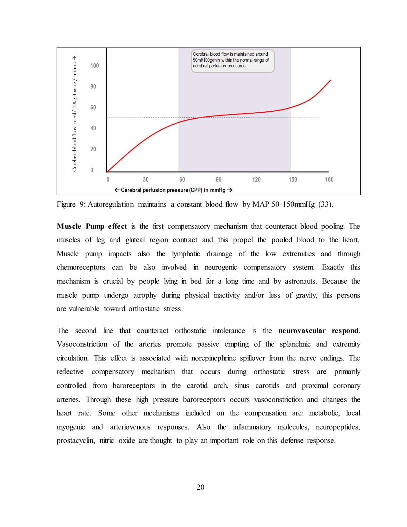
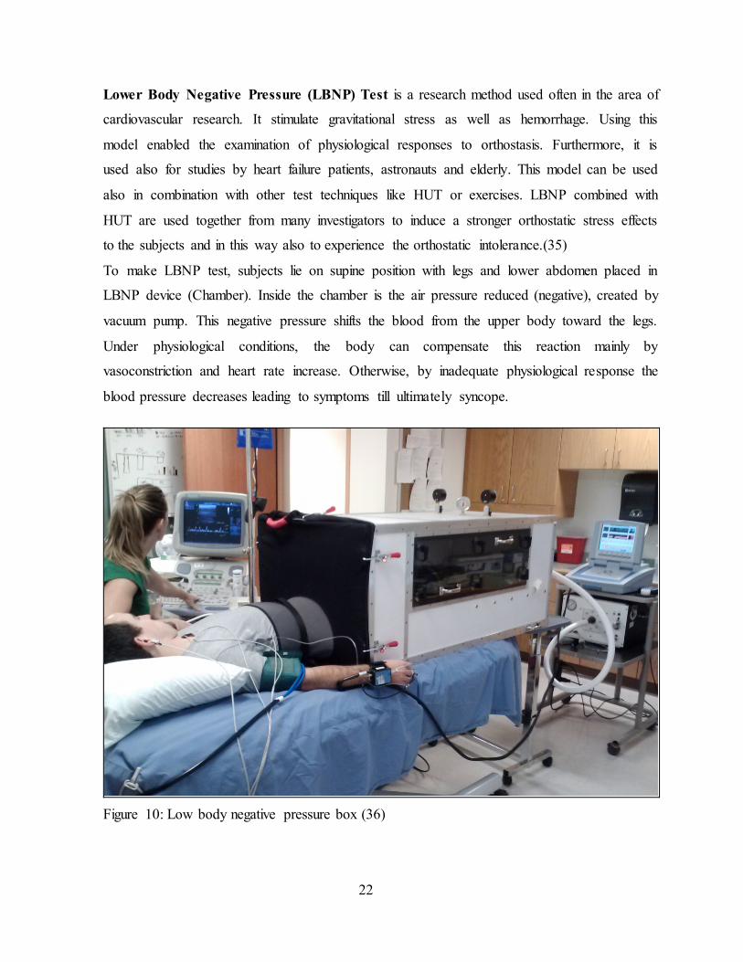
Lower Body Negative Pressure (LBNP) Test is a research method used often in the area of
cardiovascular research. It stimulate gravitational stress as well as hemorrhage. Using this
model enabled the examination of physiological responses to orthostasis. Furthermore, it is
used also for studies by heart failure patients, astronauts and elderly. This model can be used
also in combination with other test techniques like HUT or exercises. LBNP combined with
HUT are used together from many investigators to induce a stronger orthostatic stress effects
to the subjects and in this way also to experience the orthostatic intolerance.(35)
To make LBNP test, subjects lie on supine position with legs and lower abdomen placed in
LBNP device (Chamber). Inside the chamber is the air pressure reduced (negative), created by
vacuum pump. This negative pressure shifts the blood from the upper body toward the legs.
Under physiological conditions, the body can compensate this reaction mainly by
vasoconstriction and heart rate increase. Otherwise, by inadequate physiological response the
blood pressure decreases leading to symptoms till ultimately syncope.
Figure 10: Low body negative pressure box (36)
23
II. AIMS AND OBJECTIVES
Based on the literature review, I will hypothesize what happens during bed rest immobilization
with the cardiovascular system. The main question is how cardiovascular system is affected by
bed rest immobilization. Subjects that undergo bed rest have different fluid distribution, as
compared to say during standing up, the responses of the heart and the vessels will be
accordingly adjusted.
This work will also focus on current knowledge regarding blood pressure regulation as well as
effects of immobilization on the cardiovascular system. It is known that immobilization can
lead to symptoms (vertigo) by standing up. For this reason will be explored also the
mechanisms for orthostatic intolerance after that are responsible for immobilization period. An
eventually impact of bed rest length on study-results is going to be discussed.
Differences in methodologies, selection of subjects, types of interventions as well as
differences in protocols are also going to be discussed. When possible, the gender differences
correlating to bed rest responses will be also investigated.
Based on the actually knowledge that physiological changes during bed rest immobilization
occur also by space flight astronauts, I will discuss some papers that investigated the
relationship between cardiovascular system and space flight. The results of these studies are
going to be compared with bed rest studies and the similarities eventually differences are
going to be elucidated.
The aim of this thesis is to update the existing literature correlated to bed rest immobilization
and cardiovascular regulation. The knowledge gained from this work is particularly relevant
for doctors working in the area of cardiology, immobilization, syncope as well as space flight
research.
24
III. METHODOLOGY
The subject of my research was "Effects of Immobilization on the Cardiovascular System and
Orthostatic Tolerance".
To get the relevant publications for my research, I made a searching strategy. At first I
identified the essential subject by using keywords: cardiovascular system, immobilization,
orthostatic (in) tolerance. Next I found the synonyms for my keywords: "immobilization"
synonym "bed rest", "cardiovascular system" synonym "circulatory system",
"orthostatic tolerance" synonym "postural hypotension".
At last I used MESH Database to find it out which of the above mentioned terms were
represented in Pubmed.
Pubmed databases enabled me to find most of the publications relevant for my research.
To obtain my data from Pubmed, I used the strategy explored below.
Limiting criteria of my research- In order to limit my research criteria I use AND or OR or
NOT. This way I focus on my interest field. To be concrete I take some examples:
cardiovascular system AND Bed rest; cardiovascular system OR Orthostatic intolerance,
Orthostatic Intolerance AND Immobilization. Also using quote (``) for the keywords found I
helpfully in finding publications relevant to my topic.
Refining my search criteria- The above mentioned step gave me the opportunity to obtain
many publications, but not all of them were relevant to my topic. To avoid the unnecessary
studies I refined the criteria of my research. For this purpose I used the word NOT as filter:
(Cardiovascular System NOT cardiovascular system, treatment) AND bed rest.
As second filter I defined the publication years of the researched articles (1950- 2014).
Furthermore, I was interested on the articles written on English and/or Deutsch, therefore I
defined the language of the publications as another refine search criteria.
The sources of my research- To explore my thesis, I used primarily Pubmed database. I
found this database simple to use, up to data and I could find many publications related to my
topic. As alternative to Pubmed, I used also Web of Science database. I found it particularly
25
good because you can see how many times a publication has been cited before. It serves as a
filter to find out the most cited articles. Another tool of research that I found very helpful was
the list of References. Sometimes the reference of one article was the publication relevant for
my research. At last I used Internet (Google) particularly to find pictures and relevant
Information for my Introduction.
Criteria for choosing my literature:
1. Articles relevant to my topic.
2. As primary literature I used Abstracts and full-text Articles.
3. Only the publications written in the last 64 years were explored.
4. As secondary literature I used Internet as well as Textbooks.
5. English and/or German publication were explored.
After literature search, I obtained 20 articles that were relevant for my thesis. Based on the
time of bed rest period and the year of publication, I divided this articles into three groups:
1. The first group include the studies with a bed rest period of under 21 days. This articles
offer different view perspectives of the relationship between cardiovascular system and
immobilization based on the short term bed rest model.
2. Medium term bed rest studies include the studies with a bed rest period between 21 and 60
days.
3. The last group of studies examines the effects of prolonged bed rest (up to 60 days) on
cardiovascular system. An eventually impact of bed rest length on study-results is going to be
explored.
Furthermore, there are going to be discussed differences in methodologies, selection of
subjects, types of interventions as well as differences in protocols. When possible, the gender
differences correlating to bed rest will be also discussed.
Organizing and saving the References- I used Refworks to save and organize my references. I
found it very useful, especially that I directly could import my references from the databases
to my Refworks Account and from here to use these in my thesis.
26
IV. UPDATE OF THE LITERATURE
Based on my literature search, I obtained 20 articles that were relevant for my thesis.
Due to the time of bed rest period and the year of article publication, I divided this section of
my thesis into three categories:
1. The first category include the studies that offer different view perspectives of the
relationship between cardiovascular system and immobilization based on the short term bed
rest model.
2. Medium term bed rest studies were also examine. Furthermore, an eventually impact of bed
rest length on study-results is going to be explored.
3. The last category examines the effects of long term bed rest on cardiovascular system. The
correlation to differences in methods, subject selection, intervention types and protocols will
be also discussed.
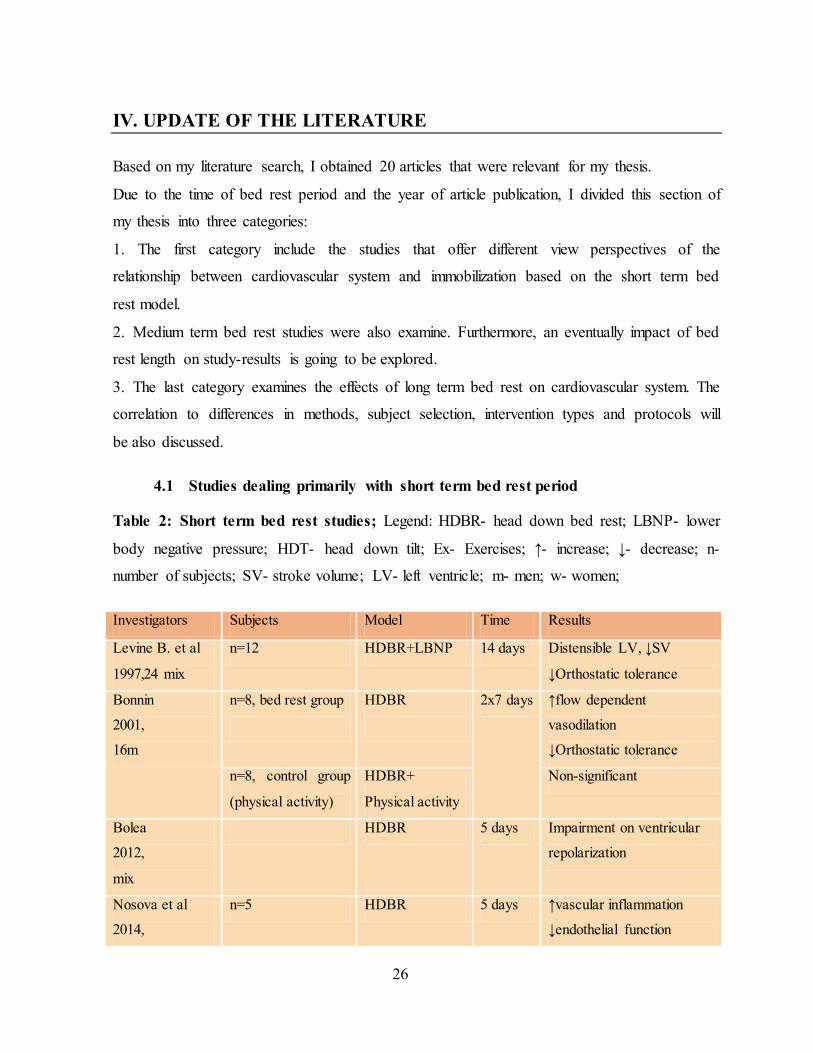
4.1 Studies dealing primarily with short term bed rest period
Table 2: Short term bed rest studies; Legend: HDBR- head down bed rest; LBNP- lower
body negative pressure; HDT- head down tilt; Ex- Exercises; ↑- increase; ↓- decrease; n-
number of subjects; SV- stroke volume; LV- left ventricle; m- men; w- women;
Investigators Subjects Model Time Results
Levine B. et al
1997,24 mix
n=12 HDBR+LBNP 14 days Distensible LV, ↓SV
↓Orthostatic tolerance
Bonnin
2001,
16m
n=8, bed rest group
HDBR
2x7 days ↑flow dependent
vasodilation
↓Orthostatic tolerance
n=8, control group
(physical activity)
HDBR+
Physical activity
Non-significant
Bolea
2012,
mix
HDBR 5 days Impairment on ventricular
repolarization
Nosova et al
2014,
n=5 HDBR 5 days ↑vascular inflammation
↓endothelial function
27
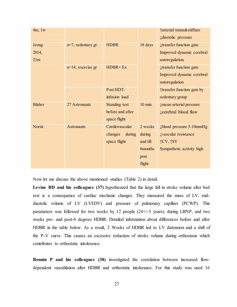
4m, 1w
↑arterial tonus&stiffnes
↓diastolic pressure
Jeong
2014,
21m
n=7, sedentary gr.
HDBR
18 days
↓transfer function gate
Improved dynamic cerebral
autoregulation
n=14, exercise gr HDBR+ Ex
↓transfer function gate
Improved dynamic cerebral
autoregulation
. Post HDT-
infusion load
↑transfer function gate by
sedentary group
Blaber 27 Astronauts Standing test
before and after
space flight
10 min. ↓mean arterial pressure
↓cerebral blood flow
Norsk Astronauts Cardiovascular
changes during
space flight
2 weeks
during
and till
6months
post
flight
↓blood pressure 5-10mmHg
↓vascular resistance
↑CV, ↑SV
Sympathetic activity high
Now let me discuss the above mentioned studies (Table 2) in detail.
Levine BD and his colleagues (37) hypothesized that the large fall in stroke volume after bed
rest is a consequence of cardiac mechanic changes. They measured the mass of LV, end-
diastolic volume of LV (LVEDV) and pressure of pulmonary capillars (PCWP). This
parameters was followed for two weeks by 12 people (24+/-5 years), during LBNP, and two
weeks pre- and post-6 degrees HDBR. Detailed information about differences before and after
HDBR in the table below. As a result, 2 Weeks of HDBR led to LV distension and a shift of
the P-V curve. This causes an excessive reduction of stroke volume during orthostasis which
contributes to orthostatic intolerance.
Bonnin P and his colleagues (38) investigated the correlation between increased flow-
dependent vasodilation after HDBR and orthostatic intolerance. For this study was used 16

28
voluntary men. Eight men was arranged into the control group and maintained its usual
physical activity and eight men underwent two periods of 7 days HDBR (-6 degrees). Using
echography and Doppler, the investigators measured blood flow velocity (BFV) and diameter
of brachial artery. The results suggested an increase of flow-dependent vasodilation of large
arteries after HDBR. As a conclusion, bed rest deconditioning enhances the flow-dependent
vasodilation and may contribute to the orthostatic intolerance.
Bolea and his colleagues (39) explored the effects of microgravity on ventricular
repolarization. Healthy adults underwent 5 days head down bed rest (HDBR). During the tilt
table test were measured: QT to RR and QT (p) to RR hysteresis. Furthermore the differences
between them have been explored. The results of the study suggested significant differences
between QT and QTp before HDBR but not after it. Furthermore, different effect of HDBR on
QT to RR and QTp to RR was evidenced. Based on the study results, impairment on
ventricular repolarization dispersion due to HDBR has been suggested.
Nosova and her colleagues (40) investigated the effects of bed rest on cardiovascular system.
They hypothesized that sedentary inactivity induce endothelial dysfunction, increase the
vascular inflammation and lead to stiffness of the arteries. To prove their hypothesis, they
took 5 healthy persons (4 men and 1 woman) those underwent 5 days of bed rest. They
measured flow- mediated vasodilatation and applanation tonometry to investigate the vascular
function. Furthermore they measured also the inflammation parameters. Isocaloric diet was
applied for the period of inactivity.
The results suggested a decrease of the endothelial function, increase of the inflammation (15-
hydroxyeicosatetraeonic acid was measured), decrease of the diastolic pressure and stiffing of
the arteries. Based on the results, study- researchers speculate that physical inactivity
encourages a vascular deconditioning state that leads to arterial stiffness and arterial tone
increase.
Jeong and his colleagues (41) explored the effects of head down bed rest in brain circulation.
21 subjects took part on the study and they were organized in two groups. First group
(sedentary group) included 7 participants which underwent 18 days bed rest. After bed rest the
29
participants became a volume infusion with dextran to restore reduced plasma volume. The
second group (exercise group) included 14 participants who performed cycling HDBR.
Furthermore, the exercise group is divided into two subgroups: the first subgroup became
dextran infusion after HDBR. By the first subgroup was performed an infusion with dextran to
load the volume (Ex+ Dex Group), while the second subgroup did not become the infusion. To
estimate the cerebral dynamic autoregulation they analyzed transfer function changes in
cerebral flow velocity and blood pressure. The results of the study suggested that after bed rest
was reduced the gain of transfer function in both groups. As study conclusion came out that in
both groups the dynamic cerebral auto regulation was improved or preserved after HDT bed
rest. Furthermore, volume loading is shown to increase the transfer function gain leading to
the suggestion that plasma volume changes are important for the dynamic cerebral
autoregulation.
Blaber and his colleagues (42) explored the regulation of cerebral blood flow on astronauts
before and after space flight. They hypothesized that there is a difference in cerebral
autoregulation before and after flight by subjects that finished and the others that did not
finished the stand test. 27 astronauts were included on the study, 19 of them finished the stand
test while 8 did not. They were part of the shuttle mission which lasted 8- 16 days. The
subjects underwent a 10 min stand test which is done 10 days before flight, 1-2 hours after
landing and 3 days after landing. Parameters that were measured included: mean blood flow
velocity of the middle cerebral artery (MCA), mean arterial pressure as well as cross spectral
power, gain, phase, and coherence. The results of the study suggested that mean arterial
pressure of the MCA was reduced by stand test. Differences between finishers and not
finishers as well as over test days, were significant. The data taken before flight suggested a
higher cerebral vasodilatation to the non-finishers compared with finishers. Furthermore is
being evidenced that on the day of landing the non-finishers suggested a significantly decrease
in cerebral mean blood flow velocity during stand test compared to finishers group. In this
study is also evidenced an interaction effect of gender from supine to stand and over the days
of study.
30
The study investigators concluded that their results indicate a mismatch of cerebral blood flow
with blood pressure as cause of presyncope in astronauts. Furthermore, also the gender is
suggested to be important in orthostatic intolerance after flight.
Norsk P (43) has written a review about the adaptive responses of cardiovascular system on
the astronauts during space flight. Based on the data he explored, a reduction of brachial
diastolic pressure by 5mmHg during the initial 2 weeks of space flight is being evidenced.
Furthermore, from the first till the sixth month of flight, a blood pressure reduction of 10
mmHg has been found. At the same time systemic vascular resistance decreased, cardiac
volume and stroke volume increased. These changes are found also on the persons being
supine on the earth. During space flight the sympathetic activity nerves sympathetic activity is
kept surprisingly high, similar to earth people seated upright. This result could not be
predicted from simulation models thus indicating that the dilatation of arterial vessels during
spaceflight is induced from another mechanism other than baroreflex induced sympathetic
decrease. Furthermore explorations are needed to find the spaceflight mechanism that causes
arterial dilatation, which can explain the high sympathetic activity, associated cardiovascular
changes and blood pressure decrease.
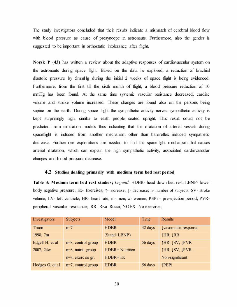
4.2 Studies dealing primarily with medium term bed rest period
Table 3: Medium term bed rest studies; Legend: HDBR- head down bed rest; LBNP- lower
body negative pressure; Ex- Exercises; ↑- increase; ↓- decrease; n- number of subjects; SV- stroke
volume; LV- left ventricle; HR- heart rate; m- men; w- women; PEPi – pre-ejection period; PVR-
peripheral vascular resistance; RR- Riva Rocci; NOEX- No exercises;
Investigators Subjects Model Time Results
Traon
1998, 7m
n=7 HDBR
(Stand+LBNP)
42 days ↓vasomotor response
↑HR, ↓RR
Edgell H. et al
2007, 24w
n=8, control group
n=8, nutrit. group
n=8, exercise gr.
HDBR
HDBR+ Nutrition
HDBR+ Ex
56 days ↑HR, ↓SV, ↓PVR
↑HR, ↓SV, ↓PVR
Non-significant
Hodges G. et al n=7, control group HDBR 56 days ↑PEPi
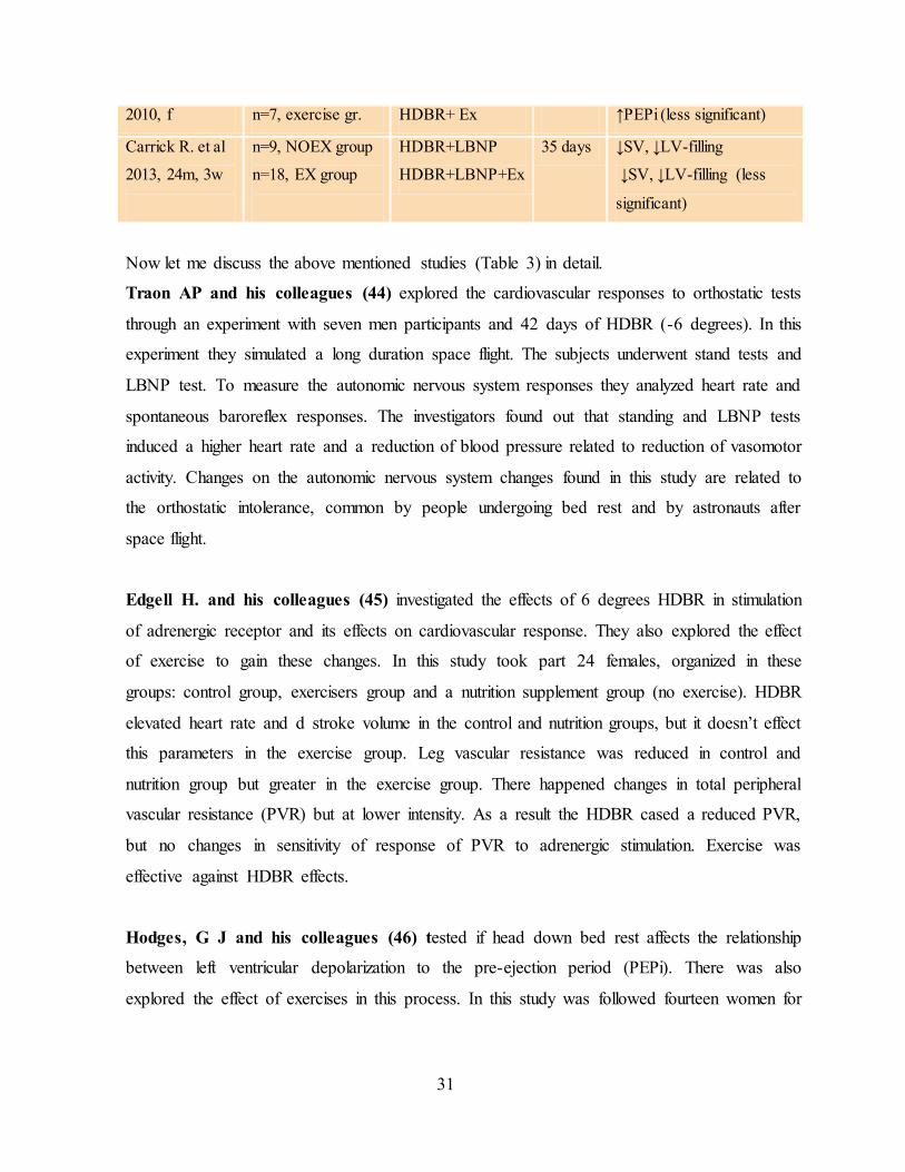
31
2010, f n=7, exercise gr. HDBR+ Ex ↑PEPi (less significant)
Carrick R. et al
2013, 24m, 3w
n=9, NOEX group
n=18, EX group
HDBR+LBNP
HDBR+LBNP+Ex
35 days ↓SV, ↓LV-filling
↓SV, ↓LV-filling (less
significant)
Now let me discuss the above mentioned studies (Table 3) in detail.
Traon AP and his colleagues (44) explored the cardiovascular responses to orthostatic tests
through an experiment with seven men participants and 42 days of HDBR (-6 degrees). In this
experiment they simulated a long duration space flight. The subjects underwent stand tests and
LBNP test. To measure the autonomic nervous system responses they analyzed heart rate and
spontaneous baroreflex responses. The investigators found out that standing and LBNP tests
induced a higher heart rate and a reduction of blood pressure related to reduction of vasomotor
activity. Changes on the autonomic nervous system changes found in this study are related to
the orthostatic intolerance, common by people undergoing bed rest and by astronauts after
space flight.
Edgell H. and his colleagues (45) investigated the effects of 6 degrees HDBR in stimulation
of adrenergic receptor and its effects on cardiovascular response. They also explored the effect
of exercise to gain these changes. In this study took part 24 females, organized in these
groups: control group, exercisers group and a nutrition supplement group (no exercise). HDBR
elevated heart rate and d stroke volume in the control and nutrition groups, but it doesn’t effect
this parameters in the exercise group. Leg vascular resistance was reduced in control and
nutrition group but greater in the exercise group. There happened changes in total peripheral
vascular resistance (PVR) but at lower intensity. As a result the HDBR cased a reduced PVR,
but no changes in sensitivity of response of PVR to adrenergic stimulation. Exercise was
effective against HDBR effects.
Hodges, G J and his colleagues (46) tested if head down bed rest affects the relationship
between left ventricular depolarization to the pre-ejection period (PEPi). There was also
explored the effect of exercises in this process. In this study was followed fourteen women for
32
56 days of HDBR. The members of this study was divided into a Control (non-exercise) and
Exercise (exercise) group, each group with 7 members.
The values of ECG and stroke volume before and after HDBR period told that there was an
increase of PEPi after HDBR, especially in the group of control. But the HDBR period had no
effect in the QRS interval and cardiac afterload.
Isoprenaline infusion reversed the HDBR-induced delay in the PEPi.
As a result HDBR cases a delay on systolic ejection onset. Because the low-dose Isoprenaline
infusion reversed this PEPi delay appears that the mechanism of delayed onset of systole
following HDBR is a consequence of the contraction of the LV and not of LV-depolarization.
Carrick Ranson and his colleagues (47) investigated the causes of upright stroke volume
reduction leading to orthostatic intolerance after bed rest exposure. The core issue of the study
was to find out if the diastolic filling and slow relaxation of left ventricle (LV) induce stroke
volume reduction or does the exercise training while in bed contribute to these changes. To
investigate this they took 27 healthy adults (24 men and 3 women) that underwent 5 weeks 6
degree head down bed rest (6° HDBR). They divided the participant into two groups: 18 of
them performed near daily ergometry (EX Group) while the other 9 lied passive on bed
(NOEX Group). To define the LV function they measured: mass of left ventricle, left
ventricular end diastolic volume (LVEDV), pressure of pulmonary capillary, stroke volume
and Doppler measures. These data were collected before and after HDBR, during bed rest,
during LV filling (used saline infusion) and during decreased LV loading (used LBNP
system).
The study results suggested that reduction of stroke volume (SV) as well as of left ventricular
end diastolic volume (LVEDV) during bed rest and LBNP were greater by the non-exercise
group compared with EX group after HDBR. By the EX group reduction was less prominent
or unaltered. Furthermore, Doppler ultrasound measures during left ventricular unloading by
LBNP after HDBR did not change significantly in either of the groups. During saline infusion
all the variables restores to pre- HDBR level on both groups. The investigators found out that
Doppler variables of dynamic left ventricular filling after HDBR were significantly reduced on
both groups. These changes were prominently by the NOEX group compared with EX group.
As study conclusion came out that upright stroke volume reduction after HDBR is much more
33
affected from changes in left ventricular loading conditions rather than diastolic suction and
ventricular relaxation.
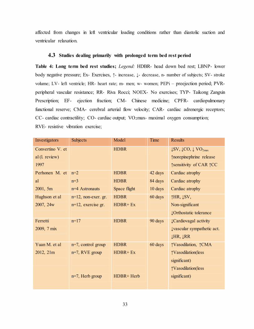
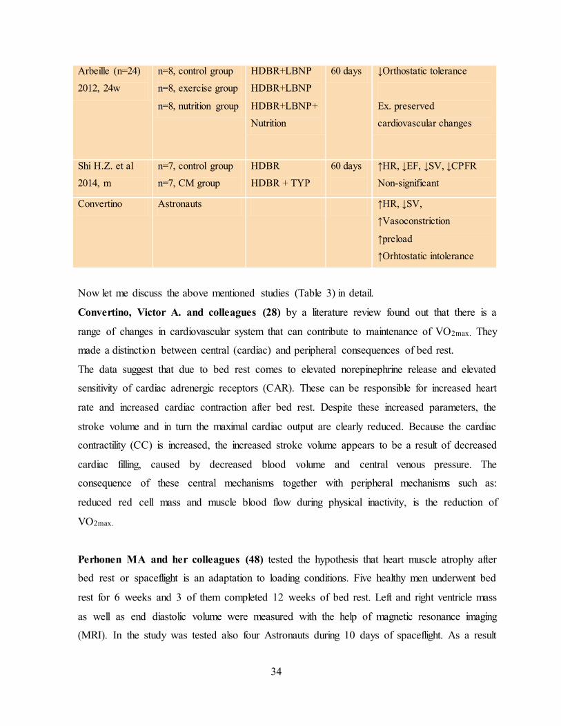
4.3 Studies dealing primarily with prolonged term bed rest period
Table 4: Long term bed rest studies; Legend: HDBR- head down bed rest; LBNP- lower
body negative pressure; Ex- Exercises, ↑- increase, ↓- decrease, n- number of subjects; SV- stroke
volume; LV- left ventricle; HR- heart rate; m- men; w- women; PEPi – preejection period; PVR-
peripheral vascular resistance; RR- Riva Rocci; NOEX- No exercises; TYP- Taikong Zangxin
Prescription; EF- ejection fraction; CM- Chinese medicine; CPFR- cardiopulmonary
functional reserve; CMA- cerebral arterial flow velocity; CAR- cardiac adrenergic receptors;
CC- cardiac contractility; CO- cardiac output; VO2max- maximal oxygen consumption;
RVE- resistive vibration exercise;
Investigators Subjects Model Time Results
Convertino V. et
al (l. review)
1997
HDBR ↓SV, ↓CO, ↓ VO2max
↑norepinephrine release
↑sensitivity of CAR ↑CC
Perhonen M. et
al
2001, 5m
n=2
n=3
n=4 Astronauts
HDBR
HDBR
Space flight
42 days
84 days
10 days
Cardiac atrophy
Cardiac atrophy
Cardiac atrophy
Hughson et al
2007, 24w
n=12, non-exer. gr.
n=12, exercise gr.
HDBR
HDBR+ Ex
60 days
↑HR, ↓SV,
Non-significant
↓Orthostatic tolerance
Ferretti
2009, 7 mix
n=17 HDBR 90 days ↓Cardiovagal activity
↓vascular sympathetic act.
↓HR, ↓RR
Yuan M. et al
2012, 21m
n=7, control group
n=7, RVE group
n=7, Herb group
HDBR
HDBR+ Ex
HDBR+ Herb
60 days ↑Vasodilation, ↑CMA
↑Vasodilation(less
significant)
↑Vasodilation(less
significant)
34
Arbeille (n=24)
2012, 24w
n=8, control group
n=8, exercise group
n=8, nutrition group
HDBR+LBNP
HDBR+LBNP
HDBR+LBNP+
Nutrition
60 days ↓Orthostatic tolerance
Ex. preserved
cardiovascular changes
Shi H.Z. et al
2014, m
n=7, control group
n=7, CM group
HDBR
HDBR + TYP
60 days
↑HR, ↓EF, ↓SV, ↓CPFR
Non-significant
Convertino Astronauts ↑HR, ↓SV,
↑Vasoconstriction
↑preload
↑Orhtostatic intolerance
Now let me discuss the above mentioned studies (Table 3) in detail.
Convertino, Victor A. and colleagues (28) by a literature review found out that there is a
range of changes in cardiovascular system that can contribute to maintenance of VO2max. They
made a distinction between central (cardiac) and peripheral consequences of bed rest.
The data suggest that due to bed rest comes to elevated norepinephrine release and elevated
sensitivity of cardiac adrenergic receptors (CAR). These can be responsible for increased heart
rate and increased cardiac contraction after bed rest. Despite these increased parameters, the
stroke volume and in turn the maximal cardiac output are clearly reduced. Because the cardiac
contractility (CC) is increased, the increased stroke volume appears to be a result of decreased
cardiac filling, caused by decreased blood volume and central venous pressure. The
consequence of these central mechanisms together with peripheral mechanisms such as:
reduced red cell mass and muscle blood flow during physical inactivity, is the reduction of
VO2max.
Perhonen MA and her colleagues (48) tested the hypothesis that heart muscle atrophy after
bed rest or spaceflight is an adaptation to loading conditions. Five healthy men underwent bed
rest for 6 weeks and 3 of them completed 12 weeks of bed rest. Left and right ventricle mass
as well as end diastolic volume were measured with the help of magnetic resonance imaging
(MRI). In the study was tested also four Astronauts during 10 days of spaceflight. As a result
35
there was determined a cardiac atrophy by all of the members in this study. The authors
suggested that this is a physiological adaptation of the heart to reduced myocardial load and
demonstrates the plasticity of cardiac muscle.
Hughson RL and his colleagues (49) studied vascular responses to 60 days of bed rest in 24
woman volunteers. They were divided into an exercise and non-exercise group to find out if
exercises are effective against bed rest consequences. They made a series of experiments and
used drugs such as nitroglycerin and isoproterenol to stimulate responses.
Finally, they found a higher heart rate in the non-exercise group after HDBR, associated with
lower stroke volume. The diameter of the leg arteries was reduced in non-exercise group.
These changes in cardiovascular system contribute to orthostatic intolerance after HDBR.
Ferretti G. and his colleagues (50) tested if head down bed rest induce impairments in
autonomic cardiovascular regulation. Seventeen healthy volunteers underwent for 90 days -6
degrees HDBR. As a measure of baroreceptor reflex activity, vascular sympathetic control,
heart rate and blood pressure, they analyzed the efferent muscle sympathetic nerve activity.
The data were obtained through noninvasive techniques in rest and simple activities like:
moderate handgrip, LBNP and active standing. Their results suggested that inactivity in
subjects (-6 degree HDBR) reduced cardio-vagal and vascular sympathetic activity. It also
reduced the blood pressure and heart rate.
Yuan M. and his colleagues (51) quantified the peripheral arterial and venous response to 60
days of HDBR. In the study took part 21 males divided into three groups each with 7 subjects:
control group, resistive vibration exercise group and Chinese Herb group.
Using echography and Doppler ultrasonography, they measured different parameters of
arteries and veins in the subjects before and after HDBR. Arterial and venous parameters were
measured 3 minutes before tilt and 1 minute after tilt.
Results suggested a vasodilation leading to significantly increased flow velocity of middle
cerebral artery after 60 days of HDBR. Furthermore, resistive vibration exercise and Chinese
herb helped against vasodilation in this areas, whereby RVE was more effective.
36
Arbeille and his colleagues (52) explored the arterial flow velocity and calf vein flow during
orthostatic intolerance test after 60 days 6°HDBR. The structures that were assessed included:
Cerebral, Femoral and Aortic arterial flow as well as Tibial and Gastrocnemius vein section.
Echography and Doppler were used to assess the vessel flow. In the study were included 24
healthy women. They were divided in 3 groups: control group (Con), exercise group against
LBNP (Ex-LBNP) and nutrition group (Nut). Before and after HDBR all participants
underwent a 10 min 80° tilt. After that, followed a LBNP till presyncope. 13 participants could
not finish the experiment because they developed presyncope symptoms, while the rest
participants finished it. The majority of the persons that did not finish the experiment
suggested a decrease on aortal flow, distension of Tibia vein and a lack of increase of
Cerebral/Femoral ratio after HDBR+LBNP correlated to orthostatic intolerance. These
abnormal changes were more evident in Control and Nutrition Groups compared with Exercise
Group.
Their study suggests that HDBR did not impact the cardiovascular changes to orthostatic
stress to the same extent in each participant. Furthermore, exercises within LBNP appear to
preserve the cardiovascular changes to tilt; nutritional intervention alone is not enough. Aortic
and cerebral/femoral ratio flows were most closely related to orthostatic intolerance.
Shi HZ and his colleagues (53) explored the changes in cardiopulmonary function induced by
6 degrees head down bed rest (HDBR) and the effects of Taikong Zangxin Prescription (TYP)
as a countermeasure. Fourteen healthy males took part in the study. They were randomly
divided into two groups (control group and Chinese medicine group) with seven members and
both groups underwent 6 degrees head down bed rest for 60 days. The members of control
group received daily placebo pills and the members of Chinese medicine (CM) group received
TYP. Before and during the study was evaluated the heart rate, cardiopulmonary functional
reserve and exercise capacity. Their results suggested that the heart rate (HR) increased
gradually during HDBR in the control group (P <0.05) but not in the CM group. The ejection
fraction (EF), stroke volume (SV), stroke volume index (SVI) and left ventricular fractional
shortening decreased over time in control group but not in the CM group. As a conclusion
there was a distinct reduction in cardiac systolic and pumping functions as well as in
cardiopulmonary functional reserve and exercise capacity for 60 days of 6 degrees HDBR in
37
the control group. The TYP improved significantly the cardiac and pumping functions in the
CM group. It appears that some traditional Chinese Medicines may indeed have important
countermeasure function. Relevant to my thesis are the results of control group, which
underwent 60 days of -6degrees HDBR. In this group was reported a gradual increase in heart
rate, reduced cardiopulmonary functional (CPFR) reserve and reduced exercise capacity.
Convertino (54) wrote another review that explored the adaptation of cardiovascular system
to spaceflight. He underlined the fact that astronauts immediately after return from spaceflight
develop an orthostatic hypotension. This process is supposed to be caused from reduction of
stroke volume and lower reserve to increase the resistance of periphery vascular vessels. As
consequence, the systemic blood pressure cannot be maintained despite heart rate increases.
Furthermore, hypo-reactivity of receptors that control smooth muscles of arteries was
suggested to impact the inadequate vasoconstriction while reduction in preload appears to
cause the stroke volume reduction. Based on systematic evaluations, Convertino underlined
also the importance of acute physical exercise as countermeasures against orthostatic
hypotension. Moreover, he suggested that the pharmacological agents which target and restore
blood volume as well as increase vasoconstriction reserve should be the focus for furthermore
explorations of countermeasures against astronauts orthostatic intolerance.
38
V. SUMMARY
Bed rest immobilization has been suggested to affect cardiovascular system. Based on the
explored studies in my thesis, there appears to be an impact of bed rest immobilization on the
cardiovascular system and orthostatic tolerance.
Studies dealing with short-term bed rest reported significant changes of the vascular wall,
such as: decrease of endothelial function, increase of the inflammation as well as stiffing of
the arteries by subjects underwent HDBR. Based on these results appears that physical
inactivity encourages a vascular deconditioning. Furthermore, these studies evidenced also a
reduction of stroke volume during orthostasis, which contribute to development of orthostatic
intolerance.
Studies dealing with medium term bed rest based on HDBR model reported an increase in
heart rate and decrease in blood pressure by Standing and LBNP tests. This effects are
suggested to be caused from reduced vasomotor response. Moreover, HDBR induced also
reduction of the total peripheral vascular resistance and delayed onset of systolic ejection that
relates on the impairment of LV contraction. Evident was also an upright stroke volume
reduction caused primarily from a decrease in LV loading.
Studies dealing with long term bed rest reported an increase in heart rate and cardiac
contraction. These changes are suggested to come from an elevated norepinephrine release as
well as elevated sensitivity of cardiac adrenergic receptors. Otherwise, stroke volume and
cardiac output are reduced. There was evidenced also a cardiac atrophy as a physiological
adaptation to reduced myocardial load demonstrating in this way the plasticity of cardiac
muscle.
In summary, the articles explored in my thesis suggest that bed rest immobilization induces
cardiovascular changes, which contribute to the development of orthostatic intolerance. As
counteracting mechanisms, exercises appear to be effective in reducing the physical inactivity
changes as well as orthostatic symptoms. Similar changes were reported by astronauts,
therefore the knowledge gained from this work is relevant also in the area of space flight
research.
39
REFERENCES
(1) Guyton, Arthur C., John E. Hall. The Heart. Textbook of Medical Physiology Philadelphia: Elsevier Saunders; 2006. p. 104-120.
(2) Figure2.jpg (JPEG-Grafik, 800 × 410 Pixel). Available at: http://websupport1.citytech.cuny.edu/faculty/ibarjis/Teaching/Anatomy and Physiology/lecture21/Lecture/Figure2.jpg. Accessed 11/14/2014, 2014.
(3) Guyton, Arthur C. and Hall,John E. Hemostasis and blood coagulation. Textbook of Medical Physiology. 11th ed. Philadelphia: Elsevier Saunders; 2006. p. 457-468.
(4) Guyton AC. Blood pressure control--special role of the kidneys and body fluids. Science 1991 Jun 28;252(5014):1813-1816.
(5) 3pqAyUDKMmp2pnCk6UTU1w.png (PNG-Grafik, 500 × 401 Pixel). Available at:
http://o.quizlet.com/3pqAyUDKMmp2pnCk6UTU1w.png. Accessed 11/14/2014, 2014.
(6) Cardiac Output. Available at: http://www.biosbcc.net/doohan/sample/htm/COandMAPhtm.htm. Accessed 11/16/2014, 2014.
(7) All About Heart Rate (Pulse). My Heart and Stroke News 2014 Tue, 30 Sep.
(8) Target Heart Rates. Fitness Basics 2014 Tue, 23 Sep.
(9) preview_html_mdf982b2.png (PNG-Grafik, 717 × 429 Pixel). Available at: http://dc347.4shared.com/doc/ONoHmyRb/preview_html_mdf982b2.png. Accessed 11/6/2014, 2014.
(10) Dampney RA, Coleman MJ, Fontes MA, Hirooka Y, Horiuchi J, Li YW, et al. Central
mechanisms underlying short- and long-term regulation of the cardiovascular system. Clin Exp Pharmacol Physiol 2002 Apr;29(4):261-268.
(11) Purves D, Augustine GJ, Fitzpatrick D, et al., editors. Autonomic Regulation of
Cardiovascular Function. In: Sunderland (MA): Sinauer Associates, editor. Neuroscience. 2nd edition; 2001.
(12) Martini, Frederic., Nath, Judi Lindsley., Bartholomew,Edwin F.,. Chapter 21: Blood Vessels and Circulation. Fundamentals of anatomy & physiology. 9th ed. San Francisco:
Benjamin Cummings; 2012. p. 729-735.
(13) Chapter 13. Available at: http://droualb.faculty.mjc.edu/Course Materials/Physiology 101/Chapter Notes/Fall 2011/chapter_13 Fall 2011.htm. Accessed 10/26/2014, 2014.
40
(14) Khurana I. Cardiovascular Regulation. In: Elsevier India, editor. Textbook of Human Physiology for Dental Students, 2/e: Elsevier India; 2012. p. 177-189.
(15) Rang HP. Pharmacology. Edinburgh; New York: Churchill Livingstone; 2003.
(16) Sagawa K. Baroreflex Control of Systemic Arterial Pressure and Vascular Bed. Comprehensive Physiology: John Wiley & Sons, Inc.; 2011.
(17) Prabhakar NR, Peng Y. Peripheral chemoreceptors in health and disease. J Appl Physiol 2003;96(1):359-366.
(18) Khurana I. Dynamics of Circulation: Pressure and Flow of Blood and Lymph. In: Kundli: Elsevier, editor. Textbook Of Medical Physiology; 2006. p. 307-343.
(19) Control of Blood Pressure. Available at: http://www.cliffsnotes.com/sciences/anatomy-
and-physiology/the-cardiovascular-system/control-of-blood-pressure. Accessed 11/14/2014, 2014.
(20) PRX_PR40AGT01SR_1.jpg (JPEG-Grafik, 450 × 297 Pixel). Available at:
http://search.cosmobio.co.jp/cosmo_search_p/search_gate2/imagedatas/PRX_/PRX_PR40AGT01SR_1.jpg. Accessed 11/15/2014, 2014.
(21) Marshall JM. Peripheral chemoreceptors and cardiovascular regulation. Physiol Rev
1994;74(3):543-594.
(22) Jost PD. Simulating human space physiology with bed rest. Hippokratia 2008 AUG;12:37-40.
(23) -6_head_down_tilt_node_full_image_2.jpg (JPEG-Grafik, 400 × 297 Pixel). Available at:
http://www.esa.int/var/esa/storage/images/esa_multimedia/images/2001/11/-6_head_down_tilt/9146762-5-eng-GB/-6_head_down_tilt_node_full_image_2.jpg. Accessed 11/13/2014, 2014.
(24) 0206-astro_full_600.jpg (JPEG-Grafik, 600 × 400 Pixel). Available at:
http://www.csmonitor.com/var/ezflow_site/storage/images/media/content/2012/0206-astro/11674896-1-eng-US/0206-astro_full_600.jpg. Accessed 11/12/2014, 2014.
(25) Creditor MC. Hazards of hospitalization of the elderly. Ann Intern Med 1993 Feb
1;118(3):219-223.
(26) Knight J, Nigam Y, Jones A. Effects of bedrest 1: cardiovascular, respiratory and haematological systems. Nurs Times 2009 Jun 2-8;105(21):16-20.
(27) The Physiological Consequences of Bed Rest - viewcontent.cgi. ;2014(5/16/2014).
41
(28) Convertino V. Cardiovascular consequences of bed rest: Effect on maximal oxygen uptake. Med Sci Sports Exerc 1997 FEB;29(2):191-196.
(29) Hinds K, Stachenfeld NS. Greater Orthostatic Tolerance in Young Black Compared With White Women. Hypertension 2010 July 01;56(1):75-81.
(30) Orthostatic Intolerance. Available at: http://emedicine.medscape.com/article/902155-
overview#aw2aab6b4. Accessed 10/31/2014, 2014.
(31) Pires PW, Dams Ramos CM, Matin N, Dorrance AM. The effects of hypertension on the cerebral circulation. American Journal of Physiology - Heart and Circulatory Physiology
2013;304(12):H1598-H1614.
(32) Cerebral Bloodflow Autoregulation. Available at: http://www.derangedphysiology.com/php/Neurocritical-care/cerebral-bloodlfow-
autoregulation.php. Accessed 11/6/2014, 2014.
(33) cerebral blood flow autoregulation.JPG (JPEG-Grafik, 849 × 538 Pixel). Available at: http://www.derangedphysiology.com/php/Neurocritical-care/images/cerebral blood flow autoregulation.JPG. Accessed 11/6/2014, 2014.
(34) Orthostatic Stress Testing & instrumentation. Available at: http://www.nymc.edu/fhp/centers/syncope/Orthostatic Stress Testing & instrumentation.htm. Accessed 11/7/2014, 2014.
(35) Esch BTA, Scott JM, Warburton DER. Construction of a lower body negative pressure
chamber. Adv Physiol Educ 2007;31(1):76-81.
(36) LBNP.jpg (JPEG-Grafik, 2560 × 1920 Pixel) - Skaliert (34%). Available at: http://cvlab.colostate.edu/images/LBNP.jpg. Accessed 11/13/2014, 2014.
(37) Levine BD, Zuckerman JH, Pawelczyk JA. Cardiac atrophy after bed-rest deconditioning:
a nonneural mechanism for orthostatic intolerance. Circulation 1997 Jul 15;96(2):517-525.
(38) Bonnin P, Ben Driss A, Benessiano J, Maillet A, Pavy le Traon A, Levy BI. Enhanced flow-dependent vasodilatation after bed rest, a possible mechanism for orthostatic intolerance
in humans. Eur J Appl Physiol 2001 Sep;85(5):420-426.
(39) Bolea J, Caiani EG, Pueyo E, Laguna P, Almeida R. Microgravity effects on ventricular response to heart rate changes. Conf Proc IEEE Eng Med Biol Soc 2012;2012:3424-3427.
(40) Nosova EV, Yen P, Chong KC, Alley HF, Stock EO, Quinn A, et al. Short-term physical inactivity impairs vascular function. J Surg Res 2014 Aug;190(2):672-682.
42
(41) Jeong SM, Hwang GS, Kim SO, Levine BD, Zhang R. Dynamic cerebral autoregulation after bed rest: effects of volume loading and exercise countermeasures. J Appl Physiol (1985) 2014 Jan 1;116(1):24-31.
(42) Blaber AP, Goswami N, Bondar RL, Kassam MS. Impairment of cerebral blood flow
regulation in astronauts with orthostatic intolerance after flight. Stroke 2011 Jul;42(7):1844-1850.
(43) Norsk P. Blood pressure regulation IV: adaptive responses to weightlessness. Eur J Appl
Physiol 2014 Mar;114(3):481-497.
(44) Traon AP, Sigaudo D, Vasseur P, Maillet A, Fortrat JO, Hughson RL, et al. Cardiovascular responses to orthostatic tests after a 42-day head-down bed-rest. Eur J Appl
Physiol Occup Physiol 1998;77(1-2):50-59.
(45) Edgell H, Zuj KA, Greaves DK, Shoemaker JK, Custaud MA, Kerbeci P, et al. WISE-2005: adrenergic responses of women following 56-days, 6 degrees head-down bed rest with or without exercise countermeasures. Am J Physiol Regul Integr Comp Physiol 2007
Dec;293(6):R2343-52.
(46) Hodges GJ, Mattar L, Zuj KA, Greaves DK, Arbeille PM, Hughson RL, et al. WISE-2005: prolongation of left ventricular pre-ejection period with 56 days head-down bed rest in
women. Exp Physiol 2010 Nov;95(11):1081-1088.
(47) Carrick-Ranson G, Hastings JL, Bhella PS, Shibata S, Levine BD. The effect of exercise training on left ventricular relaxation and diastolic suction at rest and during orthostatic stress
after bed rest. Exp Physiol 2013 Feb;98(2):501-513.
(48) Perhonen MA, Franco F, Lane LD, Buckey JC, Blomqvist CG, Zerwekh JE, et al. Cardiac atrophy after bed rest and spaceflight. J Appl Physiol (1985) 2001 Aug;91(2):645-653.
(49) Hughson RL, Shoemaker JK, Arbeille P, Dyson KS, Edgell H, Kerbeci P, et al. WISE 2005: vascular responses to 60-day bed rest in women. J Gravit Physiol 2007 Jul;14(1):P53-4.
(50) Ferretti G, Iellamo F, Pizzinelli P, Kenfack MA, Lador F, Lucini D, et al. Prolonged head
down bed rest-induced inactivity impairs tonic autonomic regulation while sparing oscillatory cardiovascular rhythms in healthy humans. J Hypertens 2009 Mar;27(3):551-561.
(51) Yuan M, Coupe M, Bai Y, Gauquelin-Koch G, Jiang S, Aubry P, et al. Peripheral arterial
and venous response to tilt test after a 60-day bedrest with and without countermeasures (ES-IBREP). PLoS One 2012;7(3):e32854.
(52) Arbeille P, Shoemaker K, Kerbeci P, Schneider S, Hargens A, Hughson R. Aortic,
cerebral and lower limb arterial and venous response to orthostatic stress after a 60-day bedrest. Eur J Appl Physiol 2012 Jan;112(1):277-284.
43
(53) Shi HZ, Li YZ, Tang ZZ, Zhong CF, Fan QC, Gao JY, et al. Impact of 60 days of 6 degrees head down bed rest on cardiopulmonary function, and the effects of Taikong Yangxin Prescription as a countermeasure. Chin J Integr Med 2014 Sep;20(9):654-660.
(54) Convertino VA. Consequences of cardiovascular adaptation to spaceflight: implications
for the use of pharmacological countermeasures. Gravit Space Biol Bull 2005 Jun;18(2):59-69.