Targeting of the tumor-associated urokinase-type ...mediatum.ub.tum.de/doc/645832/645832.pdf ·...
95
Fakultt für Medizin der Technischen Universitt München Targeting of the tumor-associated urokinase-type plasminogen activation system: recombinant single chain antibody scFv-IIIF10 directed to human urokinase receptor Angela Kirschenhofer Vollstndiger Abdruck der von der Fakultt für Medizin der Technischen Universitt München zur Erlangung des akademischen Grades eines Doktors der Medizin genehmigten Dissertation. Vorsitzender : Univ.-Prof. Dr. D. Neumeier Prüfer der Dissertation: 1. Priv.-Doz. Dr. V. Magdolen 2. Univ.-Prof. Dr. M. Schmitt Die Dissertation wurde am 9.01.2007 bei der Technischen Universitt München eingereicht und durch die Fakultt für Medizin am 18.07.2007 angenommen.
Transcript of Targeting of the tumor-associated urokinase-type ...mediatum.ub.tum.de/doc/645832/645832.pdf ·...

Fakultät für Medizin
der Technischen Universität München
Targeting of the tumor-associated urokinase-type plasminogen activation system:
recombinant single chain antibody scFv-IIIF10 directed to human urokinase receptor
Angela Kirschenhofer
Vollständiger Abdruck der von der Fakultät für Medizin der Technischen Universität
München zur Erlangung des akademischen Grades eines
Doktors der Medizin
genehmigten Dissertation.
Vorsitzender : Univ.-Prof. Dr. D. Neumeier
Prüfer der Dissertation:
1. Priv.-Doz. Dr. V. Magdolen
2. Univ.-Prof. Dr. M. Schmitt
Die Dissertation wurde am 9.01.2007 bei der Technischen Universität München eingereicht
und durch die Fakultät für Medizin am 18.07.2007 angenommen.

Acknowledgements
The experimental part of this work was performed during January 2001 and April 2003 in the
Clinical Research Group of the Women�s Hospital of the Technical University in Munich
under supervision of PD Dr. Viktor Magdolen.
I want to cordially thank PD Dr. Viktor Magdolen for providing the subject of this thesis, for
the patient and steady support in every arisen question, for the inspiring ideas when
discussing experimental problems and for being my mentor at all times.
I want to thank Prof. Dr. Manfred Schmitt, the head of the Clinical Research Group, as well
as PD Dr. Ute Reuning for their kind support in answering questions especially on the
experiments in cell biology.
Many special thanks to Volker Böttger, who kindly provided the phages and gave me the
technical support in phage display experiments; many special thanks to Prof. Dr. Achim
Krüger and Dr. Charlotte Koppitz for their kind support in animal experiments.
Sincere thanks are given to Sabine Creutzburg for her competent guidance through cloning
experiments, Christel Schnelldorfer for her friendly and competent assistance with FACS
experiments and Anke Benge for the encouragement in cell culture.
I want to thank all collegues and persons who are not mentioned here, but have been involved
in my work.
Elke Guthaus, Stefanie Neubauer and Juliane Farthmann are to thank for their always positive
attitude, the nice atmosphere at work and their friendship.
I want to thank Oliver, who was always there for me, for his patient help and support.
My dear parents and sister Constanze is to thank for their mental support. Without their
encouraging words I couldn´t have completed my dissertation.
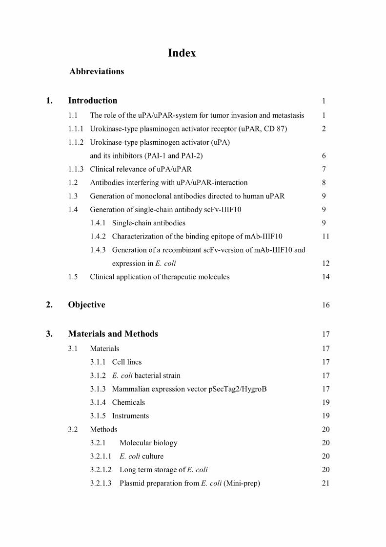
Index Abbreviations
1. Introduction 1
1.1 The role of the uPA/uPAR-system for tumor invasion and metastasis 1
1.1.1 Urokinase-type plasminogen activator receptor (uPAR, CD 87) 2
1.1.2 Urokinase-type plasminogen activator (uPA)
and its inhibitors (PAI-1 and PAI-2) 6
1.1.3 Clinical relevance of uPA/uPAR 7
1.2 Antibodies interfering with uPA/uPAR-interaction 8
1.3 Generation of monoclonal antibodies directed to human uPAR 9
1.4 Generation of single-chain antibody scFv-IIIF10 9
1.4.1 Single-chain antibodies 9
1.4.2 Characterization of the binding epitope of mAb-IIIF10 11
1.4.3 Generation of a recombinant scFv-version of mAb-IIIF10 and
expression in E. coli 12
1.5 Clinical application of therapeutic molecules 14
2. Objective 16
3. Materials and Methods 17
3.1 Materials 17
3.1.1 Cell lines 17
3.1.2 E. coli bacterial strain 17
3.1.3 Mammalian expression vector pSecTag2/HygroB 17
3.1.4 Chemicals 19
3.1.5 Instruments 19
3.2 Methods 20
3.2.1 Molecular biology 20
3.2.1.1 E. coli culture 20
3.2.1.2 Long term storage of E. coli 20
3.2.1.3 Plasmid preparation from E. coli (Mini-prep) 21
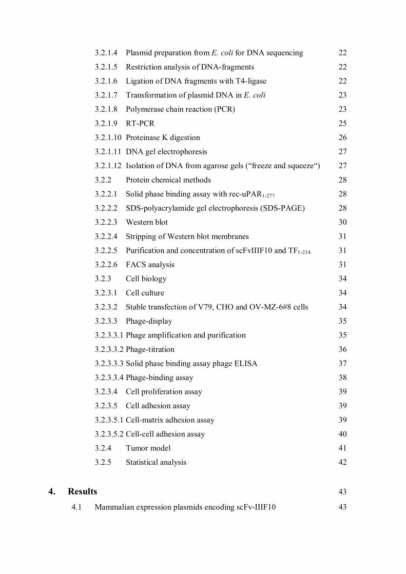
3.2.1.4 Plasmid preparation from E. coli for DNA sequencing 22
3.2.1.5 Restriction analysis of DNA-fragments 22
3.2.1.6 Ligation of DNA fragments with T4-ligase 22
3.2.1.7 Transformation of plasmid DNA in E. coli 23
3.2.1.8 Polymerase chain reaction (PCR) 23
3.2.1.9 RT-PCR 25
3.2.1.10 Proteinase K digestion 26
3.2.1.11 DNA gel electrophoresis 27
3.2.1.12 Isolation of DNA from agarose gels (�freeze and squeeze�) 27
3.2.2 Protein chemical methods 28
3.2.2.1 Solid phase binding assay with rec-uPAR1-277 28
3.2.2.2 SDS-polyacrylamide gel electrophoresis (SDS-PAGE) 28
3.2.2.3 Western blot 30
3.2.2.4 Stripping of Western blot membranes 31
3.2.2.5 Purification and concentration of scFvIIIF10 and TF1-214 31
3.2.2.6 FACS analysis 31
3.2.3 Cell biology 34
3.2.3.1 Cell culture 34
3.2.3.2 Stable transfection of V79, CHO and OV-MZ-6#8 cells 34
3.2.3.3 Phage-display 35
3.2.3.3.1 Phage amplification and purification 35
3.2.3.3.2 Phage-titration 36
3.2.3.3.3 Solid phase binding assay phage ELISA 37
3.2.3.3.4 Phage-binding assay 38
3.2.3.4 Cell proliferation assay 39
3.2.3.5 Cell adhesion assay 39
3.2.3.5.1 Cell-matrix adhesion assay 39
3.2.3.5.2 Cell-cell adhesion assay 40
3.2.4 Tumor model 41
3.2.5 Statistical analysis 42
4. Results 43
4.1 Mammalian expression plasmids encoding scFv-IIIF10 43
4.2 Generation of stable transfectants in eukaryotic cells 47
4.3 Purification and characterization of soluble scFv-IIIF10 and soluble
TF1-214 from eukaryotic cell culture supernatants 48
4.4 Detection of membrane anchored variants
of scFv-IIIF10 via M-13 phages 51
4.5 Interaction of membrane bound scFv-IIIF10 with human uPAR 52
4.6 Characterisation of the proliferation of OV-MZ-6#8 cells transfected
with soluble scFv-IIIF10 54
4.7 Determination of the adhesive capacities of the transfected
OV-MZ-6#8 cells to different ECM-Proteins 55
4.8 Effects of scFv-IIIF10 secretion on in vivo tumor growth of human
ovarian cancer cells 55
5. Discussion 57
5.1 scFv-IIIF10 as a therapeutic molecule 57
5.2 Limitations in the design and application of single chain fragments 58
5.3 Currently applied antibodies in clinical trials 60
5.4 Future prospects of antibody therapy 62
6. Summary 64
7. References 66
8. Curriculum vitae and publications 82
Abbreviations
Abbreviations
aa amino acid
Amp ampicillin
APS ammoniumperoxodisulfate
ATF aminoterminal fragment
bp base pair
BPB bromphenol-blue
BSA bovine serum albumine
CEA carcinoembryonic antigen
cDNA complementary desoxyribonucleic acid
CHO chinese hamster ovary
CMV Cytomegalovirus
DMEM Dulbecco´s modified Eagle´s medium
DMSO dimethylsulfoxide
DNA desoxyribonucleic acid
dNTP desoxyribonucleictriphosphate
E. coli Escherichia coli
e.g. exempli gratia (for example)
ECM extracellular matrix
EDTA ethylendiamin-tetra-acetic acid
EGFR epidermal growth factor receptor
ELISA enzyme linked immunosorbent assay
GFD growth factor-like domain
GPI glykosylphosphatidylinositol
FACS fluorescence activated cell sorting
FCS fetal calf serum
FDA Food and Drug Administration
FIGO Fédération Internationale de Gynécologie et d´Obstetrique
h hour
HEPES 2-{(4-(hydroxyethyl)-1-piperazin}ethansulfonic acid
HMW high molecular weight
HSV Herpes simplex virus
Abbreviations
kDa kilo dalton
KD dissociation´s constant
LB-medium Luria-Bertani-medium
LMW low molecular weight
mAb monoclonal antibody
min minute
MOPS 3-(N-morpholino)-propanesulfonic acid
MMP matrixmetalloproteinase
Ni-NTA nickel-nitrilotriacetic acid
ODx optical density at x nm
OS over all survival
p.a. per analysis
PAGE polyacrylamide gel electrophoresis
PAI plasminogen activator inhibitor
PBS phosphate buffered solution
P:C:I phenol:chloroform:isomylalcohol, 25:25:1
PCR polymerase chain reaction
PEG polyethyleneglycol
PMA phorbol-12-myristat-13-acetate
POX peroxidase labeled
PVDF polyvinylidenfluoride
RFS relapse free survival
rpm rounds per minute
RT room temperature
scFv single chain fragment
SDS sodium dodecyl sulfate
SDS-PAGE SDS-polyacrylamide gel electrophoresis
suPAR soluble urokinase-type plasminogen activator receptor
TBS tris buffered solution
TCD transmembrane domain
TEMED N,N,N`,N`-tetramethylethylendiamine
TMB 3,3´,5,5´-tetramethylbenzidine
TKI tyrosin kinase inhibitor

Abbreviations
tPA tissue type plasminogen activator
Tris N-[tris-(hydroxymethyl-)]aminomethane
U unit
uPA urokinase-type plasminogen activator
uPAR urokinase-type plasminogen activator receptor
o/n over night
wt wild type
amino acids
A Ala alanine M Met methionine
C Cys cysteine N Asn asparagine
D Asp aspartic acid P pro proline
E Glu glutamic acid Q Gln glutamine
F Phe phenylalanine R Arg arginine
G Gly glycine S Ser serine
H His histidine T Thr threonine
I Ile isoleucine V Val valine
K Lys lysine W Trp tryptophan
L Leu leucine Y Tyr tyrosine

1. Introduction
1
1. Introduction
1.1 The role of the uPA/uPAR-system for tumor invasion and metastasis
One of the principle properties of malignant cells, which distinguish them from normal or
benign cells, is their capability to cross tissue boundaries and to metastasize. Once detached
from the primary tumor, they are able to invade into the surrounding extracellular matrix
(ECM) and into blood or lymphatic vessels, followed by adhesion to and invasion through the
endothelium to finally re-implant at distant loci accompanied by neovascularization. The
degradation of the surrounding ECM is facilitated, when certain extracellular proteolytic
enzymes are present: matrix-metalloproteinases (MMPs), cysteine proteases (including
cathepsin B and L) and serine proteases such as plasmin and the urokinase-type plasminogen
activator (uPA) (overview in Andreasen et al., 1997; Danø et al., 1999; Reuning et al., 1998;
Schmitt et al,. 2000; Allgayer 2006).
The proteolytic urokinase-type plasminogen activator system encompasses the serine protease
urokinase-plasminogen activator (uPA), its receptor uPAR (CD 87) and its inhibitors PAI-1
and PAI-2 (Figure 1). In concert with other members of the serine protease family (plasmin,
tissue kallikreins, membrane type serine-proteases), matrix-metalloproteinases (MMPs) and
cysteine proteases, it mediates the pericellular proteolytic events leading to focal degradation
of the basement membrane and extracellular matrix in cancer growth, tumor cell invasion and
metastasis (Andreasen et al., 2000; Del Rosso et al., 2002; Ragno, 2006).
Binding of uPA to its tumor cell surface receptor uPAR converts the single polypeptide chain
plasminogen into its two-chain form plasmin and thereby not only focuses its plasminogen
activation function to the tumor cell, but also induces a cascade of other biological events
including cell proliferation, adhesion, migration, chemotaxis and angiogenesis (Rabbani and
Mazar, 2001; Blasi and Carmeliet, 2002; Reuning et al., 2003). This conversion can also be
catalyzed by tPA (tissue type plasminogen activator), an enzyme triggering the intravascular
fibrinolysis, and certain bacterial proteins (Andreasen et al., 1997). The proteolytic activity of
uPA is controlled by its inhibitors PAI-1 and PAI-2 (Blasi, 1997).
Due to the lack of a transmembrane domain, uPAR needs to functionally cooperate with other
transmembrane receptors in order to conduct intracellular signalling. A cross talk with the
adhesion and signalling receptors of the integrin superfamily has been reported (Chapman and
Wei, 2001). Integrins are transmembrane cell surface receptors which upon binding to ECM
1. Introduction
2
proteins exert regulatory functions in many processes such as cell adhesion, migration and
proliferation (Blasi and Carmeliet, 2002; Reuning et al., 2003). Recently it was reported, that
uPAR functionally interacts also with a G-protein coupled receptor involved in chemotaxis,
the high affinity receptor (FPR) for the fMet-Leu-Phe peptide (fMLP). fMLP is a peptide of
bacterial origin that is a strong leukocyte chemoattractant. fMLP-dependent cell migration
requires uPAR expression (Montuori et al., 2002; Le et al., 2002).
Upon binding, the enzymatically active uPA is focused to the cell surface resulting in a higher
state of uPA activity and a several fold enhanced rate of conversion of cell-surface associated
plasminogen to plasmin (Ellis et al., 1999). Plasminogen is a serineprotease present in plasma
and extracellular fluids with a high activity spectrum towards various extracellular matrix
components such as fibrin, fibronectin, laminin and collagen IV, thereby leading to ECM
degradation (Figure 1). In fact, in a variety of malignancies such as breast, ovarian,
esophageal, gastric, colorectal or hepatocellular cancer, a strong clinical value of the
plasminogen activation system in predicting relapse free and overall survival in cancer
patients has been demonstrated (Harbeck et al., 2002; Look et al., 2002).
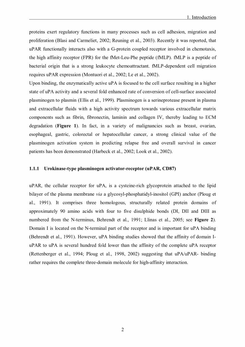
1.1.1 Urokinase-type plasminogen activator-receptor (uPAR, CD87)
uPAR, the cellular receptor for uPA, is a cysteine-rich glycoprotein attached to the lipid
bilayer of the plasma membrane via a glycosyl-phosphatidyl-inositol (GPI) anchor (Ploug et
al., 1991). It comprises three homologous, structurally related protein domains of
approximately 90 amino acids with four to five disulphide bonds (DI, DII and DIII as
numbered from the N-terminus, Behrendt et al., 1991; Llinas et al., 2005; see Figure 2).
Domain I is located on the N-terminal part of the receptor and is important for uPA binding
(Behrendt et al., 1991). However, uPA binding studies showed that the affinity of domain I-
uPAR to uPA is several hundred fold lower than the affinity of the complete uPA receptor
(Rettenberger et al., 1994; Ploug et al., 1998, 2002) suggesting that uPA/uPAR- binding
rather requires the complete three-domain molecule for high-affinity interaction.

1. Introduction
3
Figure 1. Schematic overview of the role of the uPA/uPAR-system in tumor invasion and metastasis. Binding of uPA to its tumor cell surface receptor uPAR converts the single polypeptide chain plasminogen into its two-chain form plasmin, thereby leading to degradation of the ECM, but also inducing a cascade of other biological events including cell adhesion, invasion, migration and proliferation. The proteolytic activity of uPA is controlled by its inhibitors PAI-1 and PAI-2. Using integrins as co-receptors, the uPA/uPAR-system is able to conduct intracellular signalling.
Cell membrane
DII
DIII
DI
uPAR
uPA
PAI-1
PAI-2
Plasmin
Plasminogen
ECM(Vitronectin)
α β
Integrin
Cell adhesionCell invasionCell migrationCell proliferation
Cellsig
nalling
ECM(Vitronectin)
Cell membrane
DII
DIII
DI
uPAR
uPA
PAI-1
PAI-2
Plasmin
Plasminogen
ECM(Vitronectin)
α β
Integrin
Cell adhesionCell invasionCell migrationCell proliferation
Cellsig
nalling
ECM(Vitronectin)
1. Introduction
4
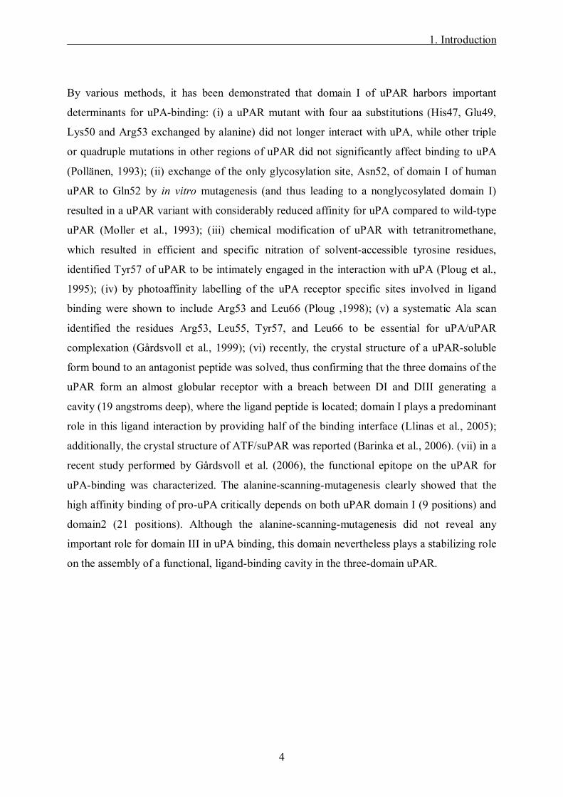
By various methods, it has been demonstrated that domain I of uPAR harbors important
determinants for uPA-binding: (i) a uPAR mutant with four aa substitutions (His47, Glu49,
Lys50 and Arg53 exchanged by alanine) did not longer interact with uPA, while other triple
or quadruple mutations in other regions of uPAR did not significantly affect binding to uPA
(Pollänen, 1993); (ii) exchange of the only glycosylation site, Asn52, of domain I of human
uPAR to Gln52 by in vitro mutagenesis (and thus leading to a nonglycosylated domain I)
resulted in a uPAR variant with considerably reduced affinity for uPA compared to wild-type
uPAR (Moller et al., 1993); (iii) chemical modification of uPAR with tetranitromethane,
which resulted in efficient and specific nitration of solvent-accessible tyrosine residues,
identified Tyr57 of uPAR to be intimately engaged in the interaction with uPA (Ploug et al.,
1995); (iv) by photoaffinity labelling of the uPA receptor specific sites involved in ligand
binding were shown to include Arg53 and Leu66 (Ploug ,1998); (v) a systematic Ala scan
identified the residues Arg53, Leu55, Tyr57, and Leu66 to be essential for uPA/uPAR
complexation (Gårdsvoll et al., 1999); (vi) recently, the crystal structure of a uPAR-soluble
form bound to an antagonist peptide was solved, thus confirming that the three domains of the
uPAR form an almost globular receptor with a breach between DI and DIII generating a
cavity (19 angstroms deep), where the ligand peptide is located; domain I plays a predominant
role in this ligand interaction by providing half of the binding interface (Llinas et al., 2005);
additionally, the crystal structure of ATF/suPAR was reported (Barinka et al., 2006). (vii) in a
recent study performed by Gårdsvoll et al. (2006), the functional epitope on the uPAR for
uPA-binding was characterized. The alanine-scanning-mutagenesis clearly showed that the
high affinity binding of pro-uPA critically depends on both uPAR domain I (9 positions) and
domain2 (21 positions). Although the alanine-scanning-mutagenesis did not reveal any
important role for domain III in uPA binding, this domain nevertheless plays a stabilizing role
on the assembly of a functional, ligand-binding cavity in the three-domain uPAR.

1. Introduction
5
Figure 2. The domain structure of the uPA receptor. uPAR consists of three structurally homologous domains and is linked to the cell surface via a C-terminal glycan lipid GPI-anchor (modified according to Ploug et al. 1994). Glycosylation sites (52NRT, 162NDT, 172NTT, 200NST; Ploug et al., 1998) are indicated by a rhombus, disulfide bonds are depicted in black. The epitope of mAb IIIF10 (aa 52-60 of uPAR) in the N-terminal domain I, which harbors main determinants for uPA binding, is indicated in grey. mAb IIIF10 binds with high affinity to both glycosylated and non-glycosylated uPAR (Luther et al., 1997).
Besides its proteolytic function, the uPA/uPAR-system has also mitogenic and chemotactic
properties. The three domains of the uPAR are joined by linker sequences, the linker region
connecting domains I and II exhibits an extreme proteolytic sensitvity and can be cleaved by
several proteolytic enzymes. Such cleaved forms (c-uPAR) lacking domain I have been
detected on the surface of different cell lines in normal and cancer tissues. An epitope has
been identified residing within the peptide region connecting DI and DII, which upon
exposure to proteolytic cleavage mimicks uPA/uPAR-mediated chemotactic activity (Andolfo
et al., 2002; Fazioli et al., 1997). Both, full length and cleaved uPAR can be shed, thus
generating soluble uPAR forms (suPAR and c-suPAR respectively). Soluble uPAR forms are
found in biological fluids in vitro and in vivo. Such variants may arise by differential splicing
or phospholipase C cleavage of the GPI-anchor (Høyer-Hansen et al., 1992; Montuori et al.,
2002, 2005).

1. Introduction
6
Moreover, uPA/uPAR-system shows cell adhesive capacity by the ability of uPAR and PAI-1
to bind to the ECM protein vitronectin. Domain II and III have been reported to bind to
vitronectin, an ECM-protein with high affinity (Waltz and Chapman, 1994).
Due to the lack of a transmembrane domain, uPAR cooperates with other transmembrane
receptors in order to conduct intracellular signalling. Hereby, the adhesion and signalling
receptors of the integrin superfamily seem to play an important role (Chapman et al., 2001).
uPAR has been reported to be able to associate with ß1-integrins as immunoprecipitates with
anti-uPAR antibodies (Wei et al., 1996). Recently it was reported, that uPAR functionally
interacts also with a G-protein coupled receptor involved in chemotaxis, the high affinity
receptor (FPR) for the fMet-Leu-Phe peptide (fMLP). fMLP is a peptide of bacterial origin
that is a strong leukocyte chemoattractant. fMLP-dependent cell migration requires uPAR
expression (Montuori et al., 2002; Le et al., 2002).
1.1.2 Urokinase type plasminogen activator (uPA) and its inhibitors (PAI-1 and PAI-2)
There are two types of plasminogen activators, the urokinase-type (uPA) and the tissue-type
(tPA). Both are capable of activating the inactive zymogen plasminogen to the active
proteinase plasmin, which can degrade extracellular matrix proteins. tPA is synthesized in
endothelial cells and plays a primary role in intravascular fibrinolysis.
uPA is a 55 kDa serine protease which is produced by various normal and cancer cells as an
inactive single-chain protein. Pro-uPA, the zymogen of uPA has a several hundred fold lower
activity than the activated two-chain uPA (Andreasen et al., 1997).
uPA consists of two disulfide bridge-linked polypeptide chains, the C-terminal serine protease
domain (B-chain) with its catalytic site, and the A-chain. The A-chain, so called
aminoterminal fragment (ATF), consists of two domains, a �growth factor-like domain� (aa 1-
46) harboring the binding site for uPAR (Appella et al., 1987) and a kringle domain (aa 47-
135), which has structural similarities to other protein domains like tPA, plasmin and
thrombin and is able to bind to uPAR.
The action of uPA on plasminogen is controlled by the inhibitors PAI-1 and PAI-2, PAI-1
being the most efficient inhibitor. Alternatively, when PAI-1 binds to uPAR-bound uPA, a
complex is formed with α2-macroglobulin/LDL-receptor-related protein (LRP), a
multifunctional transmembrane receptor, and is then rapidly endocytosed (Cubellis et al.,
1. Introduction
7
1990). Upon internalization the complex is then degraded and uPAR recycled to the cell
surface (Nykjaer et al., 1997).
1.1.3 Clinical relevance of uPA/uPAR
As early as 1988, elevated uPA levels in primary breast tumor tissue were shown to be
associated with a highly invasive phenotype and poor prognosis (Duffy et al., 1988). Jänicke
et al. (1991; 2001) were the first to describe the prognostic significance of PAI-1 in breast
cancer patients. High PAI-1 level as determined by ELISA was shown to be an independent
and significant predictor of poor prognosis.
Harbeck et al. (2002) demonstrated in a multivariate prospective analysis of 3424 primary
breast cancer patients that uPA and PAI-1 have not only a clinically relevant prognostic but
also predictive impact in primary breast cancer. This paper provides additional evidence
supporting the use of uPA/PAI-1 in the clinic by demonstrating how effects of adjuvant
systemic therapy differ in patients with high uPA/PAI-1 levels. Node-negative patients with
low uPA/PAI-1 may even be candidates for being spared the burden of adjuvant
chemotherapy. Similar findings were observed in a pooled analysis of prognostic impact of
uPA and PAI-1 in 8377 breast cancer patients (Look et al., 2002). Apart from lymph node
status, high levels of uPA and PAI-1 were the strongest predictors of both poor relapse free
survival and poor overall survival in the analysis of all patients. For lymph node-negative
breast cancer, uPA and PAI-1 measurements in primary tumors may be especially useful for
designing individualized treatment strategies.
Leissner et al. (2006) observed that high PAI-1 mRNA expression represents a strong and
independent unfavourable prognostic factor for the development of metastases and for breast
cancer specific survival in lymph node- and hormone receptor-positive breast cancer patients,
whereas uPA mRNA levels did not demonstrate significant independent prognostic value,
suggesting that PAI-1 is a stronger prognostic factor than uPA.
Elevated tumor antigen levels of uPA, PAI-1 and uPAR are associated with poor disease
outcome, high tumor grade and are conductive to tumor cell spread and metastasis (Schmitt et
al., 1997; Reuning et al., 1998; Duffy, 2002; Harbeck et al., 2002). This strong correlation
between elevated uPA, uPAR or PAI-1 values on one hand and cancer spread on the other
made the uPA system serve as a novel target for the development of new tumor biology-based
therapeutics, which either inhibit the enzymatic activity of uPA, reduce the expression of the

1. Introduction
8
components of the uPA system or block binding of uPA to uPAR (Schmitt et al., 2000; Sperl
et al., 2001; Muehlenweg et al., 2001; Reuning et al., 2003).
As tumor metastasis is one of the crucial mechanisms in patients suffering from certain
tumors, new therapeutic strategies are in development to inhibit tumor cells from invading the
ECM and metastasize.
1.2 Antibodies interfering with uPA/uPAR-interaction
A strategy to interfere with the uPA/uPAR-interaction is the use of specific blocking
antibodies directed to either uPA or uPAR. In fact, it has e.g. been demonstrated in in vitro
studies that antibodies which inhibit binding of uPA to uPAR, (i) distinctly reduced tumor cell
surface-associated plasminogen activation (Magdolen et al., 2001), (ii) inhibited uPA-
mediated stimulation of proliferation of ovarian cancer cells (Fischer et al., 1998), (iii)
disrupted the uPA-mediated activation of the ERK signalling pathway and promoted
apoptosis in breast cancer cells (Ma et al., 2001) and (iv) significantly inhibited the formation
of new micro-vascular structures in fibrin matrices by human microvascular endothelial cells
(Kroon et al., 1999). In a proof of principle-experiment, the anti-metastatic efficacy of an
antibody directed to the uPA binding domain of rat uPAR was evaluated in a syngeneic model
of rat breast cancer (Rabbani and Gladu, 2002). For this, rat breast cancer cells, which
overexpressed uPAR upon stable transfection, were inoculated into the mammary fat pad of
syngeneic female Fischer rats. Subsequently, the antibodies were topically and daily applied
for one week. The animals displayed a marked decrease in tumor growth and a significant
inhibition of metastasis to retroperitoneal and mesenteric lymph nodes as well as an obvious
delay of metastasis to lung, liver and spleen, respectively, when compared to control tumor-
bearing animals receiving the same dose of pre-immune rabbit IgG.
Bauer et al. (2005) evaluated the effect of anti-uPAR monoclonal antibodies with and without
chemotherapy on primary tumor growth, retroperitoneal invasion and hepatic metastasis in
vivo. Human pancreatic carcinoma cells were injected into the pancreatic tail of nude mice. It
was demonstrated that mice systemically treated with a combination of gemcitabine and anti-
uPAR mAb led to about 92% tumor reduction compared to the control or either agent alone
the tumor capsule remaining intact.
1. Introduction
9
1.3 Generation of monoclonal antibodies directed to human uPAR
The Clinical Research Group of the Women�s Hospital of the Technical University in Munich
together with the Institute of Pathology of the TU in Dresden have generated a series of
monoclonal antibodies (mAbs) directed against uPAR by using non-glycosylated,
recombinant human uPAR (spanning aa 1-284) expressed in E. coli as the immunogen
(Luther et al., 1997). By flow cytofluorometrical analysis, some of these mAbs (3/12) were
shown to bind to native human uPAR present on the cell surface of monocytoid U937 cells.
Interestingly, one of these mAbs, IIIF10, efficiently reduced binding of uPA to uPAR,
indicating that the epitope detected by mAb IIIF10 is located within or close to the uPA-
binding site of uPAR. Subsequent epitope mapping with overlapping synthetic peptides
(Luther et al., 1997) revealed that the epitope covers the linear sequence of amino acids (aa)
52-60 of the N-terminal domain I of uPAR (Figure 2 and 4).
The inhibitory properties of mAb IIIF10, together with the other findings described above,
strongly suggest that the mAb IIIF10-epitope, aa 52-60 of human uPAR, is located at the
uPA-binding site of uPAR.
1.4 Generation of single-chain antibody scFv-IIIF10
1.4.1 Single-chain antibodies
Although mAbs display high specificity and in vivo stability, clinical application as
therapeutic molecules, especially against solid tumors, has been rather unsuccessful which is
in part due to the inability of the mAbs to penetrate into the tumor (Reff and Heard, 2001).
Single chain antibodies (scFv), i.e. fusion proteins consisting of the antibody�s variable heavy
(VH) and light (VL) chain connected via a flexible linker, represent novel powerful agents for
the achievement of targeted therapy, since they are much smaller in size and, thus, more likely
to penetrate into the tumor mass (Figure 3).

1. Introduction
10
Figure 3: Structure of a human antibody and antibody fragments. (A) A human IgG antibody consists of two heavy and two light chains, each of the polypeptide chains bearing variable regions (VH = variable region of heavy chain, VL = variable region of light chain) being responsible for antigen binding and constant regions (CH = constant region of heavy chain, CL = constant region of light chain) being responsible for the biologic function. The combination of these chains and the amino acid sequences in addition to the six complementary determining regions (CDR) determine the antigen binding activity for a single antibody. (B) An antibody may be degraded by proteolytic enzymes into two distinct components, the Fab fragment (= fragment antigen binding) with the antigen binding site and the Fc fragment (= fragment crystallisable) which is responsible for cell attachment. A Fab fragment consists of the heavy and light chains with the antigen binding site. The two chains are held together by interaction of the CL and CH1 domains. (C) The smallest antibody component that has been generated is the single chain fragment (scFv). It consists of the variable regions only (VH and VL = fragment variable = Fv) connected via a polypeptide linker. Antigen binding pockets are indicated by an arrow (→).
Sanz et al. (2002) demonstrated as a proof of principle a direct in vivo therapeutic effect of an
anti-laminin scFv derived from a human phage-display library. This scFv inhibits
angiogenesis in the chick embryo chorioallantoic membrane assay and prevents the
establishment and growth of subcutaneous tumors in mice either when administered as bolus
protein therapy or when produced locally by gene-modified mammalian tumor cells.
BA
VL
CL
CH2
CH1
CH3
Fc
VH VL
CLCH1
C
Fab
Fv
VH
VH VL
CDR
BA
VL
CL
CH2
CH1
CH3
FcFc
VH VL
CLCH1
C
Fab
Fv
Fab
Fv
VH
VH VLVH VL
CDR

1. Introduction
11
Furthermore, modification of the scFv, e.g. fusion with additional effector functions such as a
prodrug converting enzyme, an antiangiogenic or thrombogenic factor (Helfrich et al., 2000)
or a toxin (Fan et al., 2002), can easily be achieved by recombinant technologies. scFv-
targeting of molecular processes associated with malignancies may even be utilized to
enhance the effects of conventional therapeutics such as chemotherapy and radiation or to
modulate immune response (Leath et al., 2004). 5T4 positive leukemia cells were successfully
targeted with a fusion protein consisting of an anti-5T4-scFv and human IgG1 Fc domain.
This strategy bound 5T4 positive tumor cells and provoked an antibody-dependent cell
cytotoxic immune response against the malignant cells (Myers et al., 2002).
Finally, efficient gene therapeutic approaches can be envisioned, because in vivo expression
of a therapeutic scFv molecule is much more efficient as compared to the synthesis and
correct assembly of a heteromeric mAb (Vitaliti et al., 2000). In recent years, an increasing
number of reports has in fact demonstrated that scFvs directed to various tumor-associated
target molecules (e.g. VEGF, laminin, erbB2 or mesothelin) are powerful tools to interfere
with tumor growth or block vascularization either when administered as bolus protein therapy
or when produced locally by gene-modified tumor cells (Vitaliti et al., 2000; Sanz et al.,
2002; Arafat et al., 2002; Fan et al., 2002). Also, members of tumor-associated proteolytic
systems such as cathepsin L or membrane-type serine protease 1 (MT-SP1) have been
selected as targets for the development of scFv-based therapeutic molecules (Guillaume-
Rousselet et al., 2002; Sun et al., 2003).
1.4.2 Characterization of the binding epitope of mAb IIIF10 by employing phage
based random peptide libraries
In initial experiments, which are part of another study (Kirschenhofer et al., 2003), the
binding epitope of mAb IIIF10 was characterized by employing phage-based random peptide
libraries with repertoires of hundreds of millions of unique peptide sequences (Smith and
Scott, 1993) to select mAb IIIF10 binding peptides. In a process known as biopanning, mAb
IIIF10 was incubated in solution with an aliquot of the peptide-displaying phage libraries,
antibody-bound phages were eluted, amplified and again selected for mAb IIIF10 binding.
Finally, the peptide-encoding DNA of individual mAb IIIF10-binding phages was sequenced
and the corresponding amino acid sequences were deduced. As depicted in Figure 4, amino
acids present in both phage and uPAR sequences in identical positions confirm the mAb
1. Introduction
12
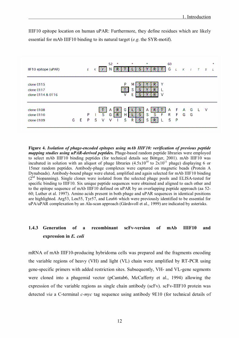
IIIF10 epitope location on human uPAR: Furthermore, they define residues which are likely
essential for mAb IIIF10 binding to its natural target (e.g. the SYR-motif).
Figure 4. Isolation of phage-encoded epitopes using mAb IIIF10: verification of previous peptide mapping studies using uPAR-derived peptides. Phage-based random peptide libraries were employed to select mAb IIIF10 binding peptides (for technical details see Böttger, 2001). mAb IIIF10 was incubated in solution with an aliquot of phage libraries (4.5x10¹⁰ to 2x10¹¹ phage) displaying 6 or 15mer random peptides. Antibody-phage complexes were captured on magnetic beads (Protein A Dynabeads). Antibody-bound phage were eluted, amplified and again selected for mAb IIIF10 binding (2nd biopanning). Single clones were isolated from the selected phage pools and ELISA-tested for specific binding to IIIF10. Six unique peptide sequences were obtained and aligned to each other and to the epitope sequence of mAb IIIF10 defined on uPAR by an overlapping peptide approach (aa 52-60; Luther et al. 1997). Amino acids present in both phage and uPAR sequences in identical positions are highlighted. Arg53, Leu55, Tyr57, and Leu66 which were previously identified to be essential for uPA/uPAR complexation by an Ala-scan approach (Gårdsvoll et al., 1999) are indicated by asterisks.
1.4.3 Generation of a recombinant scFv-version of mAb IIIF10 and
expression in E. coli
mRNA of mAb IIIF10-producing hybridoma cells was prepared and the fragments encoding
the variable regions of heavy (VH) and light (VL) chain were amplified by RT-PCR using
gene-specific primers with added restriction sites. Subsequently, VH- and VL-gene segments
were cloned into a phagemid vector (pCantab6, McCafferty et al., 1994) allowing the
expression of the variable regions as single chain antibody (scFv). scFv-IIIF10 protein was
detected via a C-terminal c-myc tag sequence using antibody 9E10 (for technical details of
1. Introduction
13
scFv production, secretion and detection see: Kay et al., 1996). To evaluate the binding
specificity of scFv-IIIF10, peptides were employed which had been used to map the epitope
of mAb IIIF10 on human uPAR (Luther et al., 1997). It was shown that only a peptide whose
sequence comprises the complete IIIF10 epitope on uPAR (51-65) is able to prevent both
scFv-IIIF10 and mAb IIIF10 from binding to recombinant uPAR. Another peptide with an
incomplete epitope sequence (encompassing aa 48-59 of uPAR) is more than 100 times less
efficient. Neither of the peptides block binding of control scFv (Z5) to its target protein
(unrelated to uPAR, data not shown here).
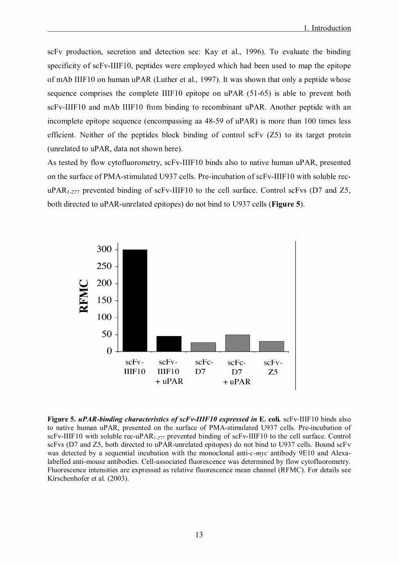
As tested by flow cytofluorometry, scFv-IIIF10 binds also to native human uPAR, presented
on the surface of PMA-stimulated U937 cells. Pre-incubation of scFv-IIIF10 with soluble rec-
uPAR1-277 prevented binding of scFv-IIIF10 to the cell surface. Control scFvs (D7 and Z5,
both directed to uPAR-unrelated epitopes) do not bind to U937 cells (Figure 5).
Figure 5. uPAR-binding characteristics of scFv-IIIF10 expressed in E. coli. scFv-IIIF10 binds also to native human uPAR, presented on the surface of PMA-stimulated U937 cells. Pre-incubation of scFv-IIIF10 with soluble rec-uPAR1-277 prevented binding of scFv-IIIF10 to the cell surface. Control scFvs (D7 and Z5, both directed to uPAR-unrelated epitopes) do not bind to U937 cells. Bound scFv was detected by a sequential incubation with the monoclonal anti-c-myc antibody 9E10 and Alexa-labelled anti-mouse antibodies. Cell-associated fluorescence was determined by flow cytofluorometry. Fluorescence intensities are expressed as relative fluorescence mean channel (RFMC). For details see Kirschenhofer et al. (2003).

1. Introduction
14
1.5 Clinical application of therapeutic molecules
In the last few years, biotechnology and drug development has made great progress.
Antibodies or antibody fragments (i.e. Fab-fragments or scFvs) in tumor therapy have entered
clinical trials and some are already commonly used in clinical settings.
Trastuzumab (Herceptin®) is a humanized mAb recognizing an epitope on the extracellular
domain of HER-2/neu (c-erbB-2), a cell-surface protein from the EGFR (epidermal growth
factor receptor) family overexpressed in ca. 25% of primary breast cancer patients.
Herceptin® is used as a common standard therapy either alone or in combination with
chemotherapy in the treatment of women with metastatic breast cancer and HER-2/neu
overexpression (Adams et al., 2005).
For radioimmune-guided surgery a radio-labeled anti-CEA scFv has been developed. CEA
(carcino-embryonic antigen) is a well characterized tumor-associated glycoprotein expressed
on endodermally derived gastrointestinal-tract neoplasms and other adenocarcinomas.
Injected intravenously before surgery, the radio-labeled anti-CEA scFv locates the tumor
tissue in the operative field (Mayer et al., 2000). Some scFvs are currently being examined in
clinical trials. In an ongoing phase I trial, 15 patients suffering from recurrent intra-abdominal
ovarian or extra-ovarian adenocarcinoma that had failed standard therapy were treated with an
intraperitoneally administered adenovirus encoding a scFv against erbB-2. Based on the
hypothesis that the intracellular expressed anti-erB-2 scFv prevents erbB-2 mediated signal
transduction and induces apoptosis, 38% of the patients had stable disease. Acceptable
toxicity was noted with no vector related toxicity experienced (Alvarez et al., 2000). Oh et al.
(2004) evaluated the tumor targeting properties of L19, a dimeric scFv2-molecule. 123I-
conjugated dimeric L19 selectively localized lung, colorectal or brain carcinomas in a phase I
trial.
As antibody therapy often bears the problem of severe side effects, i.e. the development of a
human anti-mouse immunoglobulin antibody response (HAMA) or human anti-chimeric
antibody response (HACA), other niches of new therapeutic drugs and protein engineering are
currently explored.
Novel biopharmaceuticals for the treatment of cancer such as small therapeutic molecules
have been developed and are now tested in several clinical trials. For example, gefitinib
(Iressa®, ZD 1839) is a low molecular weight, synthetic aniline-quinazoline. It is a
competitive inhibitor of the intracellular tyrosine kinase of the EGFR receptor. In phase I
1. Introduction
15
trials, oral application of gefitinib is well tolerated and active in patients with non-small-cell
lung cancer (NSCLC) and other solid tumors (Herbst et al., 2002). Erlotinib (Tarceva®, OSI-
774), is also a quinazoline-based agent which inhibits the intracellular tyrosine kinase of the
EGFR receptor. It shows promising results in phase I trials in patients with NSCLC (Hidalgo
et al., 2001). For both therapeutic molecules several phase III trial are ongoing comparing
their application in combination with chemotherapy (Martin et al., 2006).
Lapatinib (Tycerb®) is an orally-active tyrosine kinase inhibitor (TKI) that targets both c-
erbB-receptors (c-erbB-1 and -2). Its dual mode of action distinguishes it from existing TKIs
such as gefitinib and trastuzumab, which are single EGFR and HER2/neu receptor inhibitors
respectively. It is hoped, that dual TKIs may help to address the problem of drug resistance
that can arise following treatment with single receptor inhibitors. There is an actual ongoing
clinical phase III trial (so called TEACH) evaluating and comparing the efficacy and safety of
lapatinib versus placebo in women with early-stage erbB2-overexpressing breast cancer who
have completed their primary adjuvant chemotherapy and have no clinical or radiographic
evidence of disease (Moy and Goss, 2006). Additionally, in a phase III trial the efficacy and
safety of sunitinib (Sutent®), a TKI which inhibits several tyrosin kinase receptors, was
reported in patients with gastrointestinal stroma tumor after failure of imatinib, a selective
TKI (Demetri et al., 2006).
As promising drug candidates not only for cancer therapy, lipocalins have recently come in
the center of attention. Lipocalins represent a family of functional diverse, small proteins
comprising 160-180 aa residues and have naturally important biological functions in a variety
of organisms (from bacteria to humans) such as storage and transport of vitamins, steroids or
metabolic products. Because lipocalins comprise only a single, small polypeptide chain that
exhibits a simple set of four hypervariable loops, this protein family provides several benefits
for applications in biotechnology and medicine, e.g as storage proteins, carrier vehicles for
pharmaceutical compounds or as therapeutic drugs (Schlehuber and Skerra, 2005).
In the last few years, lipocalins have been recruited as a scaffold for the design of a new class
of engineered binding proteins with antibody-like ligand-binding function termed �anticalins�
(Skerra et al., 2001). Similar to the antibody-antigen-interaction, the mechanism of the
anticalin-complex formation with low molecular weight ligands gives way for the generation
of novel binding proteins with high affinity and specificity. Anticalins that specifically
recognize a tumor surface marker could be useful for drug targeting approaches (Schlehuber
and Skerra, 2005).
2. Objective
16
2. Objective
Human uPAR, which focuses uPA to the tumor cell surface, thereby leading to extracellular
matrix degradation and promoting metastasis of tumor cells, represents an attractive target for
tumor therapy. Thus, the aim of the present study was to employ a mAb IIIF10-derived
genetically engineered scFv to target the uPAR-uPA interaction. For this (i) plasmids should
be constructed for the eukaryotic expression of mAb IIIF10-derived single chain antibody
(scFv-IIIF10) in a soluble, secreted form or membrane-bound form, (ii) the binding properties
of the scFv-IIIF10 should be analyzed in vitro, (iii) human ovarian cancer cells (OV-MZ-6#8)
should be stably transfected with scFv-IIIF10-encoding expression plasmids and analysed for
the effect on primary tumor growth and spread in a xenograft nude mouse model in
comparison to the vector-transfected control.
3. Materials and Methods
17
3. Materials and Methods
3.1 Materials
3.1.1 Cell lines
The ovarian cancer cell line OV-MZ-6 was established from ascites of a patient with a serous-
papillary ovarian carcinoma FIGO IV (Möbus et al., 1992). This cell line was subcloned in
the clinical research unit of the �Frauenklinik der TU München�, characterized and further
cultured as OV-MZ-6#8 (Fischer et al., 1998; Lutz et al., 2001).
The lymphoma cell line U937 was established from the pleural effusion of a patient with
diffuse lymphoma (ATTC, Rockland, USA).
The chinese hamster ovary cells (CHO-K1) and chinese hamster lung fibroblasts (V79) were
purchased from ATTC, Rockland, USA.
3.1.2 E. coli bacterial strain
The E. coli strain XL1-Blue and K91 (Stratagene, Heidelberg) are facultative anaerob, gram-
negative rod shaped bacterial cells which have been used for cloning experiments.
3.1.3 Mammalian expression vector pSecTag2/HygroB
The vector pSecTag2/HygroB (Invitrogen) is a fusion vector encompassing 5749 base pairs
(Figure 6). Expression is driven by the strong human Cytomegalovirus (CMV) immediate-
early promoter/enhancer, secretion is supported by the murine Ig κ-chain V-J2-C signal
peptide (Boshart et al., 1985; Coloma et al., 1992). A polyadenylation signal and transcription
termination sequences of the bovine growth hormone stabilize mRNA. The ampicillin
resistance gene and hygromycin-B resistance gene ensure selection (Gritz et al., 1983).

3. Materials and Methods
18
Figure 6: Plasmid graphic map of pSecTag2/HygroB. The vector pSecTag2/HygroB was used as an expression plasmid to express scFv-IIIF10 in different eukaryotic cell lines: Promoter sequence of Cytomegalovirus (CMV), secretion is supported by the murine Ig κ-chain V-J2-C signal peptide, c-myc epitope and histidine tag easy protein detection. The ampicillin resistance gene and hygromycin-B resistance gene ensure selection.

3. Materials and Methods
19
3.1.4 Chemicals
The most chemicals used were from Sigma, Munich Germany or Merck, Darmstadt,
Germany.
3.1.5 Instruments
autoclave H+P Labortechnik GmbH, Oberschleißheim
centrifuges Biofuge Fresco and Varifuge RF,
Heraeus Instruments, Osterode; Centrifuge 5402,
Eppendorf GmbH, Hamburg
electrophoresis chambers Biorad, München
ELISA-reader Titertek Multiscan MCC/340, Labsystems,
Finnland
FACScalibur sort cytofluorometer Becton Dickinson, USA
incubator RFI-100, Infors HAT Technik, Einsbach
photometer Ultrospec Plus Spectrophotometer, Pharmacia,
Freiburg
pH meter Knick electronic measuring instruments, Berlin
drying cubicle WTC, Binder, Tuttlingen
scales BP 1200, Sartorius, Göttingen
Sartorius Basic, Sartorius, Göttingen
semi dry blotting chamber Fast Blott, Biometra, Göttingen
thermocycler Perkin-Elmer, Langen
water treatment plant Purelab Plus UF, USF, Rambach
vortexer Vortex Genie 2, Bender&Hobein AG, Basel,
Switzerland
3. Materials and Methods
20
3.2 Methods
3.2.1 Molecular Biology
3.2.1.1 E. coli culture
For generation of eukaryotic expression plasmids E. coli bacteria type XL-blue (Stratagene,
Heidelberg) were used.
E. coli is a facultative anaerob, gram-negative rod shaped bacterium. It is cultivated in full
medium on LB-agar plates or in LB-liquid medium, containing mineral- and ammonium salts,
carbohydrates and sugars. E. coli XL-blue cells display a generation time of 20 min under
optimal conditions and can grow up to 3x10⁹ cells/ml media at a temperature of 37°C.
In most cases, the antibiotic ampicillin is added at a concentration of 100 µg/ml LB-medium
for selection of the plasmid. The culture is incubated in sterile polystyrol tubes (Greiner,
Frickenhausen) at 37°C over night under 220 rpm rotation.
To isolate bacterial clones transformed with the DNA of interest, cultured bacteria are spread
on LB-agar plates containing 100 µg ampicillin/ml medium and incubated over night at 37°C.
The next day, single colonies are selected with sterile pipette tips and incubated again in LB
medium. For further control of positive clones, the probes are subjected to DNA-preparation
and gel electrophoresis.
All culture media, reaction tubes and materials are autoclaved and heat-sensitive substances
are sterile filtered.
LB-medium: 10 g/l Trypton, pH 7,0 5 g/l Bacto Yeast Extract 10 g/l NaCl in 1 l aqua dest. for agar plates add 1,5% agar
3.2.1.2 Long term storage of E. coli
For long term storage, 1.5 ml of a stationary bacterial culture is centrifuged in an Eppendorf
tube (13,000 rpm, 30 sec, RT), re-suspended in 15% glycerine and shock frozen in an ethanol
bath, then stored at �80°C.

3. Materials and Methods
21
3.2.1.3 Plasmid preparation from E. coli (Mini-prep)
For isolation of recombinant plasmid-DNA from E. coli, 5 ml of bacterial culture are grown
over night (37°C, 220 rpm). The culture is then filled in 1.5 ml Eppendorf tubes and
centrifuged at 13,000 rpm for 20 sec. 100 µl Lysis-buffer is added and vortexed for 30 sec at
maximum speed, then incubated 10 min at RT. 200 µl of freshly prepared NaOH/SDS
solution is added, carefully mixed and incubated at RT for 5 min. The solution turns clear
now. 150 µl of cooled KAc-solution is added and incubated on ice for 30 min. The solution
precipitates now. After 15 min centrifugation at 4°C 13,000 rpm, the supernatant is collected
and 400 µl phenol-chlorophorme is added (P:C:I-extraction), vortexed shortly and centrifuged
again at 13,000 rpm at RT. The upper phase is collected and mixed well with 800 µl cooled
EtOHabs. (ethyl alcohol precipitation). After incubation at RT exactly for 5 min, the solution is
centrifuged at 13,000 rpm at RT for 15 min. The supernatant is discarded and the pellet
containing the isolated plasmid DNA is dried in a SpeedVac for 5 min. The pellet is then re-
suspended in 90 µl TE and 10 µl RNAse solution (1 mg/ml) and incubated at 37°C for 15
min. 40 µl 4 M LiCl and 260 µl H2Obidest are added and P:C:I-extraction with 400 µl P:C:I is
performed. The upper phase is well mixed with 800 µl EtOH abs, incubated on ice for 5 min
and centrifuged 15 min at 13,000 rpm at 4°C. The supernatant is discarded and the pellet dried
in a SpeedVac for 5 min. For further analysis the pellet is re-suspended in 30 µl H2Obidest.
Lysis buffer: 50 mM Glucose (0.9 g/100 ml) 25 mM Tris-Cl, pH 8.0 10 mM EDTA, pH 8.0
NaOH/SDS-solution: 1% SDS 0.2 N NaOH
KAc-solution: 3 M potassium acetate, pH 4.8
RNAse-solution: 1 mg/ml in 50% glycerine 1:10 diluted in TE buffer (TE: 10 mM Tris-Cl, pH 8.0 and 1 mM EDTA, pH 8.0)
P:C:I phenol:chlorophorm:isomylalcohol, 25:25:1
3. Materials and Methods
22
3.2.1.4 Plasmid preparation from E. coli for DNA-sequencing
For sequencing, DNA was purified with the High Pure Plasmid Isolation Kit (Quiagen,
Hilden) according to the manufacturer�s manual. All sequencing was performed by Toplab in
Martinsried (Munich, Germany).
3.2.1.5 Restriction analysis of DNA-fragments
Restriction analysis is a central method in cloning experiments to produce specific DNA-
fragments of interest depending on the endonuclease being used. Restriction endonucleases
can hydrolyse DNA specifically under optimal buffer conditions, which are given by
commercial companies.
A restriction analysis is typically performed in a sterile Eppendorf tube with a reaction
volume between 10 µl and 100 µl. 1/10 Vol. 10x reaction buffer and DNA are added and
filled up to the end-volume with H2Obidest. 2 to 54 units of endonuclease per µg of DNA are
used. The added solution should not exceed 10% of the end volume, as the glycerine of the
storing buffer can disturb the reaction. Then the reaction is performed within a minimum of 1
h at 37°C.
3.2.1.6 Ligation of DNA fragments with T4-ligase
For ligation vector DNA and the DNA fragment, treated with restriction endonucleases
described above, are mixed in a 1:5 - 1:10 ratio in a reaction end volume of 21 µl. 4 µl 5x
ligase buffer and 3 µl 10 mM ATP are added. After adding 1 µl T4-DNA-ligase, the solution
is incubated at 25°C for 3.5 h. The resulting ligation product is then used for transformation in
E. coli.
5x ligation buffer 250 mM Tris/HCl, pH 7.6 50 mM MgCl2 15% polyethylene glykol (8,000 g/mol) 5 mM DTT
3. Materials and Methods
23
3.2.1.7 Transformation of plasmid DNA in E. coli
To transport DNA into bacteria, a so-called transformation is performed. Plasmid DNA is
brought to the surface of bacteria and transferred into the bacteria via heat shock. Competent
E. coli are slowly defrosted on ice for 20-30 min. 8 µl of ligation solution are mixed with 42
µl TE on ice and 100 µl competent E. coli are added and incubated on ice for 25 min, then
heat-shocked at 37°C (1 min 45 sec) and incubated on ice again for 3-5 min. 1 ml TY-media
is added and mixed gently, then incubated at 220 rpm at 37°C for 1 h and subjected to
centrifugation at 5,000 rpm for 1 min at RT. The supernatant is discarded and the pellet re-
suspended in 200 µl LB-medium. Cells are then evenly spread on LB-agar plates containing
100 µg/ml ampicillin and incubated at 37°C over night to grow selected clones containing the
plasmid of interest.
2x TY-medium: 16 g/l Trypton, pH 7.0 10 g/l yeast extract 5 g/l NaCl
3.2.1.8 Polymerase chain reaction (PCR)
PCR is a method to amplify specific DNA-fragments (templates). There are three different
steps in this process: denaturing, annealing and elongation. DNA is denatured at a
temperature of 94°C, leading to complete separation of both DNA strands. The added
oligonucleotide primers are hybridised to the single strand template-DNA with an annealing
temperature of about 55°C, depending on the primer. At a temperature optimum of 72°C, the
Taq-polymerase is then working to elongate the primers. This results in a double-strand DNA
which is an exact copy of the template. As the Taq-polymerase is working on both separated
strands of the template-DNA, the amount of template-DNA is theoretically doubled in one
single cycle. The specificity of the DNA-sequence is given by (commercially synthesized)
primers, which usually consist of 15-21 bp.
The samples are placed in a thermocycler and go through the following cycle profile, then
stored at -20 °C for further analysis:

3. Materials and Methods
24
1x denaturation template at 94°C for 5 min 10x denaturation at 94°C for 30 sec
annealing at ca. 55°C for 30 sec elongation at 72°C for ca. 1 min (ca. 1,000 bp per min)
20x denaturation at 94°C for 30 sec annealing at 55°C for 30 sec elongation at 72°C for 1 min
1x prolonged elongation at 72°C for 7 min
To amplify fragments of over 1.5 kb size, i.e. �long range PCR�, the Expand High Fidelity
PCR Kit from Roche was used according to the manufacturer`s manual.
Standardized PCR mix (end volume 50 µl):
10x buffer with MgCl2 4.5 µl dNTP 5.0 µl Primer up 5.0 µl Primer do 5.0 µl DNA (0.1 µg) x µl Taq-Polymerase (10 U/µl) 0.25 µl H20bidest x µl
The following primers were used:
TF-HIII 5´-TTG TAT AAG CTT TCA GGC ACT ACA AAT ACT GTG-3´
TF-B 5´-TTG TAT GGA TCC GCT TTC TCC TGG CCC ATA CAC-3´
scFv-HIII 5´-TTG TAT AAG CTT CAG GTG CAA CTG CAG CAG TC-3´
scFv-B 5´-TTG TAT GGA TCC CCG TTT GAT TTC CAG CTT GG-3´
TCD-do 5´-TTA TTG GAT CCA GAG AAA TAT TCT ACA TCA T-3´
TCD-up 5´-TTA TTC TCG AGT TAT GAA ACA TTC AGT GGG G-3´
GPI-do 5´-TTA TTG GAT CCA ACC ACC CAG ACC TGG ATG-3´
GPI-up 5´-TTA TTC TCG AGT TAG GTC CAG AGG AGA GTG-3´
Xho-scFv 5´-TTG TTT CTC GAG CCC GTT TGA TTT CCA GCT-3´

3. Materials and Methods
25
The following primers were used for nested PCR:
2B-DO 5´-TAC TGC TGC TCT GGG TTC CAG-3´
2B-DO2 5´-G TTC CAG GTT CCA CTG GTG AC-3´
2B UP 5´-TCG ACG GCG CTA TTC AGA TCC-3´
2B-UP2 5´-GA TCC TCT TCT GAG ATG AGT T-3´
3.2.1.9 RT-PCR
RT-PCR analysis was used to test for transcription of the expression cassette encoding
soluble, secreted scFv-IIIF10 in OV-MZ-6#8 and CHO cells. 6x10⁶ cells were treated with
Trizol® reagent (Life Technologies) according to the manufacturer�s manual. One µg RNA of
each cell line was isolated and reverse transcribed with oligo(dT) as primer using the 1st
strand cDNA Synthesis Kit for RT-PCR (AMV, Roche) and amplified by nested PCR by
scFv-IIIF10 specific primers. OV-MZ-6#8 cells stably transfected with the empty vector
pSecTag2/HygroB, OV-scFv RNA reverse transcribed without adding oligo(dT) and RT-PCR
without RNA all served as controls. A control RT-PCR of the house-keeping gene G6PDH
(glucose-6-phosphate dehydrogenase gene, h-G6PDH Housekeeping Gene Set, Roche) was
additionally performed to demonstrate the integrity of the RNA.
Standardized PCR mix (end volume 25 µl):
Tris-HCl 10 mM, pH 8.3 KCl 50 mM MgCl 1.5 mM dNTP 250 µM Primer up 1 µM Primer do 1 µM cDNA (2 µl) Taq-Polymerase (1.25 U) (Sigma-Aldrich, Inc.)

3. Materials and Methods
26
Each of the samples and controls were amplified as follows:
1st PCR
1. amplification with primers D87 + U648 2. amplification with primers D267 + U590
2nd PCR
1. amplification with primers D267 + U648 2. amplification with primers D333 + U590
The samples were placed in a thermocycler and went through the following cycle profile, then
stored at -20 °C for further analysis:
The following primers were used:
scFv D87 5´-CAC AAG CTA CGA TAT AAA TTG GG-3´
scFv D267 5´-GAA CTC TGC AGT CTA TTT CTG TG-3´
scFv D333 5´-GAC CAC GGT CAC CGT CTC CTC AG-3´
scFv U590 5´-CCT GTG AAG CGA TCA GGG ACT CC-3´
scFv U648 5´-CAG GTC TTC AGA TTG CAC ATT GC-3´
3.2.1.10 Proteinase K digestion
PCR products are digested by the so called proteinase K, to remove the Taq-polymerase still
attached to the DNA. After P:C:I-extraction, 10 µl 10x proteinase K-buffer and 2 µl
proteinase K (5 mg/ml) are added to 90 µl PCR-product and incubated for 30 min at 37°C.
The proteinase K is then deactivated at 65°C for 10 min and incubated on ice for 5 min. In the
end, the DNA is subjected to P:C:I extraction and ethyl alcohol precipitation and the product
is stored at �20°C until further analysis.
1x denaturation template at 94°C for 5 min 25x denaturation at 94°C for 30 sec
annealing at 50°C for 30 sec elongation at 72°C for 30 sec
1x prolonged elongation at 72°C for 7 min
3. Materials and Methods
27
10x proteinase K-buffer 100 mM Tris/HCL, pH 8.0 50 mM EDTA 5% (v/v) SDS
3.2.1.11 DNA gel electrophoresis
DNA gel electrophoresis is a tool to control the purity and size of the DNA-fragment. For a
1% gel, 0.3 g agarose and 30 ml TBE are mixed and heated in the microwave until the
agarose is soluble in TBE. Then the solution is cooled down and 1.5 µl ethidium-bromide is
added for visual detection. The DNA fragments, loaded with a 6x DNA loading-buffer (2 µl
on 10 µl DNA), together with a marker (1 kb or 100 bp according to the fragment�s size) will
migrate across the gel within an electric field at 80 Volt for 30 min. Large molecules/DNA-
fragments will move slower through the gel than smaller fragments and separation of the
different fragments takes place according to their size. The DNA is then visible as a band
under UV-light (316 nm) and a photo is taken for the archive.
3.2.1.12 Isolation of DNA from agarose-gels (�freeze and squeeze�)
For isolation of DNA fragments from agarose-gels, the corresponding band is cut out with a
scalpel and treated with the �freeze and squeeze�-method (Tautz 1983). The piece of gel is
incubated in 0.3 M NaAc, 0.1 mM EDTA, pH 7.0 for 20 min, then dried carefully with a
tissue paper and put in a filter-Eppendorf tube (Costar, Corning Incorp., NY, spin X
centrifuge tube filter, pore size 22 nm, cellulose acetat). After three times freezing at �80°C
for 15 min and de-freezing at 37°C for 15 min, the piece of gel is centrifuged at 9,000 rpm for
10 min at RT. The filter is discarded, the volume in the tube measured and the same volume
of P:C:I added, the tube is then vortexed and centrifuged at maximum speed for 5 min. A new
Eppendorf tube is filled with the supernatant and 1/50 volume MgCl2 in 10% acidic acid is
added. Thereafter 3x Vol. cooled 100% EtOH is added and incubated at �20°C for 20 min,
then centrifuged at maximum speed at 4°C for 10 min. The resulting pellet is washed with
70% EtOH and centrifuged at maximum speed for 5 min (4°C). The supernatant is discarded
and the pellet dried. The pellet containing the DNA from the gel can then be diluted in
H2Obidest or TE for further analysis.
3. Materials and Methods
28
TE-buffer 10 mM Tris/HCl, pH 8.0 1 mM EDTA
3.2.2 Protein chemical methods
3.2.2.1 Solid phase binding assay with rec-uPAR1-277
96-well immunoassay plates (MaxiSorpTM, Nunc, Denmark) are coated over night at 4°C with
100 µl/well of purified recombinant human soluble uPAR (rec-uPAR1-277; 0.5 µg/ml,
Magdolen et al. 1995) diluted in MOPS buffer. After washing the plates three times with
washing buffer, excess protein binding sites are blocked by incubating the plates with
blocking solution (200 µl/well) for 1 h at RT. Thereafter, the plates are incubated over night at
4°C with 100 µl/well of either cell culture supernatant as a negative control, purified fractions
of the scFv-IIIF10 or TF1-214 in serial dilutions. After washing three times with washing buffer
and incubating for 1 h with Ni2+-NTA peroxidase reacting with (His)6-tagged proteins, the
plates are washed again and peroxidase reaction is initiated by addition of 100 µl/well of
chromogenic substrate (TMB; 3,3´,5,5´-tetramethylbenzidine; KPL, Gaithersburg, Maryland,
USA). The enzymatic reaction is stopped with 0.5 M H2S04 and the absorbance measured in a
microtiter plate reader at 450 nm.
MOPS buffer MOPS 100 mM NaCl 150 mM CaCl2-2H2O 5 mM washing buffer PBS/0.05% Tween 20 blocking solution PBS/2% BSA
3.2.2.2 SDS polyacrylamide gel electrophoresis (SDS-PAGE)
SDS-PAGE is a tool to resolve proteins individually in an electric field. Using a
discontinuous buffer system that incorporates SDS in the buffer, the proteins are denatured by
heating in a buffer containing SDS and a thiol reducing agent such as ß-mercaptoethanol. The
resulting polypeptides take a negative charge and migrate across the gel to the anode resulting
in different single bands according to their size. In the stacking and resolving gel respectively,
different buffers and electrode solutions are used. Samples are compressed into a thin starting

3. Materials and Methods
29
band and proteins are finely resolved and separated individually. Using polyacrylamide gels,
one can adjust the size of the pores to the size of the analysed protein, depending on the
acrylamide concentration and the amount of polymers.
For SDS-PAGE two gels are prepared: a stacking gel (5%) and a resolving gel (12%). First,
the resolving gel is filled between two glass plates (separation distance 1.5 mm). The reaction
is initiated by addition of TEMED. Then, the stacking gel is added on top and a comb is used
to separate the slots for the different probes.
Prior to loading the gel, the probes are mixed 1:2 with SDS reducing sample buffer and
heated at 95°C for 3 min. As a standard, the Molecular Weight Standard, Low Range (Bio-
RAD, Krefeld, Germany) is used. Electrophoresis is performed in running buffer under the
constant voltage of 150 V. After that, the gel is incubated in fixation solution for 30 min
shaking, stained with Coomassie Blue G-250 for 1 h and de-stained with 10% acetic acid for
2 h.
SDS-polyacrylamide resolving gel buffer (12%)
acrylamide/Bis 40% (ml) 2.4 1.5 M Tris/HCl, pH 8.8 (ml)
2
10% SDS (µl) 80 H2Obidest (ml) 3.46 APS 10% (µl) * 50 Temed (µl) * 10
SDS-polyacrylamide stacking gel buffer (5%)
acrylamide/Bis 40% (ml) 3.6 0.5 M Tris/HCl, pH 6.8 (ml)
7.2
10% SDS (µl) 280 H2Obidest (ml) 17.2 APS 10% (µl) * 50 Temed (µl) * 10 (*) added immediately prior to pouring the gel
running buffer 14.4 g/l glycine 3.0 g/l Tris base, pH 8.3
1.0 g/l SDS

3. Materials and Methods
30
SDS reducing sample buffer 3.55 ml H2Obidest 1.25 ml 0.5 M Tris-HCl, pH 6.8 2.5 ml glycerol
2.0 ml 10 % SDS 0.5 ml ß-mercaptoethanol
SDS non-reducing sample buffer SDS reducing sample buffer without ß-mercaptoethanol
Fixation solution 40% EtOH 10% acetic acid
Coomassie staining 0.1% Coomassie Brilliant Blue G-250 10% acetic acid
3.2.2.3 Western blot
Western blotting is used to transfer proteins from a gel onto a blotting-membrane. After
immobilizing the proteins on the membrane, they can then be detected using specific
antibodies in combination with different detection methods. To detect the presence of the
different histidine-tagged variants of scFv-IIIF10 and TF in cell culture supernatant as well as
on the cell surface, semi-dry Western blotting technique is used.
The sample is mixed with an equal volume of SDS reducing sample buffer and incubated for
5 min at 95°C, then loaded into the wells of a 12% SDS polyacrylamide gel. As a standard,
BenchMarkTM Prestained Protein Ladder (Gibco, Karlsruhe, Germany) is used. After
electrophoresis the gel is incubated in 5% blotting solution and a �sandwich� consisting of
three layers of Whatman filter paper soaked with 20% methanol in 50 mM boric acid pH 9.0,
the PVDF membrane, the gel, three layers of Whatman filter paper soaked in blotting
solution, is arranged and subjected to blotting (Fast Blott, Biometra) at 50 V, maximum 5
mA/cm2 for 2 h. Then, the membrane is blocked in PBS/5% skim milk powder (Merck,
Darmstadt, Germany) for 1 h at RT to reduce unspecific binding.
Blots are probed with a mAb directed to a (His)5 epitope (Penta-His ab, Qiagen, Hilden,
Germany) diluted 1:1,000 in PBS/1% skim milk powder at 4°C over night. After washing for
2x1 min, 1x15 min, 2x5 min with PBS/1% skim milk powder, the membrane is incubated
with a peroxidase labeled secondary anti-mouse antibody (Qiagen) diluted 1:5,000 in PBS/1%
skim milk powder and washed again. Finally, the antigen-antibody reaction is visualized
using the �ECL Western Blotting Detection Reagent� (Amersham Pharmacia, Freiburg,
Germany) according to the manufacturer�s manual.

3. Materials and Methods
31
Blotting solution 5% methanol 50 mM boric acid, pH 9.0
3.2.2.4 Stripping of Western blot membranes
To subsequently apply different antibodies to Western blot membranes, membranes can be
stripped. First the membrane is incubated in 100 ml stripping solution with 830 µl ß-
mercaptoethanol at 50°C for 30 min in a shaker, then washed 6x with H2Obidest and incubated
over night at 8°C in net-gelatine. The next day, the membrane can be re-used for applying
other antibodies.
Stripping solution 65 mM Tris-HCl pH 6.8 13 ml (0.5 M) 2% SDS 20 ml (SDS 10%) H2Obidest ad 100 ml
3.2.2.5 Purification and concentration of scFv-IIIF10 and TF1-214
Purification of (His)6-tagged proteins from cell culture supernatants is performed using Ni2+-
NTA affinity chromatography. This method is based on the interaction of (His)6-tagged
proteins with the nickel ion of a tetra-dentate chelating adsorbent.
250 ml of cell culture supernatant from OV-MZ-6#8 cells, CHO-cells and V79-
hamsterfibroblasts expressing soluble (His)6-tagged scFv-IIIF10 and sTF1-214 are collected
from cell culture flasks every second day and then subjected to 1 ml Ni2+-NTA columns
(Qiagen).
Purification of the recombinant proteins is performed under native conditions. 1 ml columns
are loaded with 0.8 ml Ni2+-NTA and equilibrated with 10 ml PBS pH 7.4. The pH of the cell
culture supernatant is checked (pH=7.4) before loading the columns. After a washing step
with PBS pH 7.4, the (His)6-tagged proteins are eluted with 0.5 ml PBS/Imidazol 200 mM in
1 ml Eppendorf tubes and stored at -20°C for further analysis.
3.2.2.6 FACS analysis
Binding of purified scFv-IIIF10 from eukaryotic cells to human uPAR was tested by flow
cytofluorometry. 5x10⁶ human monocytic U937 suspension cells were stimulated with 1 mM
3. Materials and Methods
32
(final concentration) of PMA (phorbol 12-myristate 13-acetate 4-0 methyl ether, Sigma,
Taufkirchen, Germany) for 72 h at 37°C, which makes them adherent and leads to a 10-fold
over-expression of cell surface-associated human uPAR. After washing with PBS, cells were
incubated with 50 mM glycine/100 mM NaCl, pH 3.0, for 1 min at RT to dissociate
endogenous receptor-bound uPA. The acidic buffer was neutralized with an equal volume of
500 mM HEPES/100 mM NaOH, pH 7.5. After a washing step with PBS, cells were re-
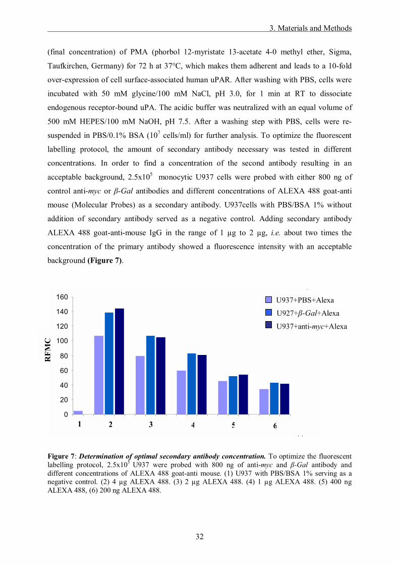
suspended in PBS/0.1% BSA (107 cells/ml) for further analysis. To optimize the fluorescent
labelling protocol, the amount of secondary antibody necessary was tested in different
concentrations. In order to find a concentration of the second antibody resulting in an
acceptable background, 2.5x10⁵ monocytic U937 cells were probed with either 800 ng of
control anti-myc or β-Gal antibodies and different concentrations of ALEXA 488 goat-anti
mouse (Molecular Probes) as a secondary antibody. U937cells with PBS/BSA 1% without
addition of secondary antibody served as a negative control. Adding secondary antibody
ALEXA 488 goat-anti-mouse IgG in the range of 1 µg to 2 µg, i.e. about two times the
concentration of the primary antibody showed a fluorescence intensity with an acceptable
background (Figure 7).
Figure 7: Determination of optimal secondary antibody concentration. To optimize the fluorescent labelling protocol, 2.5x10⁵ U937 were probed with 800 ng of anti-myc and β-Gal antibody and different concentrations of ALEXA 488 goat-anti mouse. (1) U937 with PBS/BSA 1% serving as a negative control. (2) 4 µg ALEXA 488. (3) 2 µg ALEXA 488. (4) 1 µg ALEXA 488. (5) 400 ng ALEXA 488, (6) 200 ng ALEXA 488.
0
20
40
60
80
100
120
140
160
1 2 3 4 5 6 7 8 9 10 11 12 13 14 15 16 17 18 19 20 21
1234567891011121314
1 2 3 4 5 6
U937+PBS+Alexa
U927+β-Gal+Alexa
U937+anti-myc+Alexa
RFM
C
3. Materials and Methods
33
Binding of purified scFv-IIIF10 from hamster cells to human uPAR was tested by incubating
2.5x10⁵ U937 cells with eluates of the soluble scFv-IIIF10 and sTF1-214 columns for 20 min at
RT. Anti-myc-antibody (0.8 µg, Invitrogen) directed against the C-terminal c-myc epitope of
scFv-IIIF10 followed by the fluorescence labeled secondary antibody ALEXA 488 goat-anti-
mouse (1.6 µg, Molecular Probes) was added and incubated for another 20 min at RT. Prior to
FACS analysis propidium-jodide was added in order to be able to exclude dead cells from
analysis. Single cell-associated fluorescence was then quantified by FACS analysis using the
FACScalibur cytofluorometer (Becton Dickinson). Autofluorescence of anti-myc antibody
and Alexa 488 goat-anti-mouse was determined in the absence of the purified protein. sTF1-214
eluate was used as a non-binding control protein.
For specificity testing, one sample of scFv-IIIF10 eluate was simultaneously incubated with
recombinant human pro-uPA (4 µg) for 30 min. Fluorescence intensities were expressed as
relative fluorescence mean channel (RFMC).
3. Materials and Methods
34
3.2.3 Cell biology
3.2.3.1 Cell culture
OV-MZ-6#8 cells are adherent cells and grow as a monolayer attached to the bottom of the
culture flask. They are cultured under standard conditions (37°C, 5% CO2, humid
atmosphere) in DMEM (Dublecco´s Modified Eagle Media), containing 10 mM HEPES, 10%
fetal calf serum (FCS), penicillin/streptomycin (100 µg/ml, 100 U/ml) (all from Gibco,
Karlsruhe, Germany) and 0.27 mM asparagine, 0.55 mM arginine (Sigma, St.Louis, USA).
Cells are subcultured 3 times a week. By adding splitting solution EDTA/PBS 1:20 and
incubating the adherent cells for 2 min at 37°C, the washed cells detach from the culture flask
and are then centrifuged 3 min at 940 rpm, RT. The supernatant is discarded and cells are re-
suspended in PBS. According to the amount of cells needed, cells are spread into new culture
flasks.
For experiments 70-80% confluent cells between passages 2 to 9 are used. Every second
month, cells are tested for mycoplasma contamination using PCR.
U937 cells are monocytic suspension cells cultured under standard conditions in RPMI media
containing 10 mM HEPES, 2 mM L-glutamine, 10% FCS and penicillin/streptomycin (100
µg/ml, 100 U/ml). The cells are maintained in culture by transfer of 1 ml cell suspension in 30
ml fresh culture media once a week. For FACS experiments, the cells are stimulated with
PMA which make them adherent and lead to a 10 fold over expression of uPAR on the cell
surface.
For long term storage, cell pellets of approximately 106 cells are re-suspended in 1 ml
DMSO/FCS (1:10) in kryo-tubes and shock frozen in an ethanol bath, then stored at �80°C in
nitrogen.
3.2.3.2 Stable transfection of V79, CHO and OV-MZ-6#8 cells
For stable transfection of plasmids, SuperFect� (QIAGEN, Hilden, Germany) was used.
Cells were grown in a 6-well plate until they reached 40-60% confluence. 10 µl of
SuperFect� (QIAGEN, Hilden, Germany) and 100 µl of DMEM (without serum and
antibiotics) were dissolved.

3. Materials and Methods
35
Two µg DNA of various eukaryotic expression plasmids (pSecTag2/HygroB-scFv-IIIF10,
pSecTag2/HygroB-scFv-IIIF10his, pSecTag2/HygroB-scFv-IIIF10-GPI, pSecTag2/HygroB-
scFvIIIF10-TCD, pSecTag2/HygroB-sTF and empty vector pSecTag2/HygroB) were
dissolved each in 100 µl DMEM (without serum and antibiotics), mixed well and incubated
for 10 min at RT. DNA- and SuperFect�- solution were mixed together and incubated for 10
min to allow complex formation. Then, 300 µl FCS-free cell culture medium were added to
the SuperFect�-DNA mixture and the total volume was transferred immediately to the cells
that were previously washed with PBS (Gibco, Karlsruhe, Germany). After 3 h of incubation
at 37°C, cells were washed 3 times with PBS and further cultured in 10% FCS DMEM. The
day, cells reached 80-90% confluence, the selection of stably transfected cells was started
using cell culture medium containing 250 µg/ml hygromycin B (HygroGold, InvivoGen, San
Diego, USA). Selected cells were then kept in culture for further investigation.
3.2.3.3 Phage-display
3.2.3.3.1 Phage amplification and purification
Different M13-phages (0108, 0109, 0110, 0114, 0115, 0117 binding to mAb-IIIF10 and an
irrelevant non-binding phage as a negative control) were kindly provided by Dr. Volker
Böttger, Wilex AG, Munich. For phage amplification, 1 µl of phage suspension was incubated
with 200 µl K91 E. coli bacteria, grown to late log phase (OD600nm ≈ 1), for 15 min at 37°C
without shaking followed by another 15 min with shaking at 200 rpm. The infected bacteria
were then transferred to a 50 ml Falcon tube (polypropylene) with 10 ml 2x TY medium,
supplemented with 20 µg/ml tetracycline (Smith et al., 1993) and grown for 24 h at 37°C,
gently shaking (200 rpm). The suspension was then centrifuged at 3,400xg and 4°C for 30
min and the supernatant transferred to a new 50 ml Falcon tube. 100 µl were kept for titration.
For phage purification/precipitation, 2 ml of PEG/NaCl solution were added to the 10 ml
supernatant, mixed by inverting the tube several times and incubated on ice for at least 2 h.
The suspension was then centrifuged at 3,400xg and 4°C for 30 min, the supernatant
discarded and the phage pellet re-suspended in 1 ml aqua dest. Transferred to a 1.5 ml
Eppendorf tube, suspension was spun at 10,000xg and 4°C for 10 min to remove remaining
bacteria and cellular debris. The phage supernatant was transferred to a new 1.5 ml tube and
200 µl PEG/NaCl added for a second precipitation, mixed and left on ice for 1 h, then spun at

3. Materials and Methods
36
10,000xg at 4°C for 10 min. The supernatant was poured off, the tube re-spun briefly and the
remaining supernatant removed with a pipette. The phage pellet was then re-suspended in 1
ml TBS, filtered through a 0.2 µm syringe filter and stored at 4°C.
PEG/NaCl 100 g polyethylene glycol 8,000 117 g NaCl 475 ml H2O, then autoclaved
3.2.3.3.2 Phage-titration
To determine the concentration of phage after amplification, phage titration is carried out. It is
based on counting bacterial colonies, which had been infected with a dilution series of phage,
making them grow on tetracycline selection plates. Therefore, only transforming units, i.e.
infectious phage particles, are determined.
LB-agar plates were prepared with 20 µg/ml tetracycline and dried with open lid under sterile
conditions for several hours. A grid with 1.5x1.5 cm squares (4x4 for a 9 cm petri dish) was
drawn on the bottom of each plate. 45 µl TBS were filled into each well of a 96-well
microtiterplate and 5 µl of the purified phage added to the first well. Properly mixed by
aspirating and dispensing at least ten times, 5 µl were transferred into the next well and
continued until 10 serial dilutions had been made. The pipette tip was changed to minimize
unwanted carry over of phage. Then 155 µl log phase K91 bacteria were added to each well,
mixed carefully with the phage dilution and incubated at 37°C for 30 min. The bacteria in the
wells were re-suspended and 10 µl of each dilution spotted onto the prepared agar plate.
Waiting until the liquid had completely disappeared the lid was closed and the plate incubated
face down over night at 37°C. The colonies at dilutions with well separated clones were
counted and the phage concentration of the starting material calculated (Figure 8).
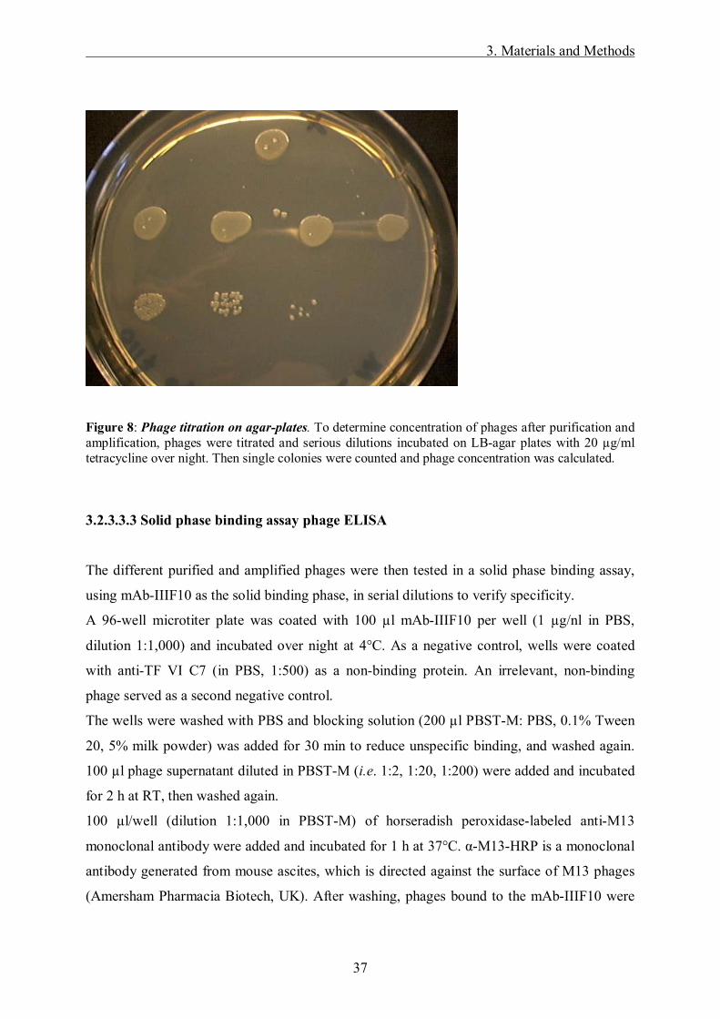
3. Materials and Methods
37
Figure 8: Phage titration on agar-plates. To determine concentration of phages after purification and amplification, phages were titrated and serious dilutions incubated on LB-agar plates with 20 µg/ml tetracycline over night. Then single colonies were counted and phage concentration was calculated.
3.2.3.3.3 Solid phase binding assay phage ELISA
The different purified and amplified phages were then tested in a solid phase binding assay,
using mAb-IIIF10 as the solid binding phase, in serial dilutions to verify specificity.
A 96-well microtiter plate was coated with 100 µl mAb-IIIF10 per well (1 µg/nl in PBS,
dilution 1:1,000) and incubated over night at 4°C. As a negative control, wells were coated
with anti-TF VI C7 (in PBS, 1:500) as a non-binding protein. An irrelevant, non-binding
phage served as a second negative control.
The wells were washed with PBS and blocking solution (200 µl PBST-M: PBS, 0.1% Tween
20, 5% milk powder) was added for 30 min to reduce unspecific binding, and washed again.
100 µl phage supernatant diluted in PBST-M (i.e. 1:2, 1:20, 1:200) were added and incubated
for 2 h at RT, then washed again.
100 µl/well (dilution 1:1,000 in PBST-M) of horseradish peroxidase-labeled anti-M13
monoclonal antibody were added and incubated for 1 h at 37°C. α-M13-HRP is a monoclonal
antibody generated from mouse ascites, which is directed against the surface of M13 phages
(Amersham Pharmacia Biotech, UK). After washing, phages bound to the mAb-IIIF10 were
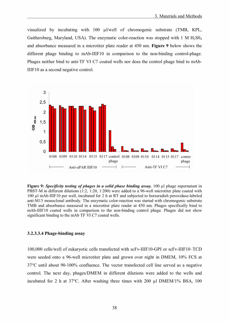
3. Materials and Methods
38
visualized by incubating with 100 µl/well of chromogenic substrate (TMB, KPL,
Gaithersburg, Maryland, USA). The enzymatic color-reaction was stopped with 1 M H2S04
and absorbance measured in a microtiter plate reader at 450 nm. Figure 9 below shows the
different phage binding to mAb-IIIF10 in comparison to the non-binding control-phage.
Phages neither bind to anti-TF VI C7 coated wells nor does the control phage bind to mAb-
IIIF10 as a second negative control.
Figure 9: Specificity testing of phages in a solid phase binding assay. 100 µl phage supernatant in PBST-M in different dilutions (1:2, 1:20, 1:200) were added to a 96-well microtiter plate coated with 100 µl mAb-IIIF10 per well, incubated for 2 h at RT and subjected to horseradish peroxidase-labeled anti-M13 monoclonal antibody. The enzymatic color-reaction was started with chromogenic substrate TMB and absorbance measured in a microtiter plate reader at 450 nm. Phages specifically bind to mAb-IIIF10 coated wells in comparison to the non-binding control phage. Phages did not show significant binding to the mAb TF VI C7 coated wells.
3.2.3.3.4 Phage-binding assay
100,000 cells/well of eukaryotic cells transfected with scFv-IIIF10-GPI or scFv-IIIF10�TCD
were seeded onto a 96-well microtiter plate and grown over night in DMEM, 10% FCS at
37°C until about 90-100% confluence. The vector transfected cell line served as a negative
control. The next day, phages/DMEM in different dilutions were added to the wells and
incubated for 2 h at 37°C. After washing three times with 200 µl DMEM/1% BSA, 100
0
0,5
1
1,5
2
2,5
3
01090108 0110 0114 01170115 controlphage
OD
450
nm
controlphage
0108 0109 0110 0114 0115 0117
Anti-TF VI C7Anti-uPAR IIIF10
0
0,5
1
1,5
2
2,5
3
01090108 0110 0114 01170115 controlphage
OD
450
nm
controlphage
0108 0109 0110 0114 0115 0117
Anti-TF VI C7Anti-uPAR IIIF10
3. Materials and Methods
39
µl/well (dilution 1:1,000) of the horseradish peroxidase labeled anti-M13 monoclonal
antibody was added and incubated for 1 h at 37°C). The wells were then washed two times
with 200 µl DMEM/1% BSA and once more with PBS. The adherent cells were visualized by
incubating with 100 µl/well of chromogenic substrate TMB. The enzymatic color reaction
was stopped with 0.5 M H2S04 and the absorbance measured in a microtiter plate reader at 450
nm.
3.2.3.4 Cell proliferation assay
The proliferation assay was performed to compare the growth rate of vector transfected
control cells and cell lines transfected with expression plasmids encoding scFv-IIIF10. 30,000
cells in 1 ml 10% FCS DMEM were seeded in triplicate into the wells of a 24-well plate and
incubated for 48 h or 96 h. After incubation cells were washed with PBS, detached from the
wells by incubation with 1% (w/v) EDTA/PBS solution and counted in a Neubauer
hemocytometer upon Trypan blue exclusion.
3.2.3.5 Cell adhesion assay
3.2.3.5.1 Cell-matrix adhesion assay
The cell-matrix adhesion assay was used to estimate any differences in the adhesive behaviour
of the scFv-IIIF10 expressing eukaryotic cell lines to different ECM proteins in comparison to
the vector transfected cell line.
Fibronectin (Becton Dickinson, Heidelberg, Germany), vitronectin (Promega) and collagen
type IV (Sigma, München, Germany) were diluted in PBS to the final concentration of 10
µg/ml. For coating, 100 µl of the protein solution were added to each well of a 96-well plate
in triplicate and incubated over night at 4°C. The next day, wells were washed two times with
PBS, blocked with PBS/2% BSA for 3 h at RT and again washed with PBS. Afterwards,
40,000 cells in 100 µl DMEM/0.5%BSA were seeded to each well, the plate was incubated
for 2 h at 37°C and washed carefully with PBS to remove non-adherent cells. Adherent cells
were then visualized by incubating with substrate solution (50 µl/well + 50 µl/well PBS) for 1
h at 37°C. The resulting color reaction was stopped with the stop solution and the absorbance

3. Materials and Methods
40
measured in a microtiter plate reader at 405 nm. Serial dilutions of cell suspension in a range
from 2,500 to 40,000 cells in 50 µl PBS served as a standard.
Substrate solution 15 mM p-nitrophenol-N-acetyl-beta-D-glucosaminide 0.5% Triton X-100 100 mM sodium acetate, pH 5.0
Stop solution 200 mM NaOH 5 mM EDTA
3.2.3.5.2 Cell-cell adhesion assay
The cell-cell adhesion assay was performed to determine the cell-cell interaction of the
membrane anchored variants of the scFv-IIIF10 expressed by eukaryotic cells in comparison
to the vector transfected control cells. 50,000 cells/100 µl were seeded into each well of a 96-
well plate and incubated over night in DMEM. The next day, the adherent cells were washed
two times with 100 µl RPMI and the scFv-IIIF10 expressing cells and the control group were
incubated with pro-uPA (10 µg/ml RPMI) in quadruplicate for 20 min, then, washed once
more. Making cell-cell interaction visible, 3x10⁶ U937 suspension cells were incubated at
37°C over night with a fluorescent lipophilic dye, the carbocyanine DIO 1:100 in RPMI
(Molecular Probes, USA). This tracer is weakly fluorescent in water but highly fluorescent
and quite photostable, when incorporated into cell membranes. Once applied to cells, the dye
diffuses laterally within the plasma membrane, resulting in staining of the entire cell. Transfer
of these probes between intact membranes is usually negligible. The next day, 100,000 DIO-
labeled U937 per 100 µl RPMI were added to each well and incubated for 3 h at 37°C,
washed two times with 100 µl RPMI/well and the extinction was measured in the fluorimeter
at 480 nm. Serial dilutions of fluorescence labeled U937 cells in a range from 5,000 to
100,000/well in 100 µl RPMI in triplicate served as a standard. With the standard dilutions
and the extinction of the probes, the amount of fluorescence labeled U937, bound to the
transfected, adherent eukaryotic cell lines, can be calculated, thereby giving an idea of U937
expressing uPAR on their cell surface and interacting with membrane anchored scFv-IIIF10.
Phenol-red-free RPMI medium was used not to interact with the fluorescent dye. The
preparations for the experiment were done under reduced exposure to bright light at all times.

3. Materials and Methods
41
Figure 10: Schematic overview of the principle of the cell-cell adhesion assay described above. DIO-labeled U937 cells overexpressing uPAR on their cell surface interact with membrane anchored variants of scFv-IIIF10 expressed on the surface of transfected eukaryotic cell lines. At the same time, there might be possible interaction between scFv-IIIF10 and uPAR expressed on the surface of transfected cell lines, thereby inhibiting scFv-IIIF10 and uPAR interaction described before. After pre-incubation with pro-uPA, cell surface associated uPAR/scFv-IIIF10 interaction can be inhibited. scFv-IIIF10 is free for interaction with DIO-labeled U937 cells.
3.2.4 Tumor model
Pathogen-free, female a-thymic (nu/nu, CD1) mice (9 weeks old), were purchased from
Charles River Laboratories (Sulzfeld, Germany). The animal experiments were performed
under semi-sterile conditions in cooperation with Prof. Dr. A. Krüger, Institute for
Experimental Oncology, Klinikum rechts der Isar, Munich.
Cell membrane
VLVH VLVH
U937
pro-upA
U937
uPAR
Cell membrane
VLVH VLVH VLVH VLVH
U937U937U937
pro-upA
U937U937U937
uPAR

3. Materials and Methods
42
Stably transfected OV-MZ-6#8 cells expressing soluble scFv-IIIF10, transfected with an
empty expression plasmid (pRcRSV) and with the trifunctional inhibitor pRcRSV-N-hTimp-
1-chCys-uPA19-31 (for further details see Krol et al. 2003) were grown until 60-80%
confluence. 7x10⁶ cells of each cell line were inoculated into the peritoneal cavity of nude
mice. After 56 days, the mice were sacrificed, all intraperitoneal organs including the tumor
removed and weighed. These data were taken further as total situs weight. All visible tumor
mass was then removed and subsequently weighed. To estimate the differences between
individual mice, the ratio of tumor mass to total situs weight was calculated. Tumor of three
mice from each group were frozen in liquid nitrogen for further detection of soluble scFv-
IIIF10. Blood and ascites were collected, centrifuged at 2,000 xg for 30 min at 4°C and stored
at �20°C for further detection.
3.2.5 Statistical analysis
Significant differences in tumour weight over total situs weight between the groups were
investigated using a Man-Whitney Rank Sum Test according to the distribution of the data. A
level of p<0.05 was considered statistically significant.
4. Results
43
4. Results
4.1 Mammalian expression plasmids encoding scFv-IIIF10
Previously, the sequence encoding scFv-IIIF10 was isolated from the mRNA of mAb IIIF10-
producing hybridoma cells. For this, the fragments encoding the variable regions of heavy
(VH) and light (VL) chain were amplified by RT-PCR using gene-specific primers with
added restriction sites. VH- and VL-gene segments were then connected via a glycine- and
serine-rich, flexible linker and expressed as a single-chain antibody (scFv) in E. coli
(Kirschenhofer et al., 2003).
Using the phagemid vector encoding scFv-IIIF10 as template, an eukaryotic expression
plasmid based on the vector pSecTag2/HygroB was generated, encoding scFv-IIIF10 as a
secreted, soluble form with a C-terminal c-myc epitope as well as a (His)6-tag. These two tags
allow for detection and/or purification of the recombinant protein. Expression is driven by the
strong viral CMV promoter, secretion is supported by the murine Ig κ-chain V-J2-C signal
peptide. In addition, two membrane-anchored variants were designed to display scFv-IIIF10
on the surface of mammalian cells: (i) One fusion harbors the C-terminal sequence of uPAR
(aa 294-335 with the Met of the pre-protein being pos. 1). This sequence contains the signals
for the attachment of a glycan lipid anchor to a newly formed C-terminus at Gly305
(corresponds to Gly283 of mature uPAR) which results in the cell surface linkage of the
recombinant protein via a glycosylphosphatidylinositol (GPI) anchor (Sevlever et al., 2000).
ii) The other variant harbors the transmembrane and the short cytoplasmic domain (TCD) of
tissue factor (TF250-295 with the Met of the pre-protein being pos. 1), the cellular receptor for
the blood coagulation factor VII. A schematic presentation of the three different scFv-IIIF10-
encoded fusion proteins is given in Figure 11A, the complete amino acid sequence of the
fusion protein encoding the soluble form of scFv-IIIF10 is depicted in Figure 11B. In a
similar manner, we also generated a pSecTag2/HygroB-based expression vector encoding the
extracellular domain of mature tissue factor TF1-214 (Magdolen et al., 1998), which served as a
control protein for monitoring expression in eukaryotic cells. A schematic overview of the
plasmid-constructions is shown in Figure 12 and 13.

4. Results
44
Figure 11: Mammalian expression plasmids encoding scFv-IIIF10. (A) ScFv-IIIF10 DNA was amplified by PCR using gene-specific primers with added restriction sites and inserted into the expression vector pSecTag2/HygroB (Invitrogen). Three different vectors were generated which encode: i) a soluble, secreted form of scFv-IIIF10 harboring a C-terminally located c-myc epitope plus a (His)6 tag; ii) scFv-IIIF10 with the C-terminal part of the unprocessed uPAR (encoding uPAR294-335 of pre-uPAR) for display of scFv-IIIF10 on the cell surface in a glycan lipid-anchored form; iii) scFv-IIIF10 with the C-terminal part of the unprocessed tissue factor (TF) (encoding TF250-295 of pre-TF) for insertion of scFv-IIIF10 into the cell surface via a transmembrane domain. (B) Complete amino acid sequence of the secreted, soluble form of scFv-IIIF10. N-terminal and C-terminal extensions (encoding the murine Ig κ-chain V-J2-C signal peptide and the c-myc epitope plus (His)6 tag, respectively) as well as the flexible linker region separating the VH- and VL-chains are indicated by a grey background. The cleavage site of the signal sequence is indicated by an arrowhead, the c-myc epitope and the (His)6 tag are boxed. Hypervariable regions within VH and VL (CDRs H1-H3 and L1-L3, respectively) are overlined.

4. Results
45
Figure 12. Construction of the expression plasmids pSecTag2/HygroB encoding soluble scFv-IIIF10 and soluble TF1-214. The vector pSecTag2/HygroB (Invitrogen) was subjected to the restriction endonucleases HindIII and BamH1 resulting in a cloning site to insert directionally the sequences of the soluble secreted form of scFv-IIIF10 and TF1-214. PCR of the templates was performed with the primers scFv-HIII/scFv-B and TFIII/TF-B and PCR-products were subjected to restriction analysis with the endonucleases HindIII and BamHI and isolated from the agarose gel via �freeze and squeeze� method. The corresponding gel is depicted above: lane 1, 1kb ladder, lane 2, TF (650 bp); lane 3, soluble scFv (735 bp); lane 4, pSecTag2/HygroB. Fragments were then ligated into the vector pSecTag2/HygroB and transformed into E. coli. DNA-preparation from E. coli and restriction analysis with HindIII and BamHI shows positive clones on the agarose gel. Verified clones were sent for DNA sequencing. Upper panel: lane 1, 1kb ladder; lane 2, negative clone; lane 3 to 5 positive clones for soluble scFv-IIIF10 (735 bp); lane 6 and 7, negative clones. Lower panel: lane 1, 1 kb ladder; lane 2 to 6, positive clones for soluble TF1-214 (650 bp); lane 7, negative clone.
pSecTag2/HygroB
pSecTag2/HygroB-scFvIIIF10
down-stream primer up-stream primer
ligation and transformation in E. coli
HindIII BamH1
PCR of the scFv-IIIF10 and sTF1-214 templateswith Taq4-Polymerase
restriction with HindIII and BamH1
HindIII BamH1
1 2 3 4
500 bp
1000 bp
3000 bp
pSecTag2/HygroB-sTF1-214
750 bp1500 bp
750 bp1500 bp3000 bp
3000 bp
1 3 5 62 4 7
pSecTag2/HygroB
pSecTag2/HygroB-scFvIIIF10
down-stream primer up-stream primer
ligation and transformation in E. coli
HindIII BamH1
PCR of the scFv-IIIF10 and sTF1-214 templateswith Taq4-Polymerase
restriction with HindIII and BamH1
HindIII BamH1
1 2 3 4
500 bp
1000 bp
3000 bp
500 bp
1000 bp
3000 bp
pSecTag2/HygroB-sTF1-214
750 bp1500 bp
750 bp1500 bp1500 bp3000 bp3000 bp
3000 bp3000 bp
1 3 5 62 4 7
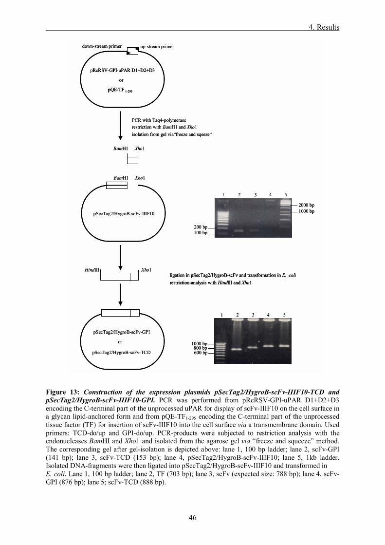
4. Results
46
Figure 13: Construction of the expression plasmids pSecTag2/HygroB-scFv-IIIF10-TCD and pSecTag2/HygroB-scFv-IIIF10-GPI. PCR was performed from pRcRSV-GPI-uPAR D1+D2+D3 encoding the C-terminal part of the unprocessed uPAR for display of scFv-IIIF10 on the cell surface in a glycan lipid-anchored form and from pQE-TF1-295 encoding the C-terminal part of the unprocessed tissue factor (TF) for insertion of scFv-IIIF10 into the cell surface via a transmembrane domain. Used primers: TCD-do/up and GPI-do/up. PCR-products were subjected to restriction analysis with the endonucleases BamHI and Xho1 and isolated from the agarose gel via �freeze and squeeze� method. The corresponding gel after gel-isolation is depicted above: lane 1, 100 bp ladder; lane 2, scFv-GPI (141 bp); lane 3, scFv-TCD (153 bp); lane 4, pSecTag2/HygroB-scFv-IIIF10; lane 5, 1kb ladder. Isolated DNA-fragments were then ligated into pSecTag2/HygroB-scFv-IIIF10 and transformed in E. coli. Lane 1, 100 bp ladder; lane 2, TF (703 bp); lane 3, scFv (expected size: 788 bp); lane 4, scFv-GPI (876 bp); lane 5; scFv-TCD (888 bp).
down-stream primer up-stream primer
restriction-analysis with HindIII and Xho1ligation in pSecTag2/HygroB-scFv and transformation in E. coli
restriction with BamH1 and Xho1isolation from gel via�freeze and sqeeze�
pRcRSV-GPI-uPAR D1+D2+D3
pQE-TF 1-295
or
Xho1HindIII
PCR with Taq4-polymerase
BamH1 Xho1
BamH1 Xho1
pSecTag2/HygroB-scFv-IIIF10
pSecTag2/HygroB-scFv-GPI
pSecTag2/HygroB-scFv-TCD
or
1 2 4 53
100 bp200 bp
1000 bp2000 bp
1 2 4 53
1000 bp800 bp600 bp
down-stream primer up-stream primer
restriction-analysis with HindIII and Xho1ligation in pSecTag2/HygroB-scFv and transformation in E. colirestriction-analysis with HindIII and Xho1ligation in pSecTag2/HygroB-scFv and transformation in E. coli
restriction with BamH1 and Xho1isolation from gel via�freeze and sqeeze�
pRcRSV-GPI-uPAR D1+D2+D3
pQE-TF 1-295
or
pRcRSV-GPI-uPAR D1+D2+D3
pQE-TF 1-295
or
Xho1HindIII
PCR with Taq4-polymerase
BamH1 Xho1
BamH1 Xho1
pSecTag2/HygroB-scFv-IIIF10
pSecTag2/HygroB-scFv-GPI
pSecTag2/HygroB-scFv-TCD
or
1 2 4 53
100 bp200 bp
1000 bp2000 bp
1 2 4 53
1000 bp800 bp600 bp

4. Results
47
4.2 Generation of stable transfectants in eukaryotic cells Plasmids encoding either of the three variants of scFv-IIIF10, soluble TF1-214 and the empty
vector pSecTag2/HygroB, respectively were transfected into Chinese hamster ovary cells
(CHO), hamster fibroblasts (V79) and the human ovarian cancer cell line OV-MZ-6#8. In
addition, OV-MZ-6#8 cells were available, which had been previously transfected with a
pRcRSV-derived expression plasmid encoding a trifunctional inhibitor directed against
different tumor-associated proteolytic systems and the vector only. These cells were later used
in in vivo experiments as controls to analyze the effects of scFv-IIIF10 expression on tumor
growth and spread (see below). The characterization of the pRcRSV-derived cell lines has
been described in detail elsewhere (Krol et al., 2003 a, b). The trifunctional recombinant
inhibitor, N-hTIMP-1-chCys-uPA19-31 is composed of the N-terminal domain of the human
matrix-metalloproteinase inhibitor TIMP-1 and a chicken cystatin variant harboring the uPAR
binding site of uPA, chCys-uPA19-31, which in addition to its inhibitory activity towards
cysteine proteases interferes with uPA/uPAR-interaction (Muehlenweg et al., 2000).
Subsequently to selection of hygromycin-resistent cells, integration of the pSecTag2/HygroB-
derived plasmids into genomic DNA was proven by PCR (Figure 14A). For this, genomic
DNA was prepared from the various transfectants and used as template for a nested PCR with
specific oligodeoxynucleotide primers directed to the DNA encoding the murine signal
sequence and the c-myc epitope of pSecTag2/HygroB, respectively. The amplification product
of the integrated vector corresponded to the expected fragment of 166 bp (not shown), the
PCR products of the other stably transfected cell lines varied between 798 bp and 971 bp,
depending on the DNA inserted (Figure 14A). In order to prove not only integration of the
pSecTag2/HygroB-derived plasmids into genomic DNA but also transcription under the
control of the strong viral CMV promoter, total RNA was isolated from the transfected cell
lines, reverse transcribed and amplified with gene specific oligodeoxynucleotides. Figure
14B depicts the results of such a RT-PCR analysis for detection of mRNA encoding the
soluble, secreted scFv-IIIF10 in CHO and OV-MZ-6#8 cells. In both cell lines a specific
product was amplified, which lacks in the vector-control as expected.

4. Results
48
Figure 14: Analysis of stably transfected cell lines by PCR. Two µg of purified DNA of either expression plasmid were transfected with the SuperFectTM Transfection Reagent into three different mammalian cell lines: CHO, V79 and OV-MZ-6#8. (A) PCR analysis to test for integration of the expression plasmids into genomic DNA of OV-MZ-6#8 cells. Genomic DNA was isolated from 2x10⁶ cells of the various transfected cell lines and used as template for a nested PCR with pSecTag2/HygroB-specific primers. Lane 1, 100 bp ladder; lane 2, amplification product from OV-scFv (expected size: 877 bp); lane 3, plasmid pSecTag2/HygroB-scFv-IIIF10 as positive control (877 bp); lane 4, OV-scFv-GPI (959 bp); lane 5, OV-scFv-TCD (971 bp); lane 6, OV-sTF (798 bp). (B) RT-PCR analysis to test for transcription of the expression cassette encoding soluble, secreted scFv-IIIF10 in OV-MZ-6#8 and CHO cells. 6x10⁶ cells were treated with Trizol reagent (Life Technologies) according to the manufacturers manual and total RNA was isolated, reverse transcribed into first/strand cDNA with random and oligo (dT) primers and amplified by scFv-IIIF10 specific primers (expected fragment: 323 bp, upper panel). Lanes 1 and 8, 100 bp ladder; lane 2, OV-scFv; lane 3, OV-MZ-6#8 cells stably transfected with the empty vector pSecTag2/HygroB; lane 4, OV-scFv RNA reverse transcribed w/o oligo(dT) (= control); lane 5, RT-PCR w/o RNA (= control); lane 6, CHO-scFv; lane 7, CHO- pSecTag2/HygroB. The lower panel shows the control RT-PCR of the house-keeping gene G6PDH to demonstrate the integrity of the RNA.
4.3 Purification and characterization of soluble scFv-IIIF10 and soluble TF1-214 from
eukaryotic cell culture supernatants
Since both, the secreted form of scFv-IIIF10 as well as sTF1-214 contain a C-terminal (His)6
tag, these proteins were initially purified from culture supernatants of the transfected
eukaryotic cell lines via Ni2+-NTA-agarose affinity chromotagraphy under native conditions.
Expression and purification of sTF was followed by applying a TF ELISA (Albrecht et al.
1992). The cells produced about 20 ng/ml (CHO cells) to 55 ng/ml sTF per 106 cells after 48
h of cultivation. The recovery rates of sTF amounted to 65 to 90% after purification. It has
previously been demonstrated that sTF1-214 expressed in E. coli displays functional activity
(Magdolen et al., 1998; Randolf et al., 1998; Albrecht et al., 2002). As tested by a soluble TF
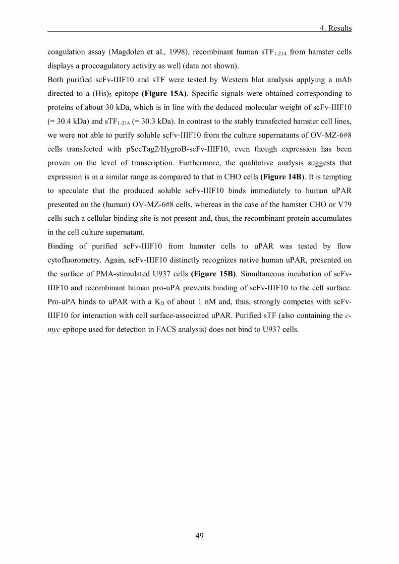
4. Results
49
coagulation assay (Magdolen et al., 1998), recombinant human sTF1-214 from hamster cells
displays a procoagulatory activity as well (data not shown).
Both purified scFv-IIIF10 and sTF were tested by Western blot analysis applying a mAb
directed to a (His)5 epitope (Figure 15A). Specific signals were obtained corresponding to
proteins of about 30 kDa, which is in line with the deduced molecular weight of scFv-IIIF10
(= 30.4 kDa) and sTF1-214 (= 30.3 kDa). In contrast to the stably transfected hamster cell lines,
we were not able to purify soluble scFv-IIIF10 from the culture supernatants of OV-MZ-6#8
cells transfected with pSecTag2/HygroB-scFv-IIIF10, even though expression has been
proven on the level of transcription. Furthermore, the qualitative analysis suggests that
expression is in a similar range as compared to that in CHO cells (Figure 14B). It is tempting
to speculate that the produced soluble scFv-IIIF10 binds immediately to human uPAR
presented on the (human) OV-MZ-6#8 cells, whereas in the case of the hamster CHO or V79
cells such a cellular binding site is not present and, thus, the recombinant protein accumulates
in the cell culture supernatant.
Binding of purified scFv-IIIF10 from hamster cells to uPAR was tested by flow
cytofluorometry. Again, scFv-IIIF10 distinctly recognizes native human uPAR, presented on
the surface of PMA-stimulated U937 cells (Figure 15B). Simultaneous incubation of scFv-
IIIF10 and recombinant human pro-uPA prevents binding of scFv-IIIF10 to the cell surface.
Pro-uPA binds to uPAR with a KD of about 1 nM and, thus, strongly competes with scFv-
IIIF10 for interaction with cell surface-associated uPAR. Purified sTF (also containing the c-
myc epitope used for detection in FACS analysis) does not bind to U937 cells.
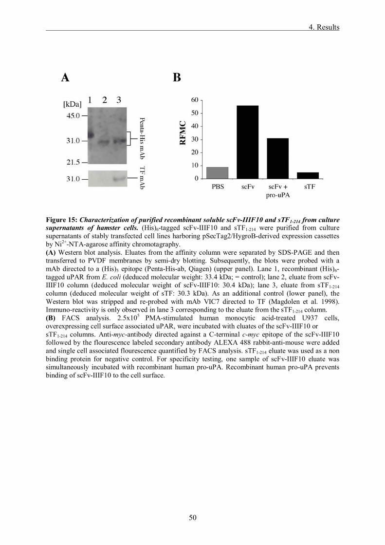
4. Results
50
Figure 15: Characterization of purified recombinant soluble scFv-IIIF10 and sTF1-214 from culture supernatants of hamster cells. (His)6-tagged scFv-IIIF10 and sTF1-214 were purified from culture supernatants of stably transfected cell lines harboring pSecTag2/HygroB-derived expression cassettes by Ni2+-NTA-agarose affinity chromotagraphy. (A) Western blot analysis. Eluates from the affinity column were separated by SDS-PAGE and then transferred to PVDF membranes by semi-dry blotting. Subsequently, the blots were probed with a mAb directed to a (His)5 epitope (Penta-His-ab, Qiagen) (upper panel). Lane 1, recombinant (His)6-tagged uPAR from E. coli (deduced molecular weight: 33.4 kDa; = control); lane 2, eluate from scFv-IIIF10 column (deduced molecular weight of scFv-IIIF10: 30.4 kDa); lane 3, eluate from sTF1-214 column (deduced molecular weight of sTF: 30.3 kDa). As an additional control (lower panel), the Western blot was stripped and re-probed with mAb VIC7 directed to TF (Magdolen et al. 1998). Immuno-reactivity is only observed in lane 3 corresponding to the eluate from the sTF1-214 column. (B) FACS analysis. 2.5x10⁵ PMA-stimulated human monocytic acid-treated U937 cells, overexpressing cell surface associated uPAR, were incubated with eluates of the scFv-IIIF10 or sTF1-214 columns. Anti-myc-antibody directed against a C-terminal c-myc epitope of the scFv-IIIF10 followed by the flourescence labeled secondary antibody ALEXA 488 rabbit-anti-mouse were added and single cell associated flourescence quantified by FACS analysis. sTF1-214 eluate was used as a non binding protein for negative control. For specificity testing, one sample of scFv-IIIF10 eluate was simultaneously incubated with recombinant human pro-uPA. Recombinant human pro-uPA prevents binding of scFv-IIIF10 to the cell surface.
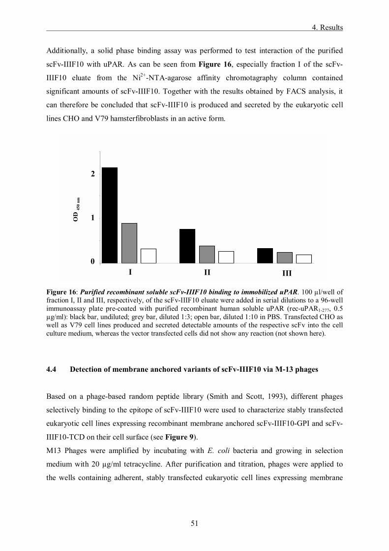
4. Results
51
Additionally, a solid phase binding assay was performed to test interaction of the purified
scFv-IIIF10 with uPAR. As can be seen from Figure 16, especially fraction I of the scFv-
IIIF10 eluate from the Ni2+-NTA-agarose affinity chromotagraphy column contained
significant amounts of scFv-IIIF10. Together with the results obtained by FACS analysis, it
can therefore be concluded that scFv-IIIF10 is produced and secreted by the eukaryotic cell
lines CHO and V79 hamsterfibroblasts in an active form.
Figure 16: Purified recombinant soluble scFv-IIIF10 binding to immobilized uPAR. 100 µl/well of fraction I, II and III, respectively, of the scFv-IIIF10 eluate were added in serial dilutions to a 96-well immunoassay plate pre-coated with purified recombinant human soluble uPAR (rec-uPAR1-277, 0.5 µg/ml): black bar, undiluted; grey bar, diluted 1:3; open bar, diluted 1:10 in PBS. Transfected CHO as well as V79 cell lines produced and secreted detectable amounts of the respective scFv into the cell culture medium, whereas the vector transfected cells did not show any reaction (not shown here).
4.4 Detection of membrane anchored variants of scFv-IIIF10 via M-13 phages
Based on a phage-based random peptide library (Smith and Scott, 1993), different phages
selectively binding to the epitope of scFv-IIIF10 were used to characterize stably transfected
eukaryotic cell lines expressing recombinant membrane anchored scFv-IIIF10-GPI and scFv-
IIIF10-TCD on their cell surface (see Figure 9).
M13 Phages were amplified by incubating with E. coli bacteria and growing in selection
medium with 20 µg/ml tetracycline. After purification and titration, phages were applied to
the wells containing adherent, stably transfected eukaryotic cell lines expressing membrane
0
0,5
1
1,5
2
2,5
1 2 3 4 5 6 7 8 9 10 11
OD
450
nm
I II III
1
2
0
OD
450
nm
0
0,5
1
1,5
2
2,5
1 2 3 4 5 6 7 8 9 10 11
OD
450
nm
I II III
1
2
0
OD
450
nm
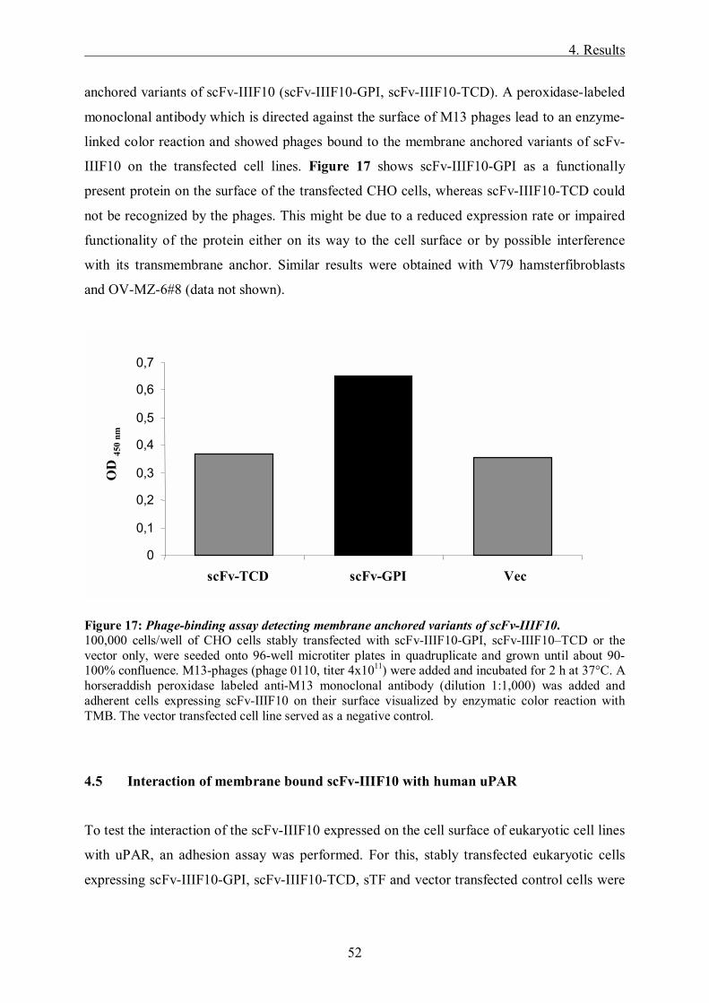
4. Results
52
anchored variants of scFv-IIIF10 (scFv-IIIF10-GPI, scFv-IIIF10-TCD). A peroxidase-labeled
monoclonal antibody which is directed against the surface of M13 phages lead to an enzyme-
linked color reaction and showed phages bound to the membrane anchored variants of scFv-
IIIF10 on the transfected cell lines. Figure 17 shows scFv-IIIF10-GPI as a functionally
present protein on the surface of the transfected CHO cells, whereas scFv-IIIF10-TCD could
not be recognized by the phages. This might be due to a reduced expression rate or impaired
functionality of the protein either on its way to the cell surface or by possible interference
with its transmembrane anchor. Similar results were obtained with V79 hamsterfibroblasts
and OV-MZ-6#8 (data not shown).
Figure 17: Phage-binding assay detecting membrane anchored variants of scFv-IIIF10. 100,000 cells/well of CHO cells stably transfected with scFv-IIIF10-GPI, scFv-IIIF10�TCD or the vector only, were seeded onto 96-well microtiter plates in quadruplicate and grown until about 90-100% confluence. M13-phages (phage 0110, titer 4x10¹¹) were added and incubated for 2 h at 37°C. A horseraddish peroxidase labeled anti-M13 monoclonal antibody (dilution 1:1,000) was added and adherent cells expressing scFv-IIIF10 on their surface visualized by enzymatic color reaction with TMB. The vector transfected cell line served as a negative control.
4.5 Interaction of membrane bound scFv-IIIF10 with human uPAR
To test the interaction of the scFv-IIIF10 expressed on the cell surface of eukaryotic cell lines
with uPAR, an adhesion assay was performed. For this, stably transfected eukaryotic cells
expressing scFv-IIIF10-GPI, scFv-IIIF10-TCD, sTF and vector transfected control cells were
OD
450n
m
0
0,1
0,2
0,3
0,4
0,5
0,6
0,7
scFv-TCD scFv-GPI Vec
OD
450
nmO
D 45
0 nm
OD
450n
mO
D45
0nm
0
0,1
0,2
0,3
0,4
0,5
0,6
0,7
scFv-TCD scFv-GPI Vec
OD
450
nm
0
0,1
0,2
0,3
0,4
0,5
0,6
0,7
scFv-TCD scFv-GPI Vec
OD
450
nmO
D 45
0 nm

4. Results
53
seeded onto 96-well plates and cultured over night. The next day, fluorescence labeled human
U937 overexpressing uPAR were added and incubated. Fluorescence of adherent U937cells
was measured in the fluorimeter and number of adherent U937 cells calculated. As the
membrane-associated uPAR expressed on the surface of the transfected cells might interact
with membrane bound scFv in its neighbourhood, wells were pre-incubated with pro-uPA
before adding U937 cells. In comparison to the wells containing the vector only or wells not
pre-incubated with pro-uPA, an increased number of fluorescence labeled U937 could be
detected (Figure 18). Vector transfected cells and sTF did not show a distinct increase of
binding of fluorescently labeled U937 cells. Similar results were obtained with V79
hamsterfibroblasts and OV-MZ-6#8 (data not shown).
Figure 18: Determination of cell-cell interaction of membrane bound scFv-IIIF10-variants. To determine the interaction of transfected CHO cells expressing membrane bound scFv-IIIF10 with uPAR expressed on the surface of U937 cells, an adhesion assay was performed. 50,000 cells were seeded into each well of a 96-well plate and incubated over night. 100,000 DIO-labeled U937 were added to the wells and incubated. Extinction was measured in the fluorimeter at 480 nm. scFv-IIIF10-GPI expressing cells pre-incubated with pro-uPA (black bar) interact with an increased number of fluorescently labeled U937 compared to not pre-incubated cells and vector only (grey bar).
0
10000
20000
30000
40000
Vector scFv-IIIF10-GPI sTF
1
3
2
4
Num
ber
of c
ells
(104 )
10000
20000
30000
40000
Vector scFv-IIIF10-GPI sTF
1
3
2
4
00
10000
20000
30000
40000
0
10000
20000
30000
40000
Vector scFv-IIIF10-GPI sTF
1
3
2
4
Num
ber
of c
ells
(104 )
10000
20000
30000
40000
10000
20000
30000
40000
Vector scFv-IIIF10-GPI sTF
1
3
2
4
0

4. Results
54
4.6 Characterization of proliferation of OV-MZ-6#8 cells transfected with
soluble scFv-IIIF10
The proliferative behavior of the stably transfected cells was examined in in vitro
proliferation assays under normal culturing conditions. Cells were subcultured in 24-well
plates with serum containing medium for 48 h and 96 h, then counted using a Neubauer
hemocytometer. As shown in Figure 19A, proliferation rates of transfected OV-MZ-6#8 cell
lines expressing scFv-IIIF10 were comparable to the rates obtained with the vector control
cell line OV-pRcRSV.
Figure 19: Characterization of OV-MZ-6#8 derived cells expressing single chain antibody directed against uPAR. (A) Proliferation of OV-MZ-6#8 derived cells, expressing single chain antibody directed against uPAR (OV-scFv-IIIF10), or transfected with vector pRc-RSV only. 30,000 cells in 1 ml DMEM, 10% (v/v) FCS were seeded into wells of a 24-well plate. After 48 and 96 h of incubation, cells were counted in a Neubauer hemocytometer in the presence of Trypan blue. Expression of single chain antibody scFv-IIIF10 had no influence on cell proliferation compared to OV-MZ-6#8 cells stably transfected with vector only. Three independent experiments were performed in triplicate. Mean values ± SD are indicated. (B) Adhesion of OV-MZ-6#8 derived cells expressing scFv-IIIF10 (OV-scFv-IIIF10) or transfected with vector only to different extracellular matrix (ECM) proteins. 30,000 cells were seeded into wells of a 96-well plate precoated with collagen type IV (Col-IV), fibronectin (Fn) or vitronectin (Vn). Two hours later non-adherent cells were washed out with PBS and remaining cells were indirectly quantified via a hexoaminidase substrate solution. The resulting color reaction was measured on a microtest plate reader at a wave length of 405 nm. Values are given as adhesion in % setting adhesion of OV-pRcRSV at 100%. Expression of scFv-IIIF10 had no influence on cell adhesion to all tested ECM proteins compared to OV-pRc-RSV, Mean values ± SD of four independent experiments performed in duplicate are indicated.
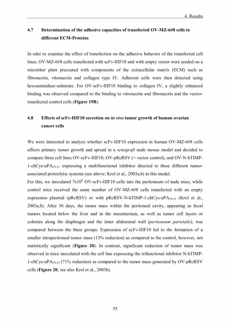
4. Results
55
4.7 Determination of the adhesive capacities of transfected OV-MZ-6#8 cells to
different ECM-Proteins
In oder to examine the effect of transfection on the adhesive behavior of the transfected cell
lines, OV-MZ-6#8 cells transfected with scFv-IIIF10 and with empty vector were seeded on a
microtiter plate precoated with components of the extracellular matrix (ECM) such as
fibronectin, vitronectin and collagen type IV. Adherent cells were then detected using
hexoaminidase-substrate. For OV-scFv-IIIF10 binding to collagen IV, a slightly enhanced
binding was observed compared to the binding to vitronectin and fibronectin and the vector-
transfected control cells (Figure 19B).
4.8 Effects of scFv-IIIF10 secretion on in vivo tumor growth of human ovarian
cancer cells
We were interested to analyze whether scFv-IIIF10 expression in human OV-MZ-6#8 cells
affects primary tumor growth and spread in a xenograft nude mouse model and decided to
compare three cell lines OV-scFv-IIIF10, OV-pRcRSV (= vector control), and OV-N-hTIMP-
1-chCys-uPA19-31 expressing a multifunctional inhibitor directed to three different tumor-
associated proteolytic systems (see above; Krol et al., 2003a,b) in this model.
For this, we inoculated 7x10⁶ OV-scFv-IIIF10 cells into the peritoneum of nude mice, while
control mice received the same number of OV-MZ-6#8 cells transfected with an empty
expression plasmid (pRcRSV) or with pRcRSV-N-hTIMP-1-chCys-uPA19-31 (Krol et al.,
2003a,b). After 56 days, the tumor mass within the peritoneal cavity, appearing as focal
tumors located below the liver and in the mesenterium, as well as tumor cell layers or
colonies along the diaphragm and the inner abdominal wall (peritoneum parietalis), was
compared between the three groups. Expression of scFv-IIIF10 led to the formation of a
smaller intraperitoneal tumor mass (13% reduction) as compared to the control, however, not
statistically significant (Figure 20). In contrast, significant reduction of tumor mass was
observed in mice inoculated with the cell line expressing the trifunctional inhibitor N-hTIMP-
1-chCys-uPA19-31 (71% reduction) as compared to the tumor mass generated by OV-pRcRSV
cells (Figure 20; see also Krol et al., 2003b).
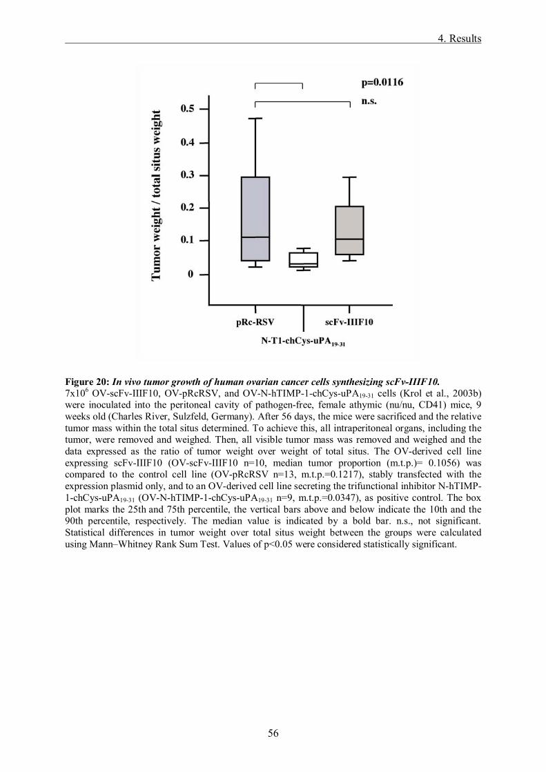
4. Results
56
Figure 20: In vivo tumor growth of human ovarian cancer cells synthesizing scFv-IIIF10. 7x10⁶ OV-scFv-IIIF10, OV-pRcRSV, and OV-N-hTIMP-1-chCys-uPA19-31 cells (Krol et al., 2003b) were inoculated into the peritoneal cavity of pathogen-free, female athymic (nu/nu, CD41) mice, 9 weeks old (Charles River, Sulzfeld, Germany). After 56 days, the mice were sacrificed and the relative tumor mass within the total situs determined. To achieve this, all intraperitoneal organs, including the tumor, were removed and weighed. Then, all visible tumor mass was removed and weighed and the data expressed as the ratio of tumor weight over weight of total situs. The OV-derived cell line expressing scFv-IIIF10 (OV-scFv-IIIF10 n=10, median tumor proportion (m.t.p.)= 0.1056) was compared to the control cell line (OV-pRcRSV n=13, m.t.p.=0.1217), stably transfected with the expression plasmid only, and to an OV-derived cell line secreting the trifunctional inhibitor N-hTIMP-1-chCys-uPA19-31 (OV-N-hTIMP-1-chCys-uPA19-31 n=9, m.t.p.=0.0347), as positive control. The box plot marks the 25th and 75th percentile, the vertical bars above and below indicate the 10th and the 90th percentile, respectively. The median value is indicated by a bold bar. n.s., not significant. Statistical differences in tumor weight over total situs weight between the groups were calculated using Mann�Whitney Rank Sum Test. Values of p<0.05 were considered statistically significant.

5. Discussion
57
5. Discussion
5.1 scFv-IIIF10 as a therapeutic molecule
The uPA/uPAR-system is an attractive target for tumor therapy to affect tumor invasion and
metastasis (Sperl et al., 2001; Reuning et al., 2003). In the present study, we have selected
mAb-IIIF10 directed to the uPA-binding site of uPAR to develop a single-chain antibody,
scFv-IIIF10, as a potentially interesting therapeutic molecule. Pro- and eukaryotic expression
plasmids were generated expressing scFv-IIIF10 either in a soluble form or attached to the
cell membrane via a GPI or a transmembrane anchor. The binding properties of the different
scFv-IIIF10 variants were analyzed in vitro. The soluble form of scFv-IIIF10 was initially
purified from cell culture supernatants of the transfected eukaryotic cell lines via Ni2+-NTA-
agarose affinity chromatography under native conditions. The purified protein was then
subjected to different proteinchemical and cell biological methods, demonstrating that soluble
scFv-IIIF10 is expressed as a functional protein binding to human uPAR. In cell-cell-adhesion
assays it was shown, that membrane anchored, GPI-linked scFv-IIIF10 (scFv-IIIF10-GPI)
expressed on the surface of eukaryotic cell lines is recognized by M13 phages selectively
binding to the epitope of scFv-IIIF10, whereas the transmembrane form scFv-IIIF10-TCD
could not be recognized by the phages. DNA-sequencing analysis confirmed the
corresponding DNA sequence encoding pSecTag2/HygroB-scFv-IIIF10-GPI and �TCD.
Additionally, integration of both membrane anchored variants (pSecTag2/HygroB-scFv-
IIIF10-GPI and �TCD) into genomic DNA was demonstrated by nested PCR (Figure 12A).
However, a reduced expression rate or failures on the level of intracellular processing and
trafficking to the cell surface could be a possible explanation for the M13 phages not being
able to recognize the transmembrane-anchored variant of scFv-IIIF10. Furthermore, an
impaired functionality of the protein may also be due to a possible interference with its
transmembrane anchor.
Subsequently, the effects of scFv-IIIF10 synthesis by ovarian cancer cells on tumor growth
and spread were studied in an OV-MZ-6#8 xenograft mouse model. In previous studies, a
significant anti-tumorigenic effect was achieved in this animal model by blocking uPA/uPAR
interaction with synthetic uPA-derived cyclic peptides (Sato et al., 2002) or soluble uPAR
(Lutz et al., 2001) as a scavenger for uPA. This illustrates the important role of the
uPA/uPAR-system protease system in this tumor model. However, secretion of the single-
5. Discussion
58
chain antibody (scFv-IIIF10) directed against the uPAR did not result in a significant
reduction of relative tumor mass. It is possible, that the rather low secretion level of scFv-
IIIF10 by the transfected cells does not result in effective and sustained levels of scFv-IIIF10
in situ to compete for uPA-binding to all uPAR molecules on the cell surface. Even though
DNA-transcription of soluble scFv-IIIF10 could be demonstrated in OV-MZ-6#8 and CHO
cells, scFv-IIIF10 was secreted in distinct amounts only from CHO cells and V79 hamster
fibroblasts.
Thus, more efficient methods to achieve effective scFv-IIIF10 concentrations in vivo, e.g. via
viral vector-mediated expression of the secretory scFv (Arafat et al., 2002; Solly et al., 2005),
may be necessary to demonstrate applicability of scFv-IIIF10 in tumor therapy. As high
concentrations of scFvs are needed to achieve therapeutic effects, more research should be
done on the possible application of scFv-IIIF10 as a tumor targeting vehicle, as for example
described in Menotti et al. (2006). This study demonstrated in a proof of principle experiment
that a herpes simplex virus recombinant carrying the insertion of a scFv to HER2/neu is able
to selectively infect HER/2neu-positive cells only. Specific targeting of viruses to tumor cell
surface molecules can be used for both oncolytic activity and visualization of tumor cells.
This is a new possibility of directing tumor cells more specifically and effectively. Occurring
side effects when applying viral vectors might even be reduced.
5.2 Limitations in the design and application of single chain fragments
There are several problems to overcome when using genetically engineered scFvs for clinical
application: affinity, specificity, instability, molecular size, biodistribution, immunogenity
and high costs in most cases. An immunoreagent like scFv has to meet several criteria for a
potential anticancer compound. First of all, selective binding and good affinity is essential for
a therapeutic effect. In general, a decrease in binding affinity of the scFvs as compared with
intact antibodies is noticed. Some antigen-antibody interaction involves both antigen-binding
sites of an intact antibody. Thus, a monovalent scFv has a lower avidity than the divalent IgG.
To increase the molecular weight and the functional affinity of an antibody to an antigen,
multimerization is an effective method. Several strategies are being used to produce
multivalent scFvs for example the addition of a flexible hinge region or a peptide linker. The
simplest approach is based on spontaneous formation of non-covalent dimers such as
diabodies or trimers (Batra et al., 2002; Hudson et al., 1999).
5. Discussion
59
Min Fang et al. (2004) described the generation and characterization of a bispecific scFv
BHL-1 that binds specifically and with good affinity to human CD3 expressed on T-cells and
a tumor-associated antigen of human ovarian carcinoma (OC183B2). It was shown that this
bispecific scFv could bridge SCOV3 tumor cells and human T-cells and mediates tumor cell
lysis in vitro and in vivo. Biodstribution has shown that scFv BHL-1 could target the tumor in
vivo with a terminal half life time of 7.7 h. It is particularly important in terms of side effects
of application, that there is no reaction in normal human tissue, which has to be determined by
immunhistochemistry. Pavoni et al. (2006) developed a human scFv against the
carcinoembryonic antigen (CEA) and demonstrated, that this scFv binds selectively and with
good affinity to the CEA epitope expressed by metastatic melanoma, colon and lung
carcinomas, but poorly or not to normal human tissue.
As the affinity for a tumor-related antigen rises, its penetration is progressively impeded, but
specific targeting improves. Adams et al. (2001) demonstrated in a study using a series of
antibody mutants of a scFv, that high affinity limits the tumor-localization and intra-tumoral
diffusion of small antibody-based molecules. mAbs with very high affinity for tumor antigens
stably bind to the first encountered tumor antigen. This could lead to the so called �binding
site barrier effect�, i.e. patchy and incomplete tumor penetration and could be associated with
suboptimal therapeutic effects when therapeutic efficacy is dependent upon uniform delivery
to tumor cells (Fujimori et al., 1990; Juweid et al., 1992). Recently, it has been observed that
quantitative tumor retention did not significantly increase with enhancements in affinity
beyond 10-9 M and resulted in lower tumor:blood ratios (Adams et al., 2001).
When applying scFvs, immunogenity can lead to severe side effects such as anaphylactic
reactions. Most of the mAbs are murine in nature and systemic administration can lead to the
development of a human anti-mouse immunoglobulin antibody response (HAMA). This risk
can be reduced, when therapeutic antibodies are humanized, e.g. chimeric antibodies as they
become less immunogenic (Smith et al., 2004).
Due to their small size, scFvs have the advantage of good tumor penetration compared with
whole antibodies, but they are cleared from the blood by the kidneys more rapidly. In several
anti-cancer studies it was shown, that scFvs derived from monoclonal antibodies could indeed
be targeted to tumors, even improve the penetrating ability for the tumor compared with
whole IgG antibodies. However, due to their short serum half-life the retention within the
tumor and absolute uptake quantity by the tumor is low in most cases (Colcher et al., 1998).

5. Discussion
60
One way to alter the pharmacology of scFv is to modify its net charge. Charge-modified
scFvs with desired isoelectric points have been prepared by inserting negatively charged
amino acids on the template of the variable region genes. This can help to overcome
undesirable elevations of renal uptake seen with most antibody fragments (Pavlinkova et al.,
1999).
5.3 Currently applied therapeutic antibodies in clinical trials
Antibody-based therapeutics have emerged as important components of therapies for an
increasing number of human malignancies and are providing an insight into the biology of
several malignancies. Some tumor antigens, so-called tumor markers, identified by mAbs, are
being used as reliable markers of disease activity in malignancies (CEA, AFP, CA 125, Ca
19-9 etc.). CEA, e.g., is a well characterized tumor-associated glycoprotein that is expressed
on endodermally derived gastrointestinal-tract neoplasms and other adenocarcinomas. It is
present on the surface of tumor cells and also shed at high levels into the circulation. In a
study with a radio-labeled anti-CEA-scFv injected intravenously before surgery,
radioimmune-guided surgery was performed to locate tumor tissue in the operative field. 82%
true-positive rates of tumor detection were found when examining the excised tissues (Mayer
et al., 2000).
Recently, more and more clinical trials are ongoing, applying therapeutic mAbs or scFvs
against tumor-associated antigens. They are used as immunologic mediators of cytotoxicity,
by blocking receptors or conjugated to a drug, toxin or radionuclide. For example, rituximab
is one of the first mAb approved by the FDA (Food and Drug Administration; USA) for
therapeutic use in human malignancy. The chimeric anti-CD20 antibody also known as
Rituxan® demonstrated in in vitro studies that it leads to cell death by multiple mechanisms.
Phase II studies demonstrated the efficacy and safety of an antibody therapy with rituximab in
patients with previously treated low-grade B-cell lymphoma. There were response rates of
46% to 48% with an acceptable safety profile (McLaughlin et al., 1998).
A commonly used antibody for targeting solid tumors is directed against HER-2/neu (c-erbB-
2). This is a cell-surface protein from the EGFR (epidermal growth factor receptor) family
overexpressed on ca. 25% of primary breast cancer as well as on other carcinomas of the
ovary, prostate, lung and gastrointestinal tract. A humanized mAb (trastuzumab) derived from
the murine mAb 4D5 was developed recognizing an epitope on the extracellular domain of
5. Discussion
61
HER-2/neu. Approximately 15% of women who were previously treated for metastatic breast
cancer and were overexpressing HER-2/neu respond to trastuzumab (Herceptin®) therapy. A
large randomized phase III trial comparing cytotoxic chemotherapy alone or with
trastuzumab, showed substantial better efficacy and a 25% increase in survival at 29 months
with combination therapy. On the basis of these results trastuzumab was approved by the
FDA in 1998 and is now used as a standard therapy either alone or in combination with
chemotherapy in the treatment of women with metastatic breast cancer and HER-2/neu
overexpression, since the beginning of 2006 also in the adjuvant setting (Cobleigh et al.,
1999; Slamon et al., 2001; Baselga et al., 2001; Adams et al., 2005; Romond et al., 2005).
One of the receptor blocking mAbs is cetuximab (Erbitux®), a murine-human chimeric anti-
EGFR-antibody blocking ligand-receptor interactions, which is used alone or in combination
with chemotherapy in patients overexpressing EGFR suffering from colorectal, lung or head
and neck cancers (Saltz et al., 2004).
An alternative to targeting a cell-surface receptor expressed on cancer cells is to target the
ligand that initiates signalling events through the receptor. An antibody that works with this
mechanism is bevazicumab (Avastin®). This is a murine-human chimeric mAb, that blocks
binding of VEGF or VEGF-A to their receptors on the vascular endothelium. VEGF is
produced by many cancers to stimulate angiogenesis. Bevacizumab is used alone as anti-
angiogenic therapy or in combination with chemotherapy in the treatment of metastatic
colorectal and lung cancer patients. In a recent phase II trial bevazicumab showed potential
benefit in combination with fluorouracil and leucovorin in patients with first-line colorectal
cancer (Willett et al., 2004). In a large randomized phase III clinical trial of standard
chemotherapy with or without bevazicumab, anti-vascular effects of this antibody-therapy
were confirmed (Kabbinavar et al., 2005).
Alemtuzumab (MabCampath®), a humanized anti-CD 52 mAb that efficiently mediates
complement fixation, is working as an immue modulator and has been approved for use in
chemotherapy-refractory chronic-lymphotic leukaemia. A phase II multicenter study of
previous treated patients with low-grade non-Hodgkin lymphoma has reported a response rate
of 20%. Alemtuzumab is also being used to deplete T-cells from allogenic transplant grafts in
patients with other hematologic malignancies (Lundin et al., 1998, 2002; Ravandi et al.,
2006).
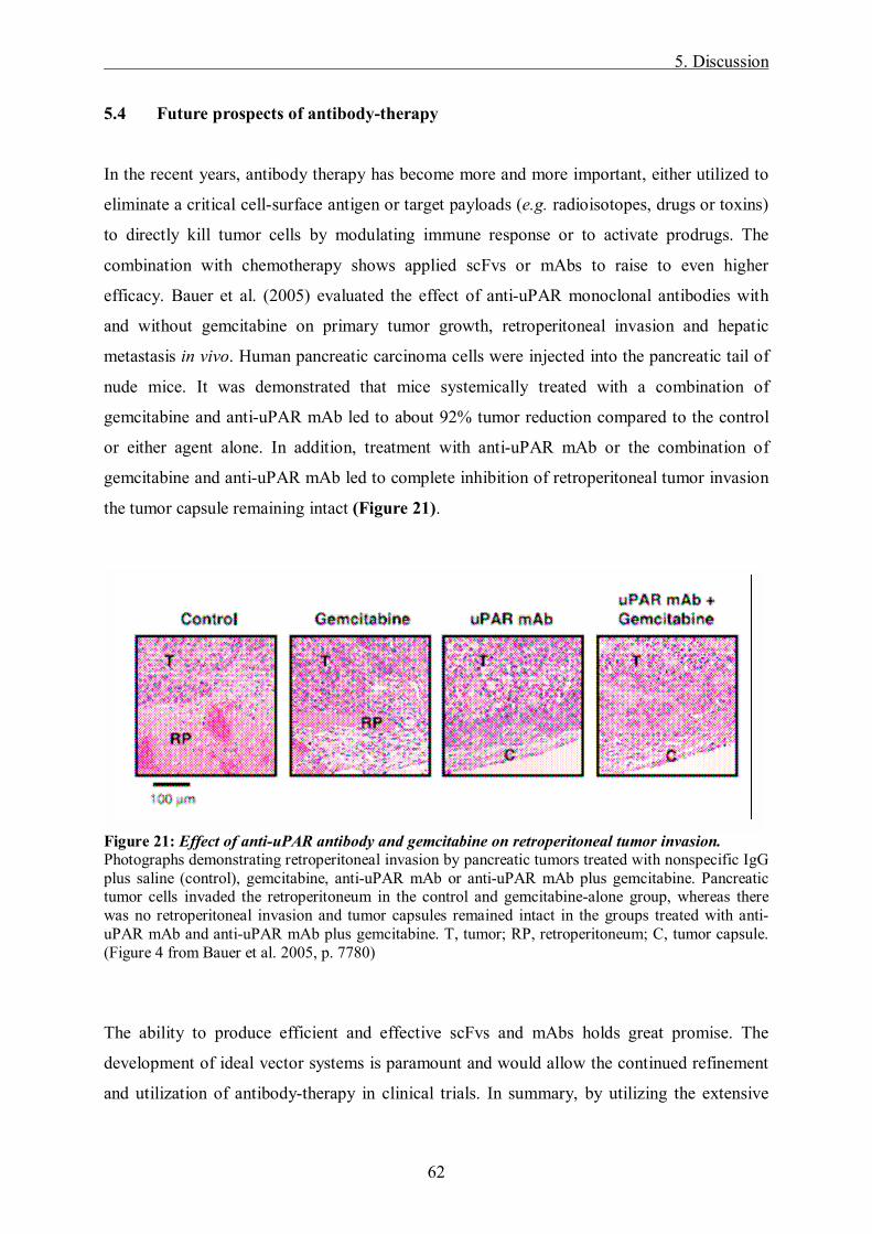
5. Discussion
62
5.4 Future prospects of antibody-therapy
In the recent years, antibody therapy has become more and more important, either utilized to
eliminate a critical cell-surface antigen or target payloads (e.g. radioisotopes, drugs or toxins)
to directly kill tumor cells by modulating immune response or to activate prodrugs. The
combination with chemotherapy shows applied scFvs or mAbs to raise to even higher
efficacy. Bauer et al. (2005) evaluated the effect of anti-uPAR monoclonal antibodies with
and without gemcitabine on primary tumor growth, retroperitoneal invasion and hepatic
metastasis in vivo. Human pancreatic carcinoma cells were injected into the pancreatic tail of
nude mice. It was demonstrated that mice systemically treated with a combination of
gemcitabine and anti-uPAR mAb led to about 92% tumor reduction compared to the control
or either agent alone. In addition, treatment with anti-uPAR mAb or the combination of
gemcitabine and anti-uPAR mAb led to complete inhibition of retroperitoneal tumor invasion
the tumor capsule remaining intact (Figure 21).
Figure 21: Effect of anti-uPAR antibody and gemcitabine on retroperitoneal tumor invasion. Photographs demonstrating retroperitoneal invasion by pancreatic tumors treated with nonspecific IgG plus saline (control), gemcitabine, anti-uPAR mAb or anti-uPAR mAb plus gemcitabine. Pancreatic tumor cells invaded the retroperitoneum in the control and gemcitabine-alone group, whereas there was no retroperitoneal invasion and tumor capsules remained intact in the groups treated with anti-uPAR mAb and anti-uPAR mAb plus gemcitabine. T, tumor; RP, retroperitoneum; C, tumor capsule. (Figure 4 from Bauer et al. 2005, p. 7780)
The ability to produce efficient and effective scFvs and mAbs holds great promise. The
development of ideal vector systems is paramount and would allow the continued refinement
and utilization of antibody-therapy in clinical trials. In summary, by utilizing the extensive

5. Discussion
63
knowledge gained in scFv pre-clinical studies, the combination of improved vectors both in
terms of targeting and oncolysis (Solly et al., 2005; Menotti et al., 2006), and an improved
ability to modulate the immune system for anti-tumoral effects, holds great promise and
provides a strategy for successful gene therapy (Leath et al., 2004).
Considering Herceptin® as a standard therapy for Her2/neu-overexpressing cancer patients,
the combination of antibody-therapy and chemotherapy shows already great success. In the
future, more targeted therapeutics have to be explored for an even more patient specific
cancer treatment. In a phase I study phage-display libraries and scFv libraries were used in
cancer patients to identify more customized tumor-targeting ligands (Krag et al., 2006). More
research has to be done in this field to explore tumor specific as well as patient specific cancer
therapies, giving hope for more possibilities in diagnostic and therapy of malignancies in the
future.
6. Summary
64
6. Summary
The cellular receptor (uPAR, CD87) for the serine protease urokinase-type plasminogen
activator (uPA) focuses uPA to the tumor cell surface, which results in extracellular matrix
degradation and modulation of migration, adhesion and proliferation of tumor cells. Thus,
uPAR represents an attractive target for tumor therapy.
In the present study, we have generated expression plasmids encoding a single chain fragment
(scFv-IIIF10) of the monoclonal antibody mAb IIIF10, which is directed to uPAR and blocks
uPA/uPAR-interaction, either as a soluble form or attached to the cell membrane via a GPI
anchor or a transmembrane domain. Plasmids encoding either of the three variants of scFv-
IIIF10, soluble TF1-214 (used as control protein), and the empty vector pSecTag2/HygroB,
respectively, were transfected into Chinese hamster ovary cells (CHO), hamster fibroblasts
(V79) and the human ovarian cancer cell line OV-MZ 6#8. scFv-IIIF10 was initially purified
from cell culture supernatants of the transfected eukaryotic cell lines via Ni2+-NTA-agarose
affinity chromatography under native conditions and analysed by Western blot. Binding of
purified scFv-IIIF10 from hamster cells to uPAR, presented on the surface of PMA-
stimulated U937 cells, was tested by flow cytofluorometry. In contrast to hamster cells, we
were not able to purify soluble scFv-IIIF10 from the culture supernatants of OV-MZ-6#8 cells
transfected with pSecTag2/HygroB-scFv-IIIF10, even though expression has been proven on
the level of transcription. In cell-cell-adhesion assays it was shown via phage display
technology, that membrane-anchored scFv-IIIF10-GPI is expressed on the surface of
eukaryotic cell lines.
Furthermore, the effects of scFv-IIIF10 synthesis by ovarian cancer cells on tumor growth and
spread were studied in an OV-MZ-6#8 xenograft mouse model. However, secretion of the
single-chain antibody (scFv-IIIF10) directed against the uPAR did not result in a significant
reduction of relative tumor mass. It is possible, that the rather low secretion level of scFv-
IIIF10 by the transfected cells does not result in effective and sustained levels of scFv-IIIF10
in situ to compete for uPA-binding to all uPAR molecules on the cell surface.

6. Summary
65
6. Zusammenfassung
Der Rezeptor (uPAR, CD87) für die Serinprotease Urokinase-Typ Plasminogen Aktivator
(uPA) fokussiert uPA an der Zelloberfläche und führt somit zum Abbau der extrazellulären
Matrix und moduliert Migration, Adhäsion und Proliferation von Tumorzellen. Auf Grund
dieser Eigenschaften ist der uPA-Rezeptor in den Mittelpunkt des Interesses für die
Entwicklung von Tumortherapeutika gerückt.
In der vorliegenden Arbeit wurden Expressionsplasmide konstruiert, die ein single chain
Fragment (scFv-IIIF10) des monoklonalen Antikörpers mAb IIIF10, der gegen uPAR
gerichtet ist und die uPA/uPAR-Interaktion blockiert, entweder als lösliche Form oder
zellgebunden (mittels eines GPI-Ankers bzw. einer Transmembrandomäne) kodieren.
Zusätzlich wurde ein Plasmid für die Expression einer löslichen Form von Tissue Factor (TF1-
214) als Kontrollprotein konstruiert. Die verschiedenen Expressionsplasmide (und auch die
Vektorkontrolle pSecTag2/HygroB) wurden in Chinese Hamster Ovary Zellen (CHO),
Hamsterfibroblasten (V79) und in die humane Ovarialtumor-Zellinie OV-MZ 6#8 transfiziert.
Um die Bindungseigenschaften der verschiedenen Transfektanten in vitro zu analysieren,
wurde anfangs löslicher scFv-IIIF10 aus dem Zellkulturüberstand transfizierter Zellen via
Ni2+-NTA-Agarose-Affinitäts-Chromatographie unter nativen Bedingungen aufgereinigt,
mittels Western Blot-Analysen nachgewiesen und die Bindungsfähigkeit von scFv-IIIF10 an
uPAR, der auf der Zelloberfläche von PMA-stimulierten monozytären U937 Zellen präsent
ist, mittels Durchfluss-Zytofluorometrie verifiziert. Obwohl die mRNA von scFv-IIIF10 auch
in OV-MZ-6#8 Zellen gezeigt werden konnte, konnte scFv-IIIF10 auf Proteinebene nur in
CHO Zellen und V79 Hamsterfibroblasten nachgewiesen werden. Die scFv-IIIF10-GPI-
Variante wurde an der Zelloberfläche von eukaryotischen Zelllinien in Zell-Zell-
Adhäsionsassays durch Phage Display Technik nachgewiesen.
Abschließend wurde in einem Tierversuch mit athymischen Nacktmäusen der Effekt von
löslichem scFv-IIIF10, exprimiert von OV-MZ-6#8 Zellen, auf Tumorwachstum und
Metastasierung untersucht. Die relative Tumormasse wurde durch scFv-IIIF10 nicht
signifikant reduziert. Durch die vermutlich eher geringe Expressionsrate scFv-IIIF10
exprimierender Ovarialtumor Zellen konnten keine ausreichenden Konzentrationen in situ
erreicht werden, um mit uPA an allen uPAR-Molekülen an der Zelloberfläche zu
konkurrieren.

7. References
66
7. References
Adams GP. Improving the tumor specificity and retention of antibody-based molecules. In
vivo 12 (1998) 11-21
Adams GP, Schier R, McCall AM, Simmons H, Horak EM, Alpaugh RK, Marks JD, Weiner
LM. High affinity restricts the localization and tumor penetration of single-chain Fv
antibody molecules. Cancer Res 61 (2001) 4750-4755
Adams GP, Weiner LM. Monoclonal antibody therapy of cancer. Nat Biotechnol 23 (2005)
1147-1157
Andreasen PA, Egelund R, Petersen HH. The plasminogen activation system in tumor growth,
invasion and metastasis. Cell Moll Life Sci 57 (2000) 25-40
Aguirre Ghiso JA, Kovalski K, Ossowski L. Tumor dormancy induced by downregulation of
urokinase receptor in human carcinoma involves integrin and MAPK signaling. J Cell
Biol 147 (1999) 89-103
Albrecht S, Luther T, Grossmann H, Flössel C, Kotzsch M, Müller M. An ELISA for tissue
factor using monoclonal antibodies. Blood Coagul Fibrinolysis 3 (1992) 263-270
Albrecht S, Magdolen V, Herzog U, Miles L, Kirschenhofer A, Baretton G, Luther T. Soluble
tissue factor interferes with angiostatin-mediated inhibition of endothelial cell
proliferation by lysine-specific interaction with plasminogen kringle domains. Thromb
Haemost 88 (2002) 1054-1059
Allgayer H. Molecular regulation of an invasion-related molecule-options for tumor staging
and clinical strategies. Eur J Cancer 42 (2006) 811-819
Altieri DC, Edgington TS. Sequential receptor cascade for coagulation proteins on
monocytes. J Biol Chem 264 (1989) 2969-2972
Alvarez RD, Barnes MN, Gomez-Navarro J, Wang M, Arafat W, Strong TV. A cancer gene
therapeutic approach utilizing an anti-erbB-2 single chain antibody-encoding
adenovirus (AD21): a phase I trial. Clin Cancer Res 6 (2000) 3081-3087
Andolfo A, English WR, Resnati M, Murphy G, Blasi F, Sidenius N. Metalloproteases cleave
the urokinase-type plasminogen activator receptor in the D1-D2 linker region and
expose epitopes not present in the intact soluble receptor. Thromb Haemost 88 (2002)
298-306

7. References
67
Andreasen P, Kjoller L, Christensen L, Duffy MJ. The urokinase plasminogen activator
system in cancer metastasis: a review. Int J Cancer 72 (1997) 1-22
Andreasen P, Sottrup-Jensen L, Kjoller L, Nykjaer A, Moestrup S, Petersen C. Receptor-
mediated endocytosis of plasminogen activators and activator/inhibitor complexes.
FEBS Lett 338 (1994) 239-245
Appella E, Robinson EA, Ullrich SJ, Stoppelli MP, Corti A, Cassani G, Blasi F. The receptor
binding sequence of urokinase. A biological function for the growth factor module for
proteases. J Biol Chem 262 (1987) 4437-4440
Arafat WO, Gomez-Navarro J, Buchsbaum DJ, Xiang J, Wang M, Casado E, Barker SD,
Mahasreshti PJ, Haisma HJ, Barnes MN, Siegal GP, Alvarez RD, Hemminki A,
Nettelbeck DM, Curiel DT. Effective single chain antibody (scFv) concentrations in
vivo via adenoviral vector mediated expression of secretory scFv. Gene Ther 9 (2002)
256-62
Barinka C, Parry G, Callahan J, Shaw D, Kuo A, Bdeir Khalil, Cines Douglas, Mazar A and
Lubkowski J. Structural basis of interaction between urokinase type plasminogen
activator and its receptor. J Mol Biol 363 (2006) 482-295
Batra SK, Jain M, Wittel UA, Chauhan SC and Colcher D. Pharmacokinetics and
biodistribution of genetically engineered antibodies. Curr Opin Biotechnol 13 (2002)
603-608
Bauer TW, Liu W, Fan F, Camp E, Yang A, Somicio R, Bucana C, Callahan J, Parry G,
Evans D, Boyd D, Mazar A, and Ellis L. Targeting of urokinase plasminogen activator
receptor in human pancreatic carcinoma cells inhibits c-Met-and insulin-like growth
factoe-I receptor mediated migration and invasion and orthotopic tumor growth in
mice. Cancer Res 65 (2005) 7775-7781
Bazan JF. Structural design and molecular evolution of a cytokine receptor superfamily. Proc
Natl Acad Sci USA 87 (1990) 6934-6938
Behrendt N, Ploug M, Patthy L, Houen G, Blasi F, Dano K. The ligand�binding domain of
the cell surface receptor for urokinase-type plasminogen activator. J Biol Chem 266
(1991) 7842-7847
Blasi F. uPA, uPAR, PAI-1: key intersection of proteolytic, adhesive and chemotactic
highways? Immunol Today 18 (1997) 415-417

7. References
68
Blasi F. Proteolysis, cell adhesion, chemotaxis, and invasiveness are regulated by the u-PA-u-
PAR-PAI-1 system. Thromb Haemost 82 (1999) 298-304
Blasi F, Carmeliet P. uPAR: a versatile signalling orchestrator. Nat Rev Mol Cell Biol 3
(2002) 932-43
Boshart M, Weber F, Jahn G, Dorsch-Häsler K, Fleckenstein B, Schaffner W. A very strong
enhancer is located upstream of an immediate early gene of human cytomegalovirus.
Cell 41 (1985) 521-530
Böttger V. Epitope-mapping with random peptide libraries. In: Antibody engineering
(Kontermann R, Dubel S eds), Springer Verlag, Berlin Heidelberg (2001) pp. 460-472
Bromberg M, Bailly M, Königsberg W. Role of protease-activated receptor 1 in tumour
metastasis promoted by tissue factor. Thromb Haemost 86 (2001) 1210-4
Bromberg M, Konigsberg W, Madison J, Pawashe A. Tissue factor promotes melanoma
metastasis by a pathway independent of blood coagulation. Proc Natl Acad Sci USA
92 (1995) 8205-8209
Chapman H, Wei Y, Simon DI, Waltz DA. Role of urokinase receptor and caveolin in
regulation of integrin signaling. Thromb Haemost 82 (1999) 291-297
Chapman H, Wei Y. Protease crosstalk with integrins: the urokinase paradigm. Thromb
Haemost 86 (2001) 124-129
Chapman H, Vavrin Z, Hibbs J. Macrophage fibrinolytic activity: identification of two
pathways of plasmin formation by intact cells and of a plasminogen activator inhibitor.
Cell 28 (1982) 653-662
Cobleigh MA, Vogel CL, Tripathy D, Robert NJ, Scholl S, Fehrenbacher L, Wolter JM, Paton
V, Shak S, Liebermann G, Slamon DJ. Multinational study of the efficacy and safety
of humanized anti-HER2 monoclonal antibody in women who have HER-2-
overexpressing metastatic breast cancer that has progressed after chemotherapy for
metastatic disease. J Clin Oncol 17 (1999) 2639-2648
Colcher D, Pavlinkova G, Beresford G, Booth BJ, Choudhury A, Batra SK. Pharmacokinetics
and biodistribution of genetically engineered antibodies. Q J Nucl Med 42 (1998) 225-
41

7. References
69
Coloma MJ, Hastings A, Wims LA, Morrison SL. Novel vectors for the expression of
antibody molecules using variable regions generated by polymerase chain reaction. J
Immunol Methods 152 (1992) 89-104
Cubellis MV, Wun TC, Blasi F. Receptor-mediated internalization and degradation of
urokinase is caused by its specific inhibitor PAI-I. EMBOJ 9 (1990) 1079-1085
Danø K, Romer J, Nielsen BS, Bjorn S, Pyke C, Rygaard J, Lund LR. Cancer invasion and
tissue remodelling-cooperation of protease systems and cell types. APMIS 107 (1999)
120-127
Del Rosso M, Fibbi G, Pucci M, D`Alessio S, Del Rosso A, Magnelli L, Chiarugi V. Multiple
pathways of cell invasion are regulated by multiple families of serine proteases. Clin
Exp Metstasis 19 (2002) 193-207
Demetri G, van Oosterom A, Garrett C, Blackstein M, Shah M, Verweij J, McArthur G,
Judson I, Heinrich M, Morgan J, Desai J, Fletcher C, Georg S, Bello C, Huang X,
Baum C, Casali P. Efficacy and safety of sunitinib in patients with advanced
gastrointestinal stromal tumor after failure of imatinib: a randomised controlled trial.
Lancet 368 (2006) 1329-1338
Deng G, Royle G, Seiffert D, Loskutoff D. The PAI-I/vitronectin interaction: two cats in a
bag? Thromb Haemost 74 (1995) 66-70
Duffy MJ, O`Grady P, Devaney D, O`Siorian L, Fennelly JJ, Lijnen HJ. Urokinase-
plasminogen activator, a marker for aggressive breast carcinoma: preliminary report.
Cancer 62 (1988) 531-533
Duffy MJ. Urokinase plasminogen activator and its inhibitor, PAI-1, as prognostic markers in
breast cancer: from pilot to level 1 evidence studies. Clin Chem 48 (2002) 1194-1197
Ehrlich H, Keijer J, Preissner K, Gebbink R, Pannekoek H. Functional interaction of
plasminogen activator inhibitor type I (PAI-1) and heparin. Biochemistry 30 (1991)
1021-2028
Ellis V, Whawell SA, Werner F, Deadman J. Assembly of urokinase receptor-mediated
plasminogen activation complexes involves direct, non-active site interactions
between urokinase and plasminogen. Biochemistry 38 (1999) 651-659

7. References
70
Fan D, Yano S, Shinohara H, Solorzano C, Van Arsdall M, Bucana CD, Pathak S, Kruzel E,
Herbst RS, Onn A, Roach JS, Onda M, Wang QC, Pastan I, Fidler IJ. Targeted therapy
against human lung cancer in nude mice by high-affinity recombinant antimesothelin
single-chain Fv immunotoxin. Mol Cancer Ther 8 (2002) 595-600
Fang M, Zhao R, Yang Z, Zhang Z, Li H, Zhang XT, Lin Q, Huang HL. Characterization of
an anti-human ovarian carcinoma x anti-human CD3 bispecific single-chain antibody
with an albumin-original interlinker. Gynecol Oncol 92 (2004) 135-146
Fazioli F, Resnati M, Sidenius N, Higashimoto Y, Appella E, Blasi F. A urokinase-sensitive
region of the human urokinase receptor is responsible for its chemotactic activity.
EMBO J 16 (1997) 7279-7286
Fischer K, Lutz V, Wilhelm O, Schmitt M, Graeff H, Heiss P, Nishiguchi T, Harbeck N,
Kessler H, Luther T, Magdolen V, Reuning U. Urokinase induces proliferation of
human ovarian cancer cells: characterization of structural elements required for growth
factor function. FEBS Lett 438 (1998) 101-105
Fujimori K, Covell DG, Fletcher JE, Weinstein JN. A modelling analysis of monoclonal
antibody percolation through tumors: a binding site barrier. J Nucl Med 31 (1990)
1191-1198
Gårdsvoll H; Danø K; Ploug M. Mapping part of the functional epitope for ligand binding on
the receptor for urokinase-type plasminogen activator by site-directed mutagenesis. J
Biol Chem 274 (1999) 37995-38003
Gårdsvoll H, Gilquin B, Le Du MH, Ménèz A, Jørgensen TJ, Ploug M. Characterization of
the functional epitope on the urokinase receptor. Complete alanine scanning
mutagenesis supplemented by chemical crosslinking. J Biol Chem 14 (2006) 19260-
19272
Gritz L, Davies J. Plasmid encoded Hygromycin B-resistance: the sequence of Hygromycin-
B-phophotransferase gene and its expression in Escherichia coli and Saccharomyces
cerevisiae. Gene 25 (1983) 179-188
Guillaume-Rousselet N, Jean D, Frade R. Cloning and characterization of anti-cathepsin L
single chain variable fragment whose expression inhibits procathepsin L secretion in
human melanoma cells. Biochem J 367 (2002) 219-227

7. References
71
Harbeck N, Kates RE, Look MP, Meijer-van Gelder ME, Klijn JG, Krüger A, Kiechle M,
Jänicke F, Schmitt M, Foekens J. Enhanced benefit from adjuvant chemotherapy in
breast cancer patients classified high risk according to urokinase-type plasminogen
activator (uPA) and plasminogen activator inhibitor type 1 (n = 3424). Cancer Res 62
(2002) 4617-4622
Helfrich W, Haisma HJ, Magdolen V, Luther T, Bom VJ, Westra J, van der Hoeven R,
Kroesen BJ, Molema G, de Leij L. A rapid and versatile method for harnessing scFv
antibody fragments with various biological effector functions. J Immunol Methods 237
(2000) 131-145
Herbst RS, Maddox AM, Rothenberg ML. Selective oral epidermal growth factor receptor-
tyrosine kinase inhibitor ZD 1839 is generally well-tolerated and has activity in non-
small-cell lung cancer and other solid tumors. Results of a phase I trial. J Clin Oncol
20 (2002) 3815-3825
Hidalgo M, Siu LL, Neumunaitis J, Rizzo J, Hammond LA, Takimoto C, Eckhardt SG,
Tolcher A, Britten CD, Denis L, Ferrante K, Von Hoff DD, Silberman S, Rowinsky
EK. Phase I and pharmacologic study of OSI-774, an epidermal growth factor receptor
tyrosine kinase inhibitor, in patients with advance solid malignancies. J Clin Oncol 19
(2001) 3267-3279
Hooper JD, Clements JA, Quigley JP, Antalis TM. Type II transmembrane serine proteases.
Insights into an emerging class of cell surface proteolytic enzymes. J Biol Chem 276
(2001) 857-860
Høyer-Hansen G, Ploug M, Behrendt N, Rønne E, Danø K. Cell surface acceleration of
urokinase-catalysed receptor cleavage. Eur J Biochem 243 (1997) 21-26
Høyer-Hansen G, Rønne E, Søberg H, Behrendt N, Ploug M, Lund LR, Ellis V, Danø K.
Urokinase plasminogen activator cleaves its cell surface receptor releasing the ligand
binding domain. J Biol Chem 267 (1992) 18224-18229
Hudson PJ. Recombinant antibodies: a novel approach to cancer diagnosis and therapy.
Expert Opin Investig Drugs 9 (2000) 1231-1242
Ichinose A, Fujikawa K, Suyama T. The activation of pro-urokinase by plasma kallikrein and
its activation by thrombin. J Biol Chem 261 (1986) 3486-3489
7. References
72
Jänicke F, Prechtl A, Thomssen C, Harbeck N, Meisner C, Untch M, Sweep CG, Graeff H,
Schmitt M, for the German Chemo N0 Study Group. Randomized adjuvant therapy
trial in high-risk lymph node negative breast cancer patients identified by urokinyse-
type plasminogen activator and plasminogen activator inhibitor type I. J Natl Cancer
Inst 93 (2001) 913-920
Jänicke F, Schmitt M, Graeff H. Clinical relevance of the urokinase-typ and tissue-type
plasminogen activators and of their type-1 inhibitor in breast cancer. Semin Thromb
Hemost 17 (1991) 30-12
Juweid M, Neumann R, Paik C, Perez-Bacete MJ, Sato J, van Osdol W, Weinstein JN.
Micropharmacology of monoclonal antibodies in solid tumors: direct experimental
evidence for binding site barrier. Cancer Res 52 (1992) 5144-5153
Kabbinavar FF, Schulz J, McCleod M, Patel T, Hamm JT, Hecht JR, Mass R, Perrou B,
Nelson B, Novotny WF. Addition of bevacizumab to bolus fluorouracil and leucovorin
in first-line metastatic colorectal cancer: results of a randomized phase II trial. J Clin
Oncol 23 (2005) 3697-3705
Kay BK, Winter J & McCafferty J. Phage display of peptides and proteins. A laboratory
manual. Academic Press (1996) London
Kirschenhofer A, Magdolen V, Schmitt M, Albrecht S, Krol J, Farthmann J, Kopitz C, Prezas
P, Krüger A, Luther T, Böttger V. Recombinant single chain antibody scFv-IIIF10
directed to human urokinase receptor. Recent Res Devel Cancer 5 (2003) 9-25
Koblinski JE, Ahram M, Sloane BF. Unraveling the role of proteases in cancer. Clin Chim
Acta 291 (2000) 113-135
Krag D, Shukla G, Shen G, Pero S, Ashikaga T, Fuller S, Weaver D, Burdette-Radoux and
Thomas C. Selection of tumor-binding ligands in cancer patients with phage display
libraries. Cancer Res 66 (2006) 7724-7733
Kristensen P, Eriksen J, Blasi F, Dano K. Two alternatively spliced mouse urokinase receptor
mRNAs with different histological localization in the gastrointestinal tract. J Cell Biol
115 (1991) 1763-1771

7. References
73
Krol J, Sato S, Rettenberger P, Assfalg-Machleidt I, Schmitt M, Magdolen V, Magdolen U.
Novel bi- and trifunctional inhibitors of tumor-associated proteolytic systems. Biol
Chem 384 (2003a) 1085-1096
Krol J, Kopitz C, Kirschenhofer A, Schmitt M, Magdolen U, Krüger A, Magdolen V
Inhibition of intraperitoneal tumor growth of human ovarian cancer cells by bi- and
trifunctional inhibitors of tumor-associated proteolytic systems. Biol Chem 384
(2003b) 1097-1102
Kroon ME, Koolwijk P, van Goor H, Weidle UH, Collen A, van der Pluijm G, van Hinsbergh
VW. Role and localization of urokinase receptor in the formation of new
microvascular structures in fibrin matrices. Am J Pathol 154 (1999) 1731-1742
Kruithof E, Baker M, Bunn C. Biological and clinical aspects of plasminogen activator
inhibitor type 2. Blood 86 (1995) 4007-4024
Le Y, Murphy P, Wang J. Formyl-peptide receptors revisited. Trends Immunol 23 (2002) 541-
548
Leath CA, Douglas JT, Curiel DT, Alvarez RD. Single-chain antibodies: A therapeutic
modality for cancer gene therapy. Int J Oncol 24 (2004) 765-771
Leissner P, Verjat T, Bachelot T, Paye M, Krause A, Puisieux A, Mougin B. Prognostic
significance of urokinase plasminogen activator and plasminogen activator inhibitor-1
mRNA expression in lymph node- and hormone receptor positive breast cancer. BMC
Cancer 6 (2006) 1-9
Llinas P, Le Du MH, Gardsvoll H, Dano K, Ploug M, Gilquin B. Crystal structure of the
human urokinase plasminogen activator receptor bound to an antagonist peptide.
EMBO J 24 (2005) 1655-1663
Look M, van Putten W, Duffy M, Harbeck N, Christensen I, Thomssen C, Kates R, Spyratos
F, Fernö M, Eppenberger-Castori S, Sweep F, Ulm K, Peyrat JP Martin P, Magdelenat
H, Brünner N, Duggn C, Lisboa B, Bendahl P, Quillien V, Daver A, Ricolleau G,
Meijer-van Gelder M, Manders P, Fiets E, Blankensein M, Broet P, Omain S,
Daxenbichler G, Windbichler G, Cufer T, Borstnar S, Kueng W, Beex L, Klijn J,
O`Higgins N, Eppenberger U, Jänicke F, Schmitt M, Foekens J. Pooled analysis of
prognostic impact of urokinase-type plasminogen activator and its inhibitor PAI-1 in
8377 breast cancer patients. J Nat Cancer Inst 94 (2002) 116-128
7. References
74
Lundin J, Kimby E, Bjorkholm M, Broliden PA, Celsing F, Hjalmar V, Mollgard L, Rebello
P, Hale G, Waldmann H, Mellstedt H, Osterborg A. Phase II trial of subcutaneous
anti-CD52 monoclonal antibody alemtuzumab (Campath-1H) as first-line treatment
for patients with B-cell chronic lymphatic leukaemia (B-CLL). Blood 100 (2002) 768-
773
Luther T, Flössel C, Weber F, Haroske G, Kotzsch M, Müller M. Expression von Tissue
Factor und Urokinase im Menschlichen Brustkrebs: Beziehungen zu
Prognosefaktoren. Verh Dtsch Path 78 (1994) 532
Luther T, Magdolen V, Albrecht S, Kasper M, Riemer C, Kessler H, Graeff H, Müller M,
Schmitt M. Epitope-mapped monoclonal antibodies as tools for functional and
morphological analyses of the human urokinase receptor in tumor tissue. Am J Pathol
150 (1997) 1231-1244
Lutz V, Reuning U, Kruger A, Luther T, von Steinburg SP, Graeff H, Schmitt M, Wilhelm
OG and Magdolen V. High level synthesis of recombinant soluble urokinase receptor
(CD87) by ovarian cancer cells reduces intraperitoneal tumor growth and spread in
nude mice. Biol Chem 382 (2001) 789-798
Ma Z, Webb DJ, Jo M, Gonias SL. Endogenously produced urokinase-type plasminogen
activator is a major determinant of the basal level of activated ERK/MAP kinase and
prevents apoptosis in MDA-MB-231 breast cancer cells. J Cell Sci 114 (2001) 3387-
3396
Magdolen V, Rettenberger P, Lopens A, Oi H, Lottspeich F, Kellermann J, Creutzburg S,
Goretzki L, Weidle U, Wilhelm O, Schmitt M, Graeff H. Expression of the human
urokinase-type plasminogen activator receptor in E. coli and Chinese hamster ovary
cells: Purification of the recombinant proteins and generation of polyclonal antibodies
in chicken. Electrophoresis 16 (1995) 813-816
Magdolen V, Albrecht S, Kotzsch M, Haller C, Bürgle M, Jacob U, Großer M, Kessler H,
Graeff H, Müller M, Schmitt M, Luther T. Immunological and functional analyses of
the extracellular domain of human tissue factor. Biol Chem 379 (1998) 157-165
Magdolen V, Bürgle M, de Prada NA, Schmiedeberg N, Riemer C, Schroeck F, Kellermann J,
Degitz K, Wilhelm OG, Schmitt M, Kessler H. Cyclo19,31[D-Cys19]-uPA19-31 is a

7. References
75
potent competitive antagonist of the interaction of urokinase-type plasminogen
activator with its receptor (CD87). Biol Chem 382 (2001) 1197-1205
Martin P, Kelly C, Carney D. Epidermal growth factor receptor-targeted agents for lung
cancer. Cancer Control 13 (2006) 129-140
Mayer A, Tsiompanou E, O´Malley D, Boxer GM, Bhatia J, Flynn AA, Chester KA,
Davidson BR, Lewis AA, Winslet MC, Dhillon AP, Hilson AJ, Begent RH.
Radioimmunoguided surgery in colorectal cancer using a genetically engineered anti-
CEA single chain Fv antibody. Clin Cancer Res 6 (2000) 1711-1719
McCafferty J, Fitzgerald KJ, Earnshaw J, Chiswell DJ, Link J, Smith R and Kenten J.
Selection and rapid purification of murine antibody fragments that bind a transition-
state analog by phage display. Appl Biochem Biotechnol 47 (1994) 157-171;
discussion 171-173
McLaughlin P, Grillo-Lopez AJ, Link BK, Levy R, Czuczman MS, Williams ME, Heyman
MR, Bence Bruckler I, White CA, Cabanillas F, Jain V, Ho AD, Lister J, Wey K, Shen
D, Dallaire BK. Rituximab chimeric anti-CD20 monoclonal antibody therapy for
relapsed indolent lymphoma: half of patients respond to a four dose treatment
program. J Clin Oncol 16 (1998) 2825-2832
Menotti L, Cerretani A and Campadelli-Fiume G. A Herpes Simplex Virus recombinant that
exhibits a single-chain antibody to HER2/neu enters cells through the mammary tumor
receptor, independently of the gD receptors. J Virol (2006) 5531�5539
Min Fang, Zhao R, Yang Z, Li H, Zhang XT, Ling Q, Huang HL. Characterization of an anti-
human ovarian carcinoma x anti-human CD3 bispecific single chain antibody with an
albumin-original interlinker. Gynecol Oncol 92 (2004) 135-146
Moller LB, Pollänen J, Ronne E, Pedersen N, Blasi, F. N-linked glycosylation of the ligand
binding domain of the human urokinase receptor contributes to the affinity for its
ligand. J Biol Chem 268 (1993) 11152-11159
Möbus V, Gerharz C, Press U, Moll R, Beck T, Mellin W, Pollow K, Knapstein P, Kreienberg
R. Morphological, immunohistochemical and biochemical characterization of 6 newly
established human ovarian carcinoma cell lines. Int J Cancer 52 (1992) 76-84.

7. References
76
Montuori N, Visconte V, Rossi G, Ragno P. Soluble and cleaved forms of the urokinase
receptor: degradation products or active molecules? Thromb Haemost 93 (2005) 192-
198
Montuori N, Carriero MV, Salzano S, Rossi G, Ragno P. The cleavage of the urokinase
receptor regulates its multiple functions. J Biol Chem 277 (2002) 46932-46939
Moy B and Goss P. Lapatinib: Current status and future directions in breast cancer.
Oncologist 11 (2006) 1047�1057
Muehlenweg B, Assfalg-Machleidt I, Parrado SG, Bürgle M, Creutzburg S, Schmitt M,
Auerswald EA, Machleidt W, Magdolen V. A novel type of bifunctional inhibitor
directed against proteolytic activity and receptor/ligand interaction. J Biol Chem 275
(2000) 33562-33566
Muehlenweg B, Sperl S, Magdolen V, Schmitt M, Harbeck N. Interference with the urokinase
plasminogen activator (uPA) system: A promising therapy concept for solid tumors.
Expert Opin Biol Ther 1 (2001) 683-691
Myers KA, Ryan MG, Stern PL. Targeting immune effector molecules to human tumor cells
through genetic delivery of 5T4-specific scFv fusion proteins. Cancer Gene Ther 9
(2002) 884-896
Nykjaer A, Conese M, Christensen EI, Olson D, Cremona O, Gliemann J, Blasi F. Recycling
of the urokinase receptor upon internalization of the uPA:serpin complexes. EMBO J
16 (1997) 2610-2620
Ny T, Mikus P. Plasminogen activator inhibitor type 2. A spontaneously polymerizing serpin
that exists in two topological forms. Adv Exp Med Biol 425 (1997) 123-30
Oh P, Li Y, Yu J, Durr E, Krasinska KM, Carver LA, Testa JE, Schnitzer JE. Subtractive
proteomic mapping of the endothelial surface in lung and solid tumors for tissue
specific therapy. Nature 429 (2004) 629-635
Ott I, Fischer EG, Miyagi Y, Mueller BM, Ruf W. A role for tissue factor in cell adhesion and
migration mediated by interaction with actin-binding protein 280. J Cell Biol 140
(1998) 1241-1253
7. References
77
Pavlinkova G, Beresford G, Booth BJ, Batra SK, Colcher D. Charge-modified single chain
antibody constructs of monoclonal antibody CC49: generation, characterization,
pharmacokinetics and biodistribution analysis. Nucl Med Biol 26 (1999) 27-34
Pavoni E, Flego M, Dupuis ML, Barca S, Petronzelli F, Anastasi AM. D�Alessio V, Pelliccia
A, Vaccaro P, Monteriu G, Ascione A, De Santis R, Felici F, Cianfriglia M and
Minenkova O. Selection, affinity maturation and characterization of a human scFv
antibody against CEA protein. BMC Cancer 6 (2006) 41
Pedersen N, Schmitt M, Ronne E, Nicoletti MI, Hoyer-Hansen G, Conese M, Giavazzi R,
Dano K, Kuhn W, Jänicke F, et al. A ligand-free, soluble urokinase receptor is present
in the ascitic fluid from patients with ovarian cancer. J Clin Invest 92 (1993) 2160-
2167
Ploug M, Rønne E, Behrendt N, Jensen AL, Blasi F, Danø K. Cellular receptor for urokinase
plasminogen activator. Carboxy-terminal processing and membrane anchoring by
glycosyl-phosphatidyl-inositol. J Biol Chem 266 (1991) 1926-1933
Ploug M, Ellis V, Danø K. Ligand interaction between urokinase-type plasminogen activator
and its receptor probed with 8-anilino-1-naphthalenesulfonate. Evidence for a
hydrophobic binding site exposed only on the intact receptor. Biochemistry 33 (1994)
8991-8997
Ploug M, Rahbek-Nielsen H, Ellis V, Roepstorff P, Danø K: Chemical modification of the
urokinase-type plasminogen activator and its receptor using tetranitromethane.
Evidence for the involvement of specific tyrosine residues in both molecules during
receptor-ligand interaction. Biochemistry 34 (1995) 12524-12534
Ploug, M. Identification of specific sites involved in ligand binding by photoaffinity labeling
of the receptor for the urokinase-type plasminogen activator. Residues located at
equivalent positions in uPAR domains I and III participate in the assembly of a
composite ligand binding site. Biochemistry 37 (1998) 16494-16505
Ploug M, Rahbek-Nielsen H, Nielsen PF, Roepstorff P, Danø K. Glycosylation profile of a
recombinant urokinase-type plasminogen activator receptor expressed in Chinese
hamster ovary cells. J Biol Chem 273 (1998) 13933-43

7. References
78
Ploug M, Gårdsvoll H, Jorgensen TJ, Lonborg Hansen L, Danø K. Structural analysis of the
interaction between urokinase-type plasminogen activator and its receptor: a potential
target for anti-invasive cancer therapy. Biochem Soc Trans 30 (2002) 177-183
Pollänen JJ. The N-terminal domain of human urokinase receptor contains two distinct
regions critical for ligand recognition. Blood 82 (1993) 2719-2729
Potempa J, Korzus E, Travis J. The serpin superfamily of proteinase inhibitors: structure,
function and regulation. J Biol Chem 269 (1994) 15957-15960
Rabbani SA, Gladu J. Urokinase receptor antibody can reduce tumor volume and detect the
presence of occult tumor metastases in vivo. Cancer Res 62 (2002) 2390-2397
Rabbani SA, Mazar AP. The role of the plasminogen activation system in angiogenesis and
metastasis. Surg Oncol Clin N Am 10 (2001) 393-415
Ragno P. The urokinase receptor: a ligand or a receptor? Story of a sociable molecule. Cell
Mol Life Sci 63 (2006) 1028-1037
Randolph G, Luther T, Albrecht S, Magdolen V, Müller W. Role of tissue factor in adhesion
of mononuclear phagocytes to and trafficking through endothelium in vitro. Blood 11
(1998) 4167-4177
Ravandi F and O'brien S. Alemtuzumab in CLL and other lymphoid neoplasms. Cancer
Invest 24 (2006) 718-725
Reff ME, Heard C. A review of modifications to recombinant antibodies: attempt to increase
efficacy in oncology applications. Crit Rev Oncol Hematol 40 (2001) 25-35
Rettenberger P, Wilhelm O, Oi H, Weidle UH, Goretzki L, Koppitz M, Lottspeich F, König
B, Pessara U, Kramer MD, Schmitt M, Magdolen V. A competitive chromogenic
assay to study the functional interaction of urokinase-type plasminogen activator with
its receptor. Biol Chem Hoppe-Seyler 376 (1995) 587-594
Reuning U, Sperl S, Kopitz C, Kessler H, Krüger A, Schmitt M and Magdolen V. Urokinase-
type plasminogen activator (uPA) and its receptor (uPAR): development of antagonists
of uPA/uPAR interaction and their effects in vitro and in vivo. Curr Pharm Des 9
(2003) 1529-1543

7. References
79
Reuning U, Magdolen V, Wilhelm O, Fischer K, Lutz V, Graeff H. Multifunctional potential
of the plasminogen activation system in tumour invasion and metastasis. Int J Oncol
13 (1998) 893-906
Romond E, Perez E, Bryant J, Suman V, Geyer C, Davidson N, Tan-Chiu E, Martino S, Paik
S, Kaufman P, Swain S, Pisansky T, Fehrenbacher L, Kutteh L, Vogel V, Visscher D,
Yothers G, Jenkins R, Brown A, Dakhil S, Mamounas E, Lingle W, Klein P, Ingle J
and Wolmark N. Trastuzumab plus adjuvant chemotherapy for operable HER2-
positive breast cancer. N Engl J Med 353 (2005) 1673-1684
Sampson MT, Kakkar AK. Coagulation proteases and human cancer. Biochem Soc Trans
(2002) 201-207
Saltz LB, Meropol NJ, Loehrer PJ, Needle MN, Kopit J, Mayer RJ. Phase II trial of
cetuximab in patients with refractory colorectal cancer that express the epidermal
growth factor receptor. J Clin Oncol 22 (2004) 1201-1208
Sanz L, Kristensen P, Blanco B, Facteau S, Russell SJ, Winter G, Alvarez-Vallina L. Single-
chain antibody-based gene therapy: inhibition of tumor growth by in situ production of
phage derived human antibody fragments blocking functionally active sites of cell-
associated matrices. Gene Ther 9 (2002) 1049-1053
Sato S, Kopitz C, Schmalix WA, Muehlenweg B, Kessler H, Schmitt M, Krüger A, Magdolen
V. High-affinity urokinase-derived cyclic peptides inhibiting urokinase/urokinase
receptor-interaction: effects on tumor growth and spread. FEBS Lett 528 (2002) 212-
216
Schlehuber S, Skerra A. Lipocalins in drug discovery: from natural ligand-binding proteins to
�anticalins�. Drug Discov Today 10 (2005) 23-33
Schmitt M, Wilhelm OG, Reuning U, Krüger A, Harbeck N, Lengyel E. The urokinase
plasminogen activator system as a novel target for tumour therapy. Fibrinolysis 6
(2000) 114-132
Schmitt M, Jänicke F, Graeff H. Tumour-associated proteases. Fibrinolysis 6 (1992) 3-26
Schmitt M, Harbeck N, Thomssen C, Wilhelm O, Magdolen V, Reuning U, Ulm K, Höfler H,
Jänicke F, Graeff H. Clinical impact of the plasminogen activation system in tumor

7. References
80
invasion and metastasis: prognostic relevance and target for therapy. Thromb Haemost
78 (1997) 285-296
Sevlever D, Chen R, Medof ME. Synthesis of the GPI anchor in paroxysmal nocturnal
hemoglobinuria and the glycophosphoinositol-linked proteins. Academic press (2000)
199-220
Skerra A. �Anticalins�: a new class of engineered ligand-binding proteins with antibody-like
properties. J Biotechnol 74 (2001) 257-275
Smith GP, Scott JK. Library of peptides and proteins displayed on filamentous phage.
Methods Enzymol 217 (1993) 228-257
Smith KA, Nelson PN, Warren P, Astley SJ, Murray PG, Greenman J.
Demystified...recombinant antibodies. J Clin Pathol 57 (2004) 912-917
Sperl S, Mueller MM, Wilhelm OG, Schmitt M, Magdolen V, Moroder L. The uPA/uPA-
receptor system as a target for tumor therapy. Drug News Perspect 14 (2001) 401-411
Solly S, Nguyen T, Weber A and Horellou P. Targeting of c-Met and urokinase expressing
human glioma cells lines by retrovirus vector displaying single-chain variable
fragment antibody. Cancer Biol Ther 4 (2005) 987-992
Sun J, Pons J, Craik CS. Potent and selective inhibition of membrane-type serine protease 1
by human single-chain antibodies. Biochemistry 42 (2003) 892-900
Taniguchi T, Kakkar AK, Tuddenham EG, Williamson RC, Lemoine NR. Enhanced
expression of urokinase receptor induced through the tissue factor-factor VIIa pathway
in human pancreatic cancer. Cancer Res 58 (1998) 4461-4467
Tautz D and Renz M. An optimized freeze-squeeze method for the recovery of DNA
fragments from agarose gels. Anal Biochem 132 (1983) 14-19
Vitaliti A, Wittmer M, Steiner R, Wyder L, Neri D, Klemenz R. Inhibition of tumor
angiogenesis by a single-chain antibody directed against vascular endothelial growth
factor. Cancer Res 60 (2000) 4311-4314
Waltz D, Chapman H. Reversible cellular adhesion to vitronectin linked to urokinase receptor
occupancy. J Biol Chem 269 (1994) 14746-14750
7. References
81
Wei Y, Lukashev M, Simon D, Bodary S, Rosenberg S, Doyle MV, Chapman HA.
Regulation of integrin function by the urokinase receptor. Science 273 (1996) 1551-
1555
Wilhelm O, Wilhelm S, Escott GM, Lutz V, Magdolen V, Schmitt M, Rifkin D, Wilson E,
Graeff H, Brunner G. Cellular glycosylphosphatidyl-specific phospholipase D
regulates urokinase receptor shedding and cell surface expression. J Cell Physiol 180
(1999) 225-235
Willett CG, Boucher Y, di Tomaso E, Duda DG, Munn LL, Tong RT, Chung DC, Sahani DV,
Kalva SP, Kozin SV, Mino M, Cohen KS, Scadden DT, Hartford AC, Fischman AJ,
Clark JW, Ryan DP, Zhu AX, Blaszkowsky LS, Chen HX, Shellito PC, Lauwers GY,
Jain RK. Direct evidence that VEGF- specific antibody bevazicumab has antivascular
effects in human rectal cancer. Nat Med 10 (2004) 145-147
8. Curriculum vitae and publications
82
8. Curriculum vitae and publications
Curriculum vitae Personal details date of birth 19.04.1975 place of birth Munich marital status single Parents
father: Dr. med. Otto Kirschenhofer, general practitioner mother: Dr. med. Elisabeth Kirschenhofer, general practitioner Education 1981-1985 Grundschule Riedering, Riedering 1985-1994 Ignaz Günther Gymnasium, Rosenheim 1994 Abitur
Medical education
11/1996-05/2005 study of medicine, Ludwig-Maximilians-Universität, München 09/1998 pre-clinical examination 04/2000 first state examination 08/2002 second state examination 05/2004 third state examination Internship
01/1999 clinical research at SmithKline Beecham Pharmaceuticals,
Munich

8. Curriculum vitae and publications
83
02/1999 department of gynaecology and obstetrics, Klinikum Rosenheim
09/1999 department of internal medicine, Hospital General Jose Parres,
Cuernavacca, Mexico 05/2000 department of anesthesia and intensive care, Allgemeines
Krankenhaus der Stadt Wien, Austria 09/2000 department of neonatology, Mercy Hospital for Women,
Melbourne, Australia 02/2001 general medical practice of Dres. E. und O. Kirschenhofer,
Riedering Clinical clerkship 02/2003-05/2003 department of gynaecology and obstetrics, Klinikum der
Universität München Großhadern, Prof. Dr. med. H. Hepp 06/2003-07/2003 department of internal medicine, Berner Klinik, Montana,
Schweiz, Dr. med. C. Vaney 08/2003-09/2003 department of internal medicine, Nepean Hospital, Sydney
University, Australia, Prof. Dr. med. N. Talley 10/2003-01/2004 department of surgery, Chirurgische Klinik Innenstadt,
Ludwig-Maximilians-Universität München, Prof. Dr. med. W. Mutschler
Specialization in gynaecology and obstetrics
08/2004-11/2006 intern at the department of gynaecology and obstetrics,
Klinikum Rosenheim, Prof. Dr. med. Th. Beck since 12/2006 intern at the department of gynaecology and obstetrics
Universitätsklinikum Großhadern, Ludwig-Maximilians-Universität München, Prof. Dr. med. K. Friese
8. Curriculum vitae and publications
84
Publications Kirschenhofer A, Magdolen V, Schmitt M, Albrecht S, Krol J, Farthmann J, Koptitz C, Prezas
P, Krüger A, Luther T, Böttger V. Recombinant single chain antibody scFv-IIIF10
directed to human urokinase receptor. Recent Res Devel Cancer 5 (2003) 9-25
Albrecht S, Magdolen V, Herzog U, Miles L, Kirschenhofer A, Baretton G, Luther T. Soluble
tissue factor interferes with angiostatin-mediated inhibition of endothelial cell
proliferation by lysine-specific interaction with plasminogen kringle domains. Thromb
Haemost 88 (2002) 1054-1059
Krol J, Kopitz C, Kirschenhofer A, Schmitt M, Magdolen U, Krüger A, Magdolen V
Inhibition of intraperitoneal tumor growth of human ovarian cancer cells by bi- and
trifunctional inhibitors of tumor-associated proteolytic systems. Biol Chem 384 (2003)
1097-1102

8. Curriculum vitae and publications
85
Lebenslauf Persönliche Daten Geburtsdatum 19.04.1975 Geburtsort München Familienstatus ledig Eltern Vater: Dr. med. Otto Kirschenhofer, Allgemeinarzt Mutter: Dr. med. Elisabeth Kirschenhofer, Allgemeinärztin Schulische Ausbildung 1981-1985 Grundschule, Riedering 1985-1994 Ignaz Günther Gymnasium, Rosenheim 1994 Abitur Medizinische Ausbildung 11/1996 Beginn des Studiums der Humanmedizin an der Ludwig-
Maximilians-Universität, München 09/1998 Physikum 04/2000 Erstes Staatsexamen 08/2002 Zweites Staatsexamen 05/2004 Drittes Staatsexamen Praktikum/Famulatur 01/1999 Praktikum in der klinischen Forschung bei SmithKline
Beecham Pharmaceuticals, München 02/1999 Famulatur im Fach Gynäkologie am Klinikum Rosenheim 09/1999 Famulatur im Fach Innere Medizin am Hospital General Jose
Parres, Cuernavacca, Mexico 05/2000 Famulatur im Fach Anästhesie und Intensivmedizin am
Allgemeinen Krankenhaus der Stadt Wien, Österreich 09/2000 Famulatur im Fach Neonatologie am Mercy Hospital for
Women, Melbourne, Australien

8. Curriculum vitae and publications
86
02/2001 Famulatur in der Gemeinschaftspraxis für Allgemeinmedizin und Sportmedizin Drs. E. und O. Kirschenhofer, Riedering
Praktisches Jahr 02/2003-05/2003 Gynäkologie am Klinikum der Universität München
Großhadern unter Prof. Dr. med. H. Hepp 06/2003-07/2003 Innere Medizin an der Berner Klinik, Montana, Schweiz unter
Dr. med. C. Vaney
08/2003-09/2003 Innere Medizin am Nepean Hospital, Sydney University, Australien unter Prof. Dr. med. N. Talley
10/2003-01/2004 Chirurgie an der Chirurgischen Klinik Innenstadt, Ludwig-
Maximilians-Universität München unter Prof. Dr. med. W. Mutschler
Gynäkologische Facharztausbildung 08/2004-11/2006 Assistenzärztin der Gynäkologie und Geburtshilfe, Klinikum
Rosenheim unter Prof. Dr. med. Th. Beck seit 12/2006 Assistenzärztin der Gynäkologie und Geburtshilfe,
Universitätsklinikum Großhadern unter Prof. Dr. med. K. Friese
8. Curriculum vitae and publications
87
Veröffentlichungen Albrecht S, Magdolen V, Herzog U, Miles L, Kirschenhofer A, Baretton G, Luther T. Soluble
tissue factor interferes with angiostatin-mediated inhibition of endothelial cell
proliferation by lysine-specific interaction with plasminogen kringle domains. Thromb
Haemost 88 (2002) 1054-1059
Kirschenhofer A, Magdolen V, Schmitt M, Albrecht S, Krol J, Farthmann J, Koptitz C, Prezas
P, Krüger A, Luther T, Böttger V. Recombinant single chain antibody scFv-IIIF10
directed to human urokinase receptor. Recent Res Devel Cancer 5 (2003) 9-25
Krol J, Kopitz C, Kirschenhofer A, Schmitt M, Magdolen U, Krüger A, Magdolen V
Inhibition of intraperitoneal tumor growth of human ovarian cancer cells by bi- and
trifunctional inhibitors of tumor-associated proteolytic systems. Biol Chem 384 (2003)
1097-1102