BIOFEEDBACK ALS CHANCE FÜR DIE...
151
BIOFEEDBACK ALS CHANCE FÜR DIE TINNITUSBEHANDLUNG Entwicklung eines biofeedbackgestützten Therapieansatzes und Evaluation unter Berücksichtigung psychophysiologischer Annahmen Dissertation zur Erlangung des Doktorgrades der Naturwissenschaften (Dr. rer. nat.) dem Fachbereich Psychologie der Philipps-Universität Marburg vorgelegt von Cornelia Weise aus Greiz Marburg/Lahn, Januar 2008
Transcript of BIOFEEDBACK ALS CHANCE FÜR DIE...

BIOFEEDBACK ALS CHANCE FÜR DIE TINNITUSBEHANDLUNG
Entwicklung eines biofeedbackgestützten Therapieansatzes und
Evaluation unter Berücksichtigung psychophysiologischer Annahmen
Dissertation
zur Erlangung des Doktorgrades der Naturwissenschaften
(Dr. rer. nat.)
dem Fachbereich Psychologie der Philipps-Universität Marburg
vorgelegt von
Cornelia Weise
aus Greiz
Marburg/Lahn, Januar 2008

Vom Fachbereich Psychologie der Philipps-Universität Marburg
als Dissertation am 22.01.2008 angenommen.
Erstgutachter: Prof. Dr. Winfried Rief
Zweitgutachter: PD Dr. Alexandra Martin
Tag der mündlichen Prüfung: 15.02.2008
Dissertation Cornelia Weise Marburg 2008 I
INHALTSVERZEICHNIS
0 Zusammenfassung und Abstract .............................................................1
0.1 Zusammenfassung ............................................................................... 1
0.2 Abstract.............................................................................................. 2
1 Theoretischer Hintergrund ......................................................................3
1.1 Tinnitus – Definition, Symptomatik und Epidemiologie............................... 3
1.2 Pathophysiologie des Tinnitus ................................................................ 4
1.3 Ätiologische Modelle ............................................................................. 5
1.3.1 Habituationsmodell ............................................................................. 5
1.3.2 Neurophysiologisches Tinnitusmodell..................................................... 6
1.3.3 Belege für die Annahmen der Modelle.................................................... 7
1.4 Diagnostik........................................................................................... 8
1.5 Behandlungsansätze............................................................................. 9
1.5.1 Primäre Tinnitusbehandlung................................................................. 9
1.5.2 Kognitiv-behaviorale Therapie .............................................................10
1.5.3 Biofeedback ......................................................................................12
1.5.4 Biofeedbackgestützte behaviorale Tinnitustherapie ................................14
2 Fragestellungen ....................................................................................17
3 Darstellung der durchgeführten Studien................................................19
3.1 Wirksamkeit einer psychophysiologischen Behandlung bei Patienten
mit chronischem, dekompensiertem Tinnitus.......................................... 19
3.1.1 Entwicklung eines Behandlungsleitfadens und Evaluation der
Akzeptanz (Studie 1) .........................................................................20
3.1.2 Evaluation der langfristigen Wirksamkeit eines biofeedback-
gestützten Ansatzes zur Behandlung des chronischen
Tinnitus (Studie 2).............................................................................22
3.2 Grundlegende Untersuchung psychophysiologischer Parameter bei
Tinnituspatienten ............................................................................... 24
3.2.1 Evaluation der Messwiederholungsstabilität psychophysiologischer
Parameter (Studie 3) .........................................................................25
3.2.2 Vergleich des psychophysiologischen Arousals bei Tinnituspatienten
und gesunden Kontrollpersonen (Studie 4) ...........................................27
4 Zusammenfassende Diskussion.............................................................29
5 Literatur ...............................................................................................35

II Dissertation Cornelia Weise Marburg 2008
6 Anhang................................................................................................. 43
Anhang A: Abdruck der Publikationen..........................................................45
(A1) Studie 1 - Weise, Heinecke & Rief (2007). Biofeedback bei
chronischem Tinnitus. Verhaltenstherapie, 17, 220-230. ........................45
(A2) Studie 2 - Weise, Heinecke & Rief (submitted-a). Biofeedback-based
behavioural treatment for chronic tinnitus – results of a randomised
controlled trial...................................................................................57
(A3) Studie 3 - Weise, Heinecke & Rief (submitted-b). Stability of physio-
logical variables in chronic tinnitus sufferers. ............................................... 83
(A4) Studie 4 - Heinecke, Weise, Schwarz & Rief (accepted). Physiological
and psychological stress reactivity in chronic tinnitus. Journal of
Behavioral Medicine. ........................................................................ 105
Anhang B: Materialien und ausgewählte Messinstrumente ............................ 131
(B1) Einverständniserklärung Patienten ..................................................... 131
(B2) Informationsblatt zur Studie ............................................................. 132
(B3) Tinnitustagebuch (adaptiert nach Kröner-Herwig, 1997) ....................... 134
(B4) Skalen zur Erfassung schmerzbezogener Kontrollüberzeugungen (FSK)
und Selbstinstruktionen (FSS) (adaptiert für Tinnitus nach Flor, 1991) ... 136
(B5) Skala zur Erfassung der Therapiezufriedenheit .................................... 138
(B6) Instruktionen für die psychophysiologische Messung ............................ 140
Anhang C: Eidesstattliche Erklärung.......................................................... 141
Anhang D: Curriculum Vitae und Publikationsverzeichnis.............................. 143
ZUSAMMENFASSUNG UND ABSTRACT
Dissertation Cornelia Weise Marburg 2008 Seite 1 von 145
0 ZUSAMMENFASSUNG UND ABSTRACT
0.1 Zusammenfassung
Chronischer Tinnitus ist mit einer Prävalenzrate von bis zu 4% ein häufiges
Symptom und kann mit einer starken Beeinträchtigung einhergehen. Aufgrund der
Vielzahl möglicher Ursachen gibt es kein eindeutiges ätiologisches Modell. Das Habi-
tuationsmodell (Hallam et al., 1984) und das Neurophysiologische Modell (Jastreboff
& Hazell, 1993) postulieren negative Bewertungsprozesse, ungünstige Aufmerksam-
keitsfokussierung und ein erhöhtes kortikales Arousal als Ursachen für die Tinnitus-
entstehung und -aufrechterhaltung. Diese Annahmen begründen den Einsatz von
kognitiv-verhaltenstherapeutischen Ansätzen und Biofeedback in der Tinnitusbehand-
lung.
Basierend auf der bisherigen Forschung wurden zwei Untersuchungsschwer-
punkte für die vorliegende Dissertation abgeleitet.
Zum einen wurde ein psychophysiologisches Behandlungskonzept entwickelt
und in einem randomisiert-kontrollierten Design an 130 Tinnituspatienten evaluiert.
Die Ergebnisse zeigten eine deutliche und langfristig stabile Verbesserung bei ver-
schiedenen Aspekten der Tinnitusbelastung, der Copingfähigkeiten und der Kon-
trollüberzeugungen. Mittlere bis hohe Effektstärken belegten die hohe klinische Rele-
vanz der Ergebnisse.
Zum anderen wurden psychophysiologische Annahmen untersucht, die den Ein-
satz von Biofeedback begründen. Die Analyse der Retest-Stabilität psychophysio-
logischer Maße ergab gute Stabilitätskoeffizienten für die EMG-Parameter. Deren Ein-
satz und Auswertung bei biofeedbackgestützten Behandlungen sind daher zu emp-
fehlen. Die Überprüfung der Annahme eines Hyperarousals bei Tinnituspatienten er-
gab, dass diese die Stressoren zwar subjektiv deutlich belastender einschätzten als
Kontrollprobanden, aber keine objektiv erhöhten physiologischen Werte zeigten.
Insgesamt wurde eine sehr gute Wirksamkeit und hohe Akzeptanz der psycho-
physiologischen Behandlung nachgewiesen. Da jedoch kein erhöhtes Arousal belegt
werden konnte, werden weitere Wirkfaktoren von Biofeedback diskutiert. Das bio-
feedbackgestützte Verfahren wird insbesondere für Patienten mit somatischem
Krankheitsmodell empfohlen, da es die Einsicht in die biopsychosoziale Bedingtheit
und Aufrechterhaltung des Tinnitus und damit den Übergang zu einer psychosomati-
schen Krankheitssichtweise erleichtert.
ZUSAMMENFASSUNG UND ABSTRACT
2 von 145 Dissertation Cornelia Weise Marburg 2008
0.2 Abstract
Chronic tinnitus is a common symptom with a prevalence of 4%. It can be ac-
companied by different problems and leads to severe suffering. Because of the large
number of possible causes, a precise etiological model does not exist. The habitua-
tion model (Hallam et al., 1984) as well as the neurophysiological tinnitus model
(Jastreboff & Hazell, 1993) assume negative appraisal processes, a dysfunctional
attention-shift and a heightened cortical arousal level as possible causes of tinnitus
development and maintenance. Based on these assumptions cognitive-behavioural
treatments and biofeedback are applied as promising treatment approaches for
chronic tinnitus.
For the present dissertation two main questions were derived from the previous
research.
First a biofeedback-based behavioural treatment approach was developed. It
was evaluated in a randomised-controlled trial on 130 patients with severe chronic
tinnitus. The results showed clear and long-term stable improvements regarding tin-
nitus distress, coping abilities and control cognitions. Medium to large effect sizes
underpinned the high clinical relevance of the achieved results.
Then psychophysiological assumptions that form the basis of the implementa-
tion of biofeedback in tinnitus treatment were investigated. The retest-reliability of
psychophysiological parameters was assessed. For electromyographic measures good
stability coefficients were detected. Additionally the assumption of tinnitus patients'
cortical hyperarousal was investigated. It was found that tinnitus patients reported a
higher subjective strain than healthy controls. However regarding the objective
physiological parameters no differences between the groups were detected.
Altogether the psychophysiological treatment developed was highly effective
and very well accepted by the patients. As the study did not show evidence for the
assumed psychophysiological hyperarousal further reasons for the efficacy of bio-
feedback are discussed. The treatment developed can especially be recommended
for patients with rather somatic illness perceptions. Through demonstrating psycho-
physiological interrelationships the treatment offers a link to biopsychosocial con-
cepts and thus enables patients to change their biomedical illness perceptions.
THEORETISCHER HINTERGRUND
Dissertation Cornelia Weise Marburg 2008 Seite 3 von 145
1 THEORETISCHER HINTERGRUND
1.1 Tinnitus – Definition, Symptomatik und Epidemiologie
Tinnitus bezeichnet die Wahrnehmung von Ohr- oder Kopfgeräuschen, denen
keine akustischen Signale aus der Umwelt zugrunde liegen und die keinen Signal-
oder Informationscharakter für den Betroffenen haben (Hallam et al., 1984).
Definitionen unterscheiden zwischen subjektivem Tinnitus, bei dem die Ohrge-
räusche nur vom Betroffenen selbst wahrgenommen werden und den seltener auftre-
tenden objektiven Ohrgeräuschen, die im oder nahe dem auditorischen System ent-
stehen und auch vom außenstehenden Untersucher gehört werden können. Diese
objektiven Ohrgeräusche werden beispielsweise durch vaskuläre Störungen, otoakus-
tische Emissionen oder Kontraktionen der Mittelohrmuskeln (Tensor tympany Syn-
drom) ausgelöst, sind aber zumeist medikamentös, angiologisch oder chirurgisch gut
behandelbar (Goebel, 2003). Die Unterscheidung zwischen subjektiven und objekti-
ven Ursachen wurde jedoch von verschiedenen Autoren kritisiert, da Ohrgeräusche
immer subjektiv wahrgenommen werden und auch subjektive Ohrgeräusche meist
ein physiologisches Korrelat aufweisen (Flor & Schwartz, 2003), weshalb im engli-
schen Sprachraum nur subjektive Ohrgeräusche als Tinnitus, objektive hingegen als
"somatosounds" bezeichnet werden (Hazell, 1995, zitiert nach Henry et al., 2005).
Des Weiteren wird der Tinnitus in den verschiedenen Definitionen hinsichtlich
seiner Dauer eingeteilt (Arnold & Ganzer, 1997; Wilhelm et al., 1995). Ab einer Dau-
er von drei Monaten gilt der Tinnitus nicht mehr als akut, sondern als subakut. Hal-
ten die Ohrgeräusche länger als sechs Monate an, so werden sie als chronisch be-
zeichnet. Einige Autoren gehen jedoch davon aus, dass der Tinnitus bereits nach we-
nigen Tagen und Wochen als chronisch zu bezeichnen sei (Pilgramm et al., 1999).
Da nur ein geringer Teil der Tinnitusbetroffenen auch substanziell leidet, wird
der chronische Tinnitus ebenfalls hinsichtlich der Sekundärsymptomatik differenziert.
Zenner (1998) setzt dabei den kompensierten Tinnitus mit einem weitgehenden Feh-
len einer Sekundärsymptomatik gleich, bei dem der Betroffene die Ohrgeräusche
zwar wahrnimmt, sich dadurch jedoch nicht wesentlich beeinträchtigt fühlt. Der de-
kompensierte Tinnitus ist demgegenüber durch einen hohen Leidensdruck, massive
Auswirkungen auf private und berufliche Lebensbereiche sowie deutliche sekundäre
Symptome wie Hilflosigkeitsgefühle, Verzweiflung, Schlaf- und Konzentrationsstö-
rungen, Angst und Depressionen gekennzeichnet.
Die hohe Komorbidität psychischer Störungen konnte in verschiedenen Studien
gezeigt werden (Halford & Anderson, 1991; Zöger et al., 2001). Danach ist der
THEORETISCHER HINTERGRUND
4 von 145 Dissertation Cornelia Weise Marburg 2008
Schweregrad der Tinnitusbelastung signifikant mit dem Auftreten komorbider de-
pressiver Störungen und Angststörungen korreliert. Hiller und Goebel (2001) unter-
suchten den zeitlichen Verlauf komorbider Störungen. Die Studie zeigte, dass psychi-
sche Störungen bei Tinnituspatienten zu nahezu gleichen Proportionen sowohl voran-
gehend und prädisponierend (primär) als auch als nachfolgende Komplikation (se-
kundär) auftreten.
Neben psychischen Symptomen tragen auch assoziierte physische Probleme wie
Hörminderung, Beeinträchtigung des Sprachverständnisses und des Richtungshörens
sowie Schwindel und Hyperakusis zur Dekompensation des Tinnitusleidens bei. In
einer repräsentativen deutschen Befragung (Pilgramm et al., 1999) gaben 53% der
Tinnitusbetroffenen eine Hörminderung auf dem betroffenen Ohr und 44% das
gleichzeitige Vorliegen einer Hyperakusis an.
In verschiedenen epidemiologischen Studien aus dem englischsprachigen Raum
zeigten sich Prävalenzraten von 10 bis 14,5% für anhaltenden Tinnitus beziehungs-
weise von 0,5 bis 5% für sehr schwer belastenden Tinnitus (Coles, 1984; Davis & El
Refaie, 2000). Für Deutschland konnten in einer repräsentativen Untersuchung der
Deutschen Tinnitus-Liga e.V. ähnliche Prävalenzraten gezeigt werden (Pilgramm et
al., 1999). Danach leiden 2% der Bevölkerung unter chronischem und schwer beein-
trächtigendem Tinnitus.
1.2 Pathophysiologie des Tinnitus
Tinnitus ist ein Symptom einer Funktionsstörung im Hörsystem. Eine genaue
und eindeutige Ursachenzuschreibung ist aufgrund der hohen Komplexität dieses
Systems jedoch nicht möglich. Zudem lässt sich bei den meisten Betroffenen mehr
als ein Ursachenfaktor finden (Feldmann, 1998).
Zu den häufigsten Ursachen gehören akute oder chronische lärmbedingte Hör-
schäden, die durch plötzliche Knall- oder Explosionstraumata oder aber durch lang-
jährige überhöhte Schalleinwirkung auf das Ohr entstehen können. Weiterhin kann
Tinnitus als Folge einer Altersschwerhörigkeit, eines Hörverlustes im Hochtonbereich,
eines Hörsturzes oder einer medikamentösen Schädigung auftreten (Delb et al.,
2002).
Neben Störungen des Hörsystems wurde der Einfluss muskulärer Faktoren auf
die Tinnitusverursachung diskutiert. Aufgrund fehlender eindeutiger wissenschaftli-
cher Belege kann er jedoch bis heute nicht als gesichert bezeichnet werden. Nach
neueren Studien ist eine Tinnitusbeeinflussung durch Funktionsstörungen im Bereich
der Halswirbelsäule vor allem dann wahrscheinlich, wenn bestimmte Kopfbewegun-
THEORETISCHER HINTERGRUND
Dissertation Cornelia Weise Marburg 2008 Seite 5 von 145
gen eine Veränderung der Tinnitusfrequenz oder -intensität hervorrufen (Biesinger,
2001; Folmer & Griest, 2003; Sanchez et al., 2002). Auch der Einfluss von Verände-
rungen im Kieferbereich wurde von verschiedenen Studien untersucht. Dabei zeigten
sich bei Tinnituspatienten erhöhte Inzidenzraten von Bruxismus und temporomandi-
bulären Störungen (Bernhardt et al., 2004; Camparis et al., 2005; Neuhauser, 2001;
Parker & Chole, 1995; Reisshauer et al., 2006).
1.3 Ätiologische Modelle
Aufgrund der Vielzahl möglicher physiologischer und psychophysiologischer
Tinnitusursachen existiert kein eindeutiges und global anwendbares Tinnitusmodell
(Goebel, 2003). Vielmehr wurden verschiedene mehrdimensionale Tinnitusmodelle
entwickelt, um der Differenziertheit des Tinnitus gerecht zu werden und eine Grund-
lage für die verschiedenen Behandlungsansätze zu bilden. Dazu zählen zwei grundle-
gende Modelle der Arbeitsgruppen um Hallam (Hallam et al., 1984) und Jastreboff
(Jastreboff & Hazell, 1993), die im Folgenden dargestellt werden.
1.3.1 Habituationsmodell
Das Habituationsmodell von Hallam (Hallam, 1987; Hallam et al., 1984) geht
davon aus, dass die Akzeptanz des Ohrgeräuschs über kürzere oder längere Zeit die
"normale" Reaktion darstellt, während Belastung und Beeinträchtigung durch den
Tinnitus eine Folge gestörter Habituationsprozesse sind. Hallam beschreibt sechs
Klassen von Variablen, die die Habituation an die Ohrgeräusche beeinträchtigen kön-
nen: (1) Die Charakteristik des Geräuschs (z. B. Intensität, Qualität, Frequenz, Auf-
tretensmuster oder Vorhersagbarkeit), da angenommen wird, dass für Geräusche,
die beispielsweise sehr variabel und irregulär auftreten, eine längere Gewöhnungs-
phase erforderlich ist. (2) Die individuelle Hörschwelle und die relative Lautstärke
anderer Stimuli, durch die der Tinnitus mehr oder weniger maskiert werden kann.
Daneben ist auch die Menge konkurrierender Aufmerksamkeitsprozesse relevant,
durch die andere Wahrnehmungen in den Vordergrund gerückt werden und die Kon-
zentration auf die Ohrgeräusche verringert werden kann. (3) Die individuelle Wich-
tigkeit und Bewertung des Tinnitus kann den Habituationsprozess ebenfalls beein-
flussen, da angenommen wird, dass ein Stimulus umso mehr Aufmerksamkeit erhält,
je wichtiger oder aversiver er von der Person eingestuft wird. (4) Das kortikale Erre-
gungsniveau hat eine zentrale Bedeutung im Habituationsmodell. Es wird postuliert,
dass mit steigendem kortikalem Arousal die Aufmerksamkeit stärker fokussiert und
damit der Habituationsprozess gestört oder verlangsamt wird. (5) Auch die individu-
THEORETISCHER HINTERGRUND
6 von 145 Dissertation Cornelia Weise Marburg 2008
ellen Verhaltensmuster des Tinnitusbetroffenen, wie beispielsweise der Informations-
verarbeitungsstil oder eine generell erhöhte Ablenkbarkeit, können den Habituati-
onsprozess beeinflussen. (6) Störungen im zentralen Nervensystem und Schädigun-
gen der Nervenbahnen werden ebenfalls als beeinflussende Faktoren im Aufmerk-
samkeits- und Habituationsprozess angenommen.
Verschiedene Studien und klinische Beobachtungen konnten Belege für das Hal-
lam'sche Modell finden. So zeigten epidemiologische Studien, dass nur ein geringer
Teil der Tinnitusbetroffenen unter den Ohrgeräuschen auch massiv leidet, während
sich ein Großteil daran gewöhnt (Coles, 1984). Des Weiteren löst der Tinnitus bei
den Betroffenen anfangs oftmals negative Assoziationen und Befürchtungen aus, was
zu einem erhöhten psychophysiologischen Arousal führt und damit der Habituation
entgegenwirkt. Eine bereits entwickelte Gewöhnung kann auch dann wieder zurück-
gehen, wenn kritische Lebensereignisse auftreten oder das Ohr durch starke Lärm-
einwirkung erneut geschädigt wird und damit eine somatische Tinnitusverstärkung
auftritt (Goebel, 2003). Nach Hallam (1987) besteht die Ursache für die fehlende
Habituation vor allem in der immer wieder stattfindenden Orientierungsreaktion und
damit der permanenten Aufmerksamkeitsfokussierung auf den Tinnitus. Stimulus-
spezifische Merkmale des Tinnitus spielen demgegenüber eine geringere Rolle, da
nachgewiesen werden konnte, dass sich Patienten mit kompensiertem und dekom-
pensiertem Tinnitus hinsichtlich Kontinuität, Frequenz und Lautheit des Tinnitus nicht
signifikant voneinander unterscheiden (Tyler & Baker, 1983).
1.3.2 Neurophysiologisches Tinnitusmodell
Aufbauend auf dem Habituationsmodell sowie auf verschiedenen tierexperimen-
tellen Untersuchungen wurde von Jastreboff und Hazell (1993) das neurophysiologi-
sche Modell der Tinnitusgenerierung entwickelt. Danach entsteht Tinnitus in einem
dreistufigen Prozess aus Generierung, Entdeckung sowie Wahrnehmung und Bewer-
tung, bei dem das limbische System und das autonome Nervensystem beteiligt sind.
Im ersten Schritt wird der Tinnitus durch eine periphere Schädigung unter Be-
teiligung der Gehörschnecke oder des Hörnervs generiert. Im zweiten Schritt finden
dann subkortikale Signalentdeckungsprozesse statt, bei denen das Signal vor dem
Hintergrund der neuronalen Spontanaktivität dekodiert, herausgefiltert und verstärkt
oder abgeschwächt wird. Signale werden dabei nur dann erkannt, wenn sie zum ge-
gebenen Zeitpunkt für den Betroffenen bedeutsam sind, während unbedeutende Ge-
räusche bereits nach kurzer Zeit herausgefiltert werden. Die Differenzierung zwi-
schen wichtigen und unwichtigen Signalen geschieht dabei in Abhängigkeit von Lern-
erfahrungen und situativen Bedingungen wie beispielsweise der aktuellen Stimmung
THEORETISCHER HINTERGRUND
Dissertation Cornelia Weise Marburg 2008 Seite 7 von 145
und des Arousalniveaus. Wird ein Reizmuster als persönlich bedeutsam eingestuft, so
ist eine Dekodierung auch dann möglich, wenn das Grundsignal im auditorischen
System relativ schwach ausgeprägt ist. Im dritten Schritt wird der akustische Reiz
dann im auditiven Kortex in der Hirnrinde, der für die Verarbeitung von Tönen ver-
antwortlich ist, wahrgenommen und bewertet.
Bei der Entdeckung und Wahrnehmung des Tinnitus spielt die Verbindung zum
limbischen System eine besondere Rolle. Es wird angenommen, dass der Tinnitus,
wenn er mit negativen Empfindungen assoziiert wird, zu einem bedrohlichen Reiz
wird. Aufgrund dieser Bedrohlichkeit wird die Durchlässigkeit des subkortikalen Fil-
ters erhöht, so dass das Signal leichter erkannt wird. Aus der Verknüpfung des Sig-
nals mit einer negativen emotionalen Reaktion resultieren unter Beteiligung des au-
tonomen Nervensystems physiologische Angst- oder Anspannungsreaktionen und
führen zum Belastungsempfinden. Findet dieser Prozess über längere Zeit statt, so
wird das Geräusch immer schon subkortikal erkannt und negative Reaktionen laufen
ohne die Bewertung des auditiven Kortex statt und sind damit willentlich nicht mehr
beeinflussbar.
1.3.3 Belege für die Annahmen der Modelle
Beide ätiologische Modelle basieren auf drei Hauptannahmen, die zum Teil be-
reits empirisch nachgewiesen werden konnten: (1) die negative Bewertung des Tin-
nitus, (2) die Aufmerksamkeitsfokussierung und (3) das erhöhte kortikale Arousal.
Die Auswirkung von negativen Bewertungen auf die Tinnitusbelastung konnte
beispielsweise von Kirsch und Kollegen (1989a) belegt werden. Sie zeigten, dass sich
Tinnituspatienten mit geringeren Copingfähigkeiten und stärker ausgeprägten dys-
funktionalen Kognitionen durch die Ohrgeräusche auch deutlich belasteter fühlten.
Auch Budd und Pugh (1995, 1996) fanden Belege, dass sich Tinnitusbetroffene mit
passiven und maladaptiven Copingstrategien vom Tinnitus stärker beeinträchtigt
fühlten und schlechter daran gewöhnen konnten.
Beide Tinnitusmodelle betonen zudem die Bedeutung von Aufmerksamkeits-
prozessen. Werden die wahrgenommenen Geräusche als negativ, persönlich bedeut-
sam oder bedrohlich bewertet, so wird in der Folge die Aufmerksamkeit darauf ge-
richtet. Rief und Kollegen (2004) fanden Belege dafür, dass eine Aufmerksamkeitsfo-
kussierung auf den Tinnitus zu einer erhöhten wahrgenommenen Tinnitusbelastung
führen kann. Auch in einer experimentellen Studie von Cuny und Kollegen (2004)
zeigte sich, dass Patienten mit dekompensiertem Tinnitus in einem Aufmerksam-
keitslenkungstest eine schlechtere Leistung erbrachten als Patienten mit weniger be-
einträchtigendem Tinnitus. Die Autoren folgerten aus diesem Ergebnis, dass die Ab-
THEORETISCHER HINTERGRUND
8 von 145 Dissertation Cornelia Weise Marburg 2008
lenkung von einem sehr beeinträchtigenden Tinnitus deutlich schwieriger ist und der
Habituationsprozess deshalb nur schwer oder verzögert einsetzen kann.
Schließlich wird in beiden Modellen die Rolle eines erhöhten kortikalen Arousals
bei der Tinnitusgewöhnung herausgehoben. Diese Annahme wurde jedoch kaum un-
tersucht und konnte von bisherigen Studien nicht eindeutig belegt werden. Rief und
Kollegen (2004) fanden in einer experimentellen Studie bei Tinnituspatienten erste
Hinweise auf eine erhöhte Herzrate und eine leicht erhöhte Muskelanspannung unter
Stress. Studien zur Cortisolaktivität von der Arbeitsgruppe um Hébert (Hébert &
Lupien, 2007; Hébert et al., 2004) zeigten chronisch erhöhte Cortisolspiegel bei
Patienten mit hoher Tinnitusbelastung im Vergleich zu Tinnituspatienten mit
geringerer Belastung. Zudem konnten sie nachweisen, dass Tinnituspatienten sowie
Patienten mit anderen körperlichen stress-assoziierten Störungen auf einen
Stresstest eine verspätete und abgestumpfte Cortisolreaktivität zeigten.
1.4 Diagnostik
Bei der Diagnostik des Tinnitus sind je nach Chronifizierungsstatus verschiede-
ne Verfahren indiziert.
Beim akuten Tinnitus steht eine ausführliche ohren- und allgemeinärztliche Dia-
gnostik im Vordergrund, um notwendige Sofortbehandlungen einleiten zu können.
Dabei werden mögliche otologische Ursachen wie Hörminderung, Hörsturz, Morbus
Menière oder Knalltraumata erfasst. Differentialdiagnostisch sollte ebenfalls geprüft
werden, ob eine Hyperakusis vorliegt. Zudem kann neben der Erstellung eines Au-
diogramms eine umfassende Tinnitusanalyse stattfinden, bei der Frequenz, Intensität
oder der minimale Maskierungslevel festgestellt werden. Im weiteren Verlauf können
bei Bedarf auch orthopädische bzw. kieferorthopädische Untersuchungen durchge-
führt und spezielle Untersuchungsverfahren wie Computertomographie, Magnetreso-
nanztomographie oder Hirnstammaudiometrie eingesetzt werden.
Neben den ausführlichen somatischen Untersuchungen wird insbesondere beim
chronischen Tinnitus eine psychologische Tinnitusdiagnostik durchgeführt, die die
Grundlage für die Auswahl eines geeigneten Therapieverfahrens bildet. Als Standard-
instrumente gelten dabei zum einen das Strukturierte Tinnitus-Interview (Goebel &
Hiller, 2001), das als Fremdbeurteilungsverfahren sowohl eine genaue Tinnitus-
anamnese erhebt als auch ätiologische Faktoren des Tinnitus und tinnitusassoziierte
psychische Beeinträchtigungen erfasst. Zum anderen wird als Selbstbeurteilungsfra-
gebogen häufig der vom englischen Original (Hallam, 1996) adaptierte Tinnitusfrage-
bogen von Goebel und Hiller (1998) eingesetzt. Dieser erfasst die verschiedenen mit
THEORETISCHER HINTERGRUND
Dissertation Cornelia Weise Marburg 2008 Seite 9 von 145
Tinnitus assoziierten Problembereiche auf den sechs Subskalen "Emotionale und Kog-
nitive Belastung", "Penetranz des Tinnitus", "Hörprobleme", "Schlafstörungen" und
"Somatische Beschwerden". Über den Gesamtscore kann eine Unterteilung der glo-
balen Tinnitusbelastung in vier Schweregrade (leicht, mittelgradig [kompensiert],
schwer, sehr schwer [dekompensiert]) erfolgen. Um die vorliegende Tinnitusbe-
lastung nicht zu überschätzen, empfehlen Kirsch und Kollegen (1987) den zusätzli-
chen Einsatz eines Tinnitustagebuchs.
Ergänzend zur psychologischen Tinnitusdiagnostik sollte bei schwer belasteten
Patienten zusätzlich das Vorliegen komorbider psychischer Störungen untersucht
werden (Härter et al., 2004). Dazu können strukturierte Interviews wie beispielswei-
se die Internationalen Diagnose Checklisten für ICD-10 (Hiller et al., 1995) einge-
setzt werden. Die Erfassung psychischer Komorbidität ist ein essentieller Baustein für
die Planung einer individuellen, multimodalen Tinnitusbehandlung.
1.5 Behandlungsansätze
Die Wahl eines adäquaten Behandlungsansatzes ist abhängig vom zeitlichen
Verlauf und Schweregrad des Tinnitus (Lenarz, 2001). Die Ziele der in den verschie-
denen Stadien eingesetzten Therapieansätze wurden dabei aus den oben beschrie-
benen ätiologischen Modellen abgeleitet.
1.5.1 Primäre Tinnitusbehandlung
Der akute Tinnitus wird hörsturzäquivalent zuerst medikamentös mit einer va-
soaktiven Infusion (kombiniert mit Cortison) behandelt, um die Innenohrdurchblu-
tung zu fördern. Bei Therapieresistenz und deutlicher Hörminderung kann zusätzlich
die hyperbare Sauerstofftherapie eingesetzt werden. Operative Therapien sind bei-
spielsweise bei Vorliegen einer Otosklerose oder eines Akustikus Neurinoms indiziert.
Zudem sollte bei persistierenden Ohrgeräuschen baldmöglichst ein erstes Counseling
stattfinden, bei dem ein Verständnis für den Tinnitus entwickelt und die Notwendig-
keit der Akzeptanz vermittelt wird.
Beim noch akuten oder bereits subakuten Tinnitus sollte außerdem eine weiter-
führende Diagnostik möglicher Begleitfaktoren (z.B. Funktionsstörungen der Halswir-
belsäule und des Kiefergelenks) erfolgen. Zu den therapeutischen Möglichkeiten zäh-
len physiotherapeutische Übungen, Manualtherapie und die Behandlung des Kiefer-
gelenks und des Kauapparats, beispielsweise durch die Anpassung einer Beißschiene.
Ein wichtiger therapeutischer Ansatz, der bereits im Akut- oder Subakutstadium
aber auch bei chronischem Tinnitus eingesetzt wird, ist die Anpassung von Hörgerä-
THEORETISCHER HINTERGRUND
10 von 145 Dissertation Cornelia Weise Marburg 2008
ten oder Noisern. Bei Patienten mit Hörminderung kann mit Hörgeräten der akusti-
sche Input von außen erhöht und damit die Ablenkung der Aufmerksamkeit vom Tin-
nitus erleichtert werden. Tinnitusnoiser ähneln Hörgeräten, verstärken aber nicht die
Umgebungsgeräusche, sondern erzeugen ein gleichmäßiges in Frequenz und Pegel
dem Tinnitus entsprechendes, möglichst breitbandiges und emotional indifferentes
Rauschsignal. Dadurch soll die akustische Hintergrundaktivität erhöht werden, um
die Detektion des Tinnitus zu erschweren, womit wiederum die Habituation der emo-
tionalen Reaktion erleichtert werden soll (Delb et al., 2002).
Gerade wenn der Tinnitus auch nach einigen Monaten persistiert, suchen Be-
troffene nach weiteren therapeutischen Möglichkeiten. Es existiert ein großer Markt
verschiedenster Behandlungen, die jedoch meist nicht wissenschaftlich fundiert sind.
Zudem führen die verschiedenen Therapieversuche aufgrund der ständigen Aufmerk-
samkeitslenkung auf den Tinnitus bei den meisten Patienten eher zu einer Steige-
rung der Tinnitusbelastung. Zur Behandlung des chronischen Tinnitus sollten daher
nur solche Verfahren eingesetzt werden, deren Wirksamkeit belegt ist. Dazu zählen
die Tinnitus-Retraining-Therapie, bei der eine apparativ-akustische Therapie mit psy-
chologischer Beratung oder Psychotherapie kombiniert wird oder kognitiv-verhaltens-
therapeutische Ansätze und Entspannungsverfahren, die im Folgenden dargestellt
werden.
1.5.2 Kognitiv-behaviorale Therapie
Kognitiv-verhaltenstherapeutische Ansätze werden vor allem dann eingesetzt,
wenn der Tinnitus trotz primärer Therapien persistiert und mit massiver Belastung
und deutlicher Sekundärsymptomatik einhergeht. Ziele für die verhaltenstherapeuti-
schen Ansätze wurden aus den ätiologischen Modellen abgeleitet. Danach sollten die
vielfältigen Faktoren berücksichtigt werden, die eine Habituation an den Tinnitus be-
einträchtigen können. Das primäre Ziel ist dabei, den Teufelskreis aus Aufmerksam-
keitszuwendung, dysfunktionaler Bewertung, Stressreaktion und Tinnitusverschlim-
merung zu unterbrechen.
Beginnend mit einer umfassenden Psychoedukation zu Tinnitus, Hörsystem,
möglichen Ursachen des Tinnitus sowie zu Zusammenhängen zwischen Kognitionen
und Emotionen soll ein erstes Störungsverständnis erreicht werden. Eines der wich-
tigsten Therapieziele ist die Veränderung dysfunktionaler Bewertungsprozesse, da
diese einen wesentlichen Anteil an der Aufrechterhaltung der Tinnitusbelastung ha-
ben. Dabei sollen irrationale, dysfunktionale und katastrophisierende Annahmen und
Bewertungen infragegestellt und modifiziert werden. Auf diese Weise soll eine höhere
Akzeptanz des Tinnitus erreicht und die generelle Gelassenheit und Stresstoleranz
THEORETISCHER HINTERGRUND
Dissertation Cornelia Weise Marburg 2008 Seite 11 von 145
gesteigert werden. Ein weiteres wichtiges Therapieelement ist die Erarbeitung der
Rolle von Aufmerksamkeit. Patienten sollen erkennen, dass jede Wahrnehmung
deutlicher wahrgenommen wird, wenn sie in den Fokus der Aufmerksamkeit gerückt
wird. Zudem werden mögliche Techniken zur Aufmerksamkeitsumlenkung wie bei-
spielsweise der bewusste Einsatz anderer Sinne, die Überdeckung des Tinnitus durch
Außengeräusche oder die Notwendigkeit positiver Aktivitäten erarbeitet. Schließlich
werden häufig Entspannungstechniken vermittelt, um das allgemeine Erregungsni-
veau und die Stressreaktivität zu verringern und damit die Habituation zu erleich-
tern.
Basierend auf den Ergebnissen früherer Studien heben einigen Autoren
(Andersson et al., 1995; Kirsch et al., 1989b; Scott et al., 1985) folgende Therapie-
ziele hervor: (1) Verbesserung von Copingfertigkeiten, (2) Verbesserung der Ent-
spannungsfähigkeit, (3) Steigerung der Selbstwirksamkeitserwartungen und Verrin-
gerung von Kontrollverlusterleben sowie (4) Verringerung von negativen Kognitio-
nen. Verschiedene neuere Studien konnten die Wirksamkeit solcher verhaltensthera-
peutischer Ansätze nachweisen. Das von Kröner-Herwig und Kollegen (1997) entwi-
ckelte Tinnitusbewältigungstraining (TBT) wurde in mehreren kontrollierten Studien
evaluiert (Kröner-Herwig et al., 2003; Kröner-Herwig et al., 1995; Schilkowsky et
al., 1997). Dabei zeigten sich deutliche Verbesserungen mit mittleren bis großen Ef-
fektstärken hinsichtlich der globalen Tinnitusbelastung, der empfundenen Kontrol-
lierbarkeit des Tinnitus und der Selbstwirksamkeit. Keine oder nur geringe Verände-
rungen zeigten sich jedoch bei der subjektiven Tinnituslautheit. Ebenso konnte eine
Studie von Hiller und Haerkötter (2005), in der eine kognitiv-verhaltenstherapeu-
tische Therapie mit und ohne Noiseranpassung untersucht wurde, signifikante Ver-
besserungen hinsichtlich globaler Tinnitusbelastung, dysfunktionalen Gedanken und
depressiven Symptomen zeigen. Jedoch konnte kein additiver Effekt für die Noiser-
anwendung nachgewiesen werden. Die Arbeitsgruppe um Andersson untersuchte die
Wirksamkeit einer kosteneffektiven verhaltenstherapeutischen Intervention, die via
Internet (Andersson et al., 2002) oder in Form eines Selbsthilfemanuals (Kaldo et
al., 2007) angeboten wurde. Für die internet-basierte Therapie zeigte sich trotz der
sehr hohen Dropout-Rate (51%) eine deutliche Verringerung der Tinnitusbelastung
und der Tinnitusbeeinträchtigung sowie in der Depressivität. Auch die Anwendung
eines Selbsthilfemanuals führte im Vergleich zu einer Wartekontrollgruppe zu einer
deutlichen Verbesserung von Tinnitusbelastung, Depressivität und Angst. Zudem
konnte auch eine Verringerung der subjektiven Tinnituslautheit in der Behandlungs-
nicht aber in der Wartekontrollgruppe nachgewiesen werden.
THEORETISCHER HINTERGRUND
12 von 145 Dissertation Cornelia Weise Marburg 2008
Insgesamt wurde die Wirksamkeit kognitiv-verhaltenstherapeutischer Ansätze
gut belegt, was sich auch in den Ergebnissen verschiedener Reviews und Meta-Ana-
lysen widerspiegelt (Andersson & Lyttkens, 1999; Martinez Devesa et al., 2007).
Verbesserungen beziehen sich primär auf die globale Tinnitusbelastung und die Le-
bensqualität, während Veränderungen bezüglich der subjektiven Tinnituslautheit
oftmals nur kurz nach Ende der Therapie, nicht aber zum Follow-up nachgewiesen
werden konnten. Die Ergebnisse zeigten auch hinsichtlich der Veränderung depressi-
ver Symptome kein einheitliches Bild.
1.5.3 Biofeedback
Das Rational für den Einsatz von Biofeedback und Entspannungsverfahren wur-
de ebenfalls aus den ätiologischen Modellen abgeleitet. Nach dem Habituationsmodell
(Hallam et al., 1984) kann ein erhöhtes kortikales Arousal den Habituationsprozess
beeinträchtigen. Das Neurophysiologische Modell (Jastreboff & Hazell, 1993) nimmt
an, dass mit erhöhtem Arousal die Wahrscheinlichkeit steigt, einen Tinnitus aus dem
Muster neuronaler Hintergrundaktivität zu entdecken. Um das erhöhte Arousal zu
reduzieren, werden im Rahmen multimodaler Therapieansätze verhaltenstherapeuti-
sche Strategien häufig mit Entspannungstrainings kombiniert. Bereits seit den
1970er Jahren wurden auch biofeedbackgestützte Entspannungstrainings für die Be-
handlung von Tinnitus eingesetzt. Am häufigsten werden dabei elektromyographi-
sche (EMG) oder peripherphysiologische Parameter (z.B. Hauttemperatur oder elek-
trodermale Aktivität) gemessen und rückgemeldet.
Ziel von Biofeedback ist die Messung und visuelle oder auditive Rückmeldung
physiologischer Prozesse, die in der Regel nicht bewusst wahrgenommen werden. Mit
Hilfe der Rückmeldung werden erreichte Veränderungen positiv verstärkt, so dass
sich die Interozeptionsfähigkeit des Betroffenen verbessert. Er lernt über "trial and
error", unbewusst ablaufende Prozesse durch Entspannung, Haltungsveränderungen
oder gedankliche Prozesse zu beeinflussen. Dadurch entwickelt sich Selbstkontrolle
über körperliche Funktionen, die vorher als unbeeinflussbar angenommen wurden.
Die Selbstwirksamkeitserwartungen und die internalen Kontrollüberzeugungen kön-
nen auf diese Weise verbessert werden. Aufgrund der hohen Augenscheinvalidität
und der aktiven Patientenrolle führt Biofeedback zu positiven Behandlungserwartun-
gen und erhöht die Therapiemotivation (Flor & Schwartz, 2003; Rief & Birbaumer,
2006).
Biofeedback wird bei Tinnitus nicht nur aufgrund des angenommenen erhöhten
generellen Arousals eingesetzt, sondern auch, weil eine erhöhte Muskelanspannung
im Kopf- und Schulterbereich angenommen wird, die den Tinnitus auslösen oder ver-
THEORETISCHER HINTERGRUND
Dissertation Cornelia Weise Marburg 2008 Seite 13 von 145
stärken kann (Bernhardt et al., 2004; Biesinger, 2001; Folmer & Griest, 2003;
Reisshauer et al., 2006). Wenngleich die Zusammenhänge zwischen Verspannungen
im Bereich der Halswirbelsäule und Auftreten des Tinnitus nicht geklärt sind, so trägt
nach Biesinger die gezielte Entspannung dieser Bereiche "wesentlich zur Kompensa-
tion der Ohrgeräusche" bei (Biesinger, 2001, S. 280).
Die ersten Studien zur Wirksamkeit von Biofeedback zeigten vielversprechende
Ergebnisse mit Besserungsraten von 80% für die globale Tinnitusbelastung (Carmen
& Svihovec, 1984; Grossan, 1976; House et al., 1977), jedoch konnten diese Ergeb-
nisse von späteren kontrollierteren Studien nicht repliziert werden. Im Review von
Andersson und Kollegen (1995) wurden sogar Belege dafür erbracht, dass die Wirk-
samkeit umso schwächer wurde, je kontrollierter die Studien waren. Eine der ersten
kontrollierten Studien untersuchte die Wirksamkeit eines Frontalis-EMG-Biofeedbacks
im Vergleich zu einer Kontrollgruppe (Haralambous et al., 1987). Diese Studie konn-
te trotz einer signifikanten Verringerung der EMG-Level keine Verbesserungen hin-
sichtlich Tinnitusbelastung, Lautheit, Schlafstörungen oder Depressivität nachweisen
und damit die Wirksamkeit von Biofeedback bei Tinnitus nicht belegen. Eine Studie
von Kirsch und Kollegen (1987) untersuchte die Wirksamkeit von Frontalis-EMG- und
Temperaturbiofeedback im Vergleich zu progressiver Muskelentspannung bei Tinni-
tuspatienten. Die Ergebnisse zeigten eine Divergenz in Abhängigkeit von der Mes-
sung: die globalen Maße belegten eine hohe Zufriedenheit mit der Behandlung und
substantielle Verbesserungen hinsichtlich Tinnitusbelastung und Copingfertigkeiten,
während sich in Tagebucheinschätzungen der Tinnitusbelastung wenig oder gar keine
Verbesserungen zeigten. Obwohl sich in beiden Studien (Haralambous et al., 1987;
Kirsch et al., 1987) die EMG-Level durch die Behandlung signifikant veränderten,
ging dies nicht mit einer Veränderung der Tinnitusbelastung einher.
Belege für die Wirksamkeit von Biofeedback konnten von einigen späteren kon-
trollierten Studien erbracht werden. Podoshin und Kollegen (1991) verglichen eine
EMG-Biofeedbackbehandlung mit Akupunktur und einer medikamentösen Behand-
lung. Die Behandlung mit Biofeedback führte dabei zu einer deutlicheren Reduktion
der Tinnitusbelastung als die beiden Vergleichsbehandlungen. Eine Studie von
Erlandsson und Kollegen (1991), in der Biofeedback mit einer kieferorthopädischen
Behandlung verglichen wurde, zeigte eine leichte Verbesserung hinsichtlich der Tinni-
tusintensität für beide Behandlungsbedingungen.
Die Effektivitätsstudien weisen sehr gemischte Ergebnisse auf und sind zudem
durch starke methodische Probleme gekennzeichnet, wodurch ihre Aussagekraft be-
grenzt ist. So fehlen in vielen Studien standardisierte Verbesserungskriterien, ausrei-
THEORETISCHER HINTERGRUND
14 von 145 Dissertation Cornelia Weise Marburg 2008
chend große Stichproben, adäquate Kontrollgruppen oder die Randomisierung der
Teilnehmer. Des Weiteren unterscheiden sich die Studien stark hinsichtlich der Güte,
Dauer und Darstellungsgenauigkeit der durchgeführten Biofeedbacktherapie. Zudem
ist fraglich, inwieweit die Auswertung physiologischer Veränderungen zulässig ist, da
bisher keine Studie die Reliabilität psychophysiologischer Messungen bei Tinnituspa-
tienten untersucht hat. Studien zur Stabilität physiologischer Messungen liegen ledig-
lich für gesunde Kontrollprobanden oder für andere Störungsgebiete vor (Arena et
al., 1989; Elert et al., 1998; Schell et al., 2002; Waters et al., 1987). Es kann daher
bei der Behandlungsevaluation nicht sicher festgestellt werden, ob physiologische
Veränderungen tatsächlich auf die Behandlung zurückzuführen oder lediglich Folge
von Messartefakten sind. Aufgrund der bisherigen Studienlage und der dargestellten
methodischen Mängel ist die Wirksamkeit von Biofeedback bei Tinnitus noch nicht
ausreichend belegt.
1.5.4 Biofeedbackgestützte behaviorale Tinnitustherapie
Die Ergebnisse bisheriger Studien zeigten, dass Biofeedback dann am erfolg-
reichsten ist, wenn es nicht als Monotherapie, sondern im Rahmen einer multimoda-
len Therapie eingesetzt wird (Kroymann et al., 2006). Dies lässt sich zum einen da-
mit begründen, dass viele Patienten von tinnitusverstärkenden Sekundärsymptomen
wie Depressivität, Schlafstörungen oder Angst berichten, diese jedoch durch eine
reine Biofeedbacktherapie nicht behandelt werden können. Zum anderen zeigten
Studien, dass die bloße Verringerung physiologischer Anspannung nicht zwangsläufig
auch zu einer Reduktion der Tinnitusbelastung führt, so dass ein reines Biofeedback-
training wenig erfolgversprechend ist (Haralambous et al., 1987).
Reviews zur Wirksamkeit psychologischer Therapien belegten jedoch auch die
Relevanz von Copingfertigkeiten im Umgang mit der Tinnitusbelastung und betonten
die Wichtigkeit, Strategien zur Verbesserung des Copings in die Therapie zu integrie-
ren (Andersson et al., 1995; Kirsch et al., 1989b). Das Erlernen gezielter Entspan-
nung mittels Biofeedback kann dabei als eine besonders relevante Copingstrategie
bezeichnet werden, um das chronisch erhöhte Arousallevel und damit die ständige
Aufmerksamkeit auf den Tinnitus zu reduzieren (Scott et al., 1985).
Entspannung als wichtige Copingstrategie kann jedoch kosteneffektiver ohne
Biofeedback in einen kognitiv-verhaltenstherapeutischen Ansatz integriert werden.
Allerdings wird ein rein psychotherapeutisches Verfahren von Tinnituspatienten mit
stark organischem Krankheitsmodell oftmals als diskrepant erlebt. Viele Betroffene
nehmen als Grund für die Verursachung, aber auch für die Aufrechterhaltung des
Tinnitus eher somatische als psychosomatische Ursachen an. Ein rein psychothera-

THEORETISCHER HINTERGRUND
Dissertation Cornelia Weise Marburg 2008 Seite 15 von 145
peutisches Vorgehen besitzt daher keinerlei Augenscheinvalidität für die Patienten
und kann zu fehlender Psychotherapiemotivation oder auch zu fehlender Compliance
führen. Eine repräsentative Umfrage in Deutschland unterstützte die Annahme, dass
viele Tinnituspatienten stark somatisch orientiert sind (Pilgramm et al., 1999). In
dieser Umfrage gaben 81% der Tinnitusbetroffenen an, dass sie eine medizinische
Behandlung aufgesucht haben, während sich nur 2% für eine Psychotherapie ent-
schieden.
Die Schwierigkeit, somatisch orientierte Patienten zur Psychotherapie zu moti-
vieren, zeigte sich auch im Bereich der somatoformen Störungen. Wickramasekera
(1989) schlug daher für die Behandlung von Patienten mit chronischen Rücken-
schmerzen zusätzlich zum psychotherapeutischen Vorgehen den Einsatz von Bio-
feedback vor. Damit kann das somatische Modell des Patienten berücksichtigt und
der Einfluss von Stress, Kognitionen und Emotionen auf körperliche Faktoren an-
schaulich demonstriert werden. Durch diese Verdeutlichung psychophysiologischer
Zusammenhänge kann der Übergang zu einem psychosomatischen Krankheitsver-
ständnis gefördert werden. Belege für die Annahmen von Wickramasekera fanden
zwei randomisiert-kontrollierte Studien von Nanke und Rief (2000, 2003). Bei Patien-
ten mit somatoformen Störungen nahmen durch den additiven Einsatz von Biofeed-
back katastrophisierende Bewertungen ab und Symptome wurden eher auf psycho-
soziale Faktoren attribuiert.
Die Wirksamkeit einer biofeedbackgestützten behavioralen Intervention wurde
bisher nur von einer Pilotstudie untersucht. Rief und Kollegen (2005) kombinierten
verhaltenstherapeutische Strategien mit Biofeedback und evaluierten die Wirksam-
keit dieses Ansatzes in einem randomisiert-kontrollierten Design an 42 Tinnituspa-
tienten. Die Ergebnisse zeigten signifikante und langfristige Verbesserungen hinsicht-
lich Tinnitusbelastung, Kontrollierbarkeit des Tinnitus, Selbstwirksamkeit und Le-
benszufriedenheit. Aufgrund der geringen Stichprobengröße können die Ergebnisse
jedoch nur als vorläufig bezeichnet werden. Da bisher keine weiteren Studien existie-
ren, kann die Wirksamkeit einer solchen kombinierten Therapie nicht eingeschätzt
werden. Zwar wird Biofeedback im stationären Setting als Teilelement multimodaler
Therapieprogramme eingesetzt, jedoch können solche Programme aufgrund der Viel-
zahl der parallel laufenden Therapieangebote nur schwer evaluiert werden und sind
nicht mit einer Behandlung, wie sie bei Rief und Kollegen (2005) im ambulanten Set-
ting durchgeführt wurde, vergleichbar.
Es kann angenommen werden, dass die Kombination von Biofeedback mit er-
probten verhaltenstherapeutischen Elementen in der Behandlung von Tinnitus sehr

THEORETISCHER HINTERGRUND
16 von 145 Dissertation Cornelia Weise Marburg 2008
erfolgversprechend ist. Jedoch sollte die Wirksamkeit eines solchen Ansatzes nicht
nur an einer größeren Stichprobe, sondern auch bei Patienten mit stark dekompen-
siertem Tinnitus evaluiert werden.

FRAGESTELLUNGEN
Dissertation Cornelia Weise Marburg 2008 Seite 17 von 145
2 FRAGESTELLUNGEN
Auf Basis der Erkenntnisse vorausgegangener Studien wurden im Rahmen der
vorliegenden publikationsbasierten Dissertation zwei grundlegende Fragestellungen
untersucht:
(1) Wie wirksam ist eine psychophysiologische Behandlung bei Patienten mit chro-
nischem, dekompensiertem Tinnitus?
Überprüfung der Akzeptanz und möglicher Nebenwirkungen eines entwickel-
ten psychophysiologischen Therapiemanuals, in dem gut evaluierte verhal-
tenstherapeutische Strategien in Kombination mit Biofeedback eingesetzt
werden (Studie 1)
Evaluation der langfristigen Wirksamkeit des entwickelten Therapieansatzes
in einem randomisiert-kontrollierten Design hinsichtlich tinnitusspezifischer
und psychopathologischer Variablen (Studie 2)
(2) Welche Rolle spielen psychophysiologische Grundlagen bei der biofeedbackge-
stützten Tinnitusbehandlung?
Evaluation der Messwiederholungsstabilität über drei Monate bei EMG-Para-
metern, elektrodermaler Aktivität und Hauttemperatur von Tinnituspatienten
(Studie 3)
Untersuchung von Stabilitätsunterschieden zwischen Absolut- und Diffe-
renzwerten
Bewertung der Reliabilität im Hinblick auf die Frage, ob Veränderungen in
psychophysiologischen Parametern auf die zugrunde liegende Intervention
attribuiert werden können
Vergleich des psychophysiologischen Arousals bei Tinnituspatienten und ge-
sunden Kontrollpersonen (Studie 4)
Erfassung und Analyse des Basisniveaus des psychophysiologischen Arou-
sals sowie der psychophysiologischen Stressreaktivität
Untersuchung der subjektiv empfundenen Belastung während verschiede-
ner Stresstests

DARSTELLUNG DER DURCHGEFÜHRTEN STUDIEN
Dissertation Cornelia Weise Marburg 2008 Seite 19 von 145
3 DARSTELLUNG DER DURCHGEFÜHRTEN STUDIEN
Im Folgenden werden die im Rahmen der Dissertation durchgeführten Studien
zusammenfassend beschrieben.
3.1 Wirksamkeit einer psychophysiologischen Behandlung bei Patienten
mit chronischem, dekompensiertem Tinnitus
Verschiedene epidemiologische Studien belegen, dass meist nur weniger als ein
Viertel aller Betroffenen mit chronischem, dekompensiertem Tinnitus die Ohrgeräu-
sche auch als stark belastend erlebt (Davis & El Refaie, 2000; Pilgramm et al.,
1999). Warum sich einige Tinnituspatienten nicht an den Tinnitus gewöhnen, versu-
chen ätiologische Modelle zu erklären. Nach dem Modell von Hallam und Kollegen
(1984) entsteht eine Tinnitusbelastung dann, wenn der Habituationsprozess durch
geringe Copingfähigkeiten oder ein erhöhtes psychophysiologisches Arousal beein-
trächtigt ist. Auch nach dem neurophysiologischen Tinnitusmodell (Jastreboff &
Hazell, 1993) kann eine verstärkte Tinnituswahrnehmung und -belastung durch ein
erhöhtes kortikales Arousal und dysfunktionale Bewertungsprozesse ausgelöst wer-
den.
Aufbauend auf den Annahmen dieser Modelle wurden in der Behandlung des
chronischen, dekompensierten Tinnitus kognitiv-verhaltenstherapeutische Ansätze
zur Veränderung negativer Bewertungsprozesse sowie zur Verbesserung der Coping-
fertigkeiten eingesetzt und evaluiert. Zur Reduktion erhöhter Arousallevel wurden
Entspannungsverfahren und Biofeedback genutzt. In einer Pilotstudie von Rief und
Kollegen (2005) wurden kognitiv-behaviorale Strategien mit Biofeedback kombiniert.
Diese Behandlung führte zu einer deutlichen Verbesserung der Tinnitusbelastung so-
wie zu einer hohen Zufriedenheit der Patienten mit der Therapie. Nach den ersten
Ergebnissen wird angenommen, dass ein solcher psychophysiologischer Behand-
lungsansatz für die Behandlung des chronischen Tinnitus und insbesondere für Pati-
enten mit somatischem Krankheitsmodell sehr erfolgversprechend ist.
Aufbauend auf dieser Untersuchung wurde im Rahmen der Studien 1 und 2 die-
ser Dissertation ein psychophysiologisch orientiertes Therapiemanual entwickelt und
an einer größeren Stichprobe stark belasteter Tinnituspatienten evaluiert.
DARSTELLUNG DER DURCHGEFÜHRTEN STUDIEN
20 von 145 Dissertation Cornelia Weise Marburg 2008
3.1.1 Entwicklung eines Behandlungsleitfadens und Evaluation der Akzeptanz (Stu-
die 1)
Zitation: Weise, C., Heinecke, K. & Rief, W. (2007). Biofeedback bei chronischem Tinni-tus – Behandlungsleitfaden und vorläufige Ergebnisse zu Wirksamkeit und Ak-zeptanz. Verhaltenstherapie, 17(4), 220-230.
Ziel der Studie
Viele Tinnitusbetroffene weisen ein stark somatisches Krankheitsmodell auf, so
dass ein effektives Behandlungsmanual neben psychischen Faktoren auch somati-
sche Aspekte der Tinnitusbelastung berücksichtigen sollte. Ziel der vorliegenden Stu-
die war es daher, ein psychophysiologisch orientiertes Therapiemanual zu entwi-
ckeln, dessen Akzeptanz und Wirksamkeit zu evaluieren und durch eine vollständige
Publikation des Leitfadens eine breite Anwendung im ambulanten Bereich zu ermögli-
chen.
Die 12 Sitzungen umfassende psychophysiologische Behandlung setzt sich zu-
sammen aus Elementen gut evaluierter kognitiv-behavioraler Therapieansätze (Delb
et al., 2002; Goebel et al., 2001; Kröner-Herwig, 1997) und solchen Biofeedback-
techniken, die sich in der Praxis und einzelnen Studien als hilfreich erwiesen haben
(Kroymann et al., 2006; Martin & Rief, 2006). Biofeedback hat dabei zum einen eine
eigenständige Funktion, indem muskuläre Verspannungen oder eine erhöhte Reakti-
vität diagnostiziert und gezielt beeinflusst werden. Zum anderen wirkt Biofeedback
unterstützend, indem es die theoretisch erarbeiteten psychophysiologischen Zusam-
menhänge anschaulich demonstriert.
Methode
Untersucht wurden 65 Tinnituspatienten mit chronischem, dekompensiertem
Tinnitus (Grad 3 oder 4 im Tinnitusfragebogen, Goebel & Hiller, 1998), die
randomisiert zur Interventions- oder Wartekontrollgruppe zugeordnet wurden.
Während Patienten der Interventionsgruppe die etwa 3-monatige psychophysiologi-
sche Behandlung erhielten, warteten Patienten der Kontrollgruppe für eine vergleich-
bare Zeit auf den Beginn der Behandlung. Die Zufriedenheit der Patienten mit der
Behandlung und das Auftreten unerwünschter Nebenwirkungen wurden mit einer
selbst entwickelten Skala erfasst (s. Anhang B5). Die Wirksamkeit der Behandlung
hinsichtlich der Tinnitusbelastung wurde sowohl mit dem Tinnitusfragebogen (Goebel
& Hiller, 1998) als auch einem Tinnitustagebuch (adaptiert nach Kröner-Herwig,
1997, siehe Anhang B3) erhoben. Darin schätzten die Patienten eine Woche lang
täglich sowohl die Tinnitusbelastung und die subjektive Tinnituslautheit (auf visuellen
Analogskalen) als auch die Dauer der Tinnituswahrnehmung (in Stunden) ein.
DARSTELLUNG DER DURCHGEFÜHRTEN STUDIEN
Dissertation Cornelia Weise Marburg 2008 Seite 21 von 145
Zur Berechnung der Intergruppenunterschiede in der Tinnitusbelastung wurden
multivariate Varianzanalysen mit Messwiederholung mit den Faktoren Zeit (prä, post)
und Gruppe (Interventions- und Kontrollgruppe) und nachfolgend univariate Varianz-
analysen berechnet. Die Behandlungszufriedenheit in der Interventionsgruppe wurde
auf deskriptiver Ebene untersucht.
Die klinische Relevanz der Ergebnisse wurde durch die Berechnung von Inter-
gruppen-Effektstärken untersucht.
Ergebnisse
Die Ergebnisse zeigten eine deutliche Verringerung der Tinnitusbelastung in der
behandelten Gruppe, nicht aber in der Wartekontrollgruppe. Sowohl für den Gesamt-
score des Tinnitusfragebogens, als auch für sämtliche Unterskalen konnten signifi-
kante Wechselwirkungen (Zeit x Gruppe) nachgewiesen werden. Zudem konnten sig-
nifikante Verbesserungen in der Interventionsgruppe auch für die Tagebuchein-
schätzungen hinsichtlich wahrgenommener Tinnituslautheit, Tinnitusbeeinträchtigung
und -kontrollierbarkeit gezeigt werden.
Für die Skala Zufriedenheit zeigte sich bei einem Skalenrange von 1-6 ein Ska-
lenmittelwert von 5,07. Damit schätzten Patienten der Interventionsgruppe die Zu-
friedenheit mit der Behandlung als sehr hoch ein. Zudem konnte ein weitgehendes
Fehlen unerwünschter Nebenwirkungen belegt werden. Beispielsweise verneinten
90% der Patienten das Item "Die Ohrgeräusche sind durch die Therapie noch
schlimmer und belastender geworden"
Diskussion
Die entwickelte psychophysiologische Behandlung führte zu einer deutlichen
Verringerung der Tinnitusbelastung, die sowohl in globalen Maßen als auch in Tage-
bucheinschätzungen nachgewiesen werden konnte. Zudem zeigten sich eine hohe
Akzeptanz der Therapie bei den Patienten sowie das weitgehende Fehlen uner-
wünschter Nebenwirkungen. Die mittleren bis hohen Effektstärken bestätigten die
klinische Relevanz der Verbesserungen. Insgesamt bestätigen die erzielten Resultate
die Ergebnisse der oben beschriebenen Pilotstudie (Rief et al., 2005) sowie der wei-
teren Studien, in denen kognitiv-behaviorale Ansätze evaluiert wurden (Andersson et
al., 2002; Hiller & Haerkötter, 2005; Kröner-Herwig et al., 2003).

DARSTELLUNG DER DURCHGEFÜHRTEN STUDIEN
22 von 145 Dissertation Cornelia Weise Marburg 2008
3.1.2 Evaluation der langfristigen Wirksamkeit eines biofeedbackgestützten Ansatzes
zur Behandlung des chronischen Tinnitus (Studie 2)
Zitation: Weise, C., Heinecke, K. & Rief, W. (submitted-a) Biofeedback-based behav-ioural treatment – results of a randomised controlled trial.
Ziel der Studie
Die vorläufigen Ergebnisse zur Wirksamkeit der im Rahmen von Studie 1 entwi-
ckelten psychophysiologischen Behandlung belegten eine gute Akzeptanz des Ansat-
zes und eine deutliche Verringerung der Tinnitusbelastung. Die Effektivität der entwi-
ckelten psychophysiologischen Behandlung wurde daher in der vorliegenden Studie
an der Gesamtstichprobe evaluiert. Dazu wurden primäre und sekundäre Outcome-
maße definiert, um Aussagen über die Wirkung der Behandlung auf verschiedene
Symptombereiche zu ermöglichen. Mit der Studie sollte zudem untersucht werden,
ob die erzielten Veränderungen über den Katamnesezeitraum von sechs Monaten
aufrechterhalten werden konnten.
Methode
130 Patienten mit chronischem, dekompensiertem Tinnitus wurden randomi-
siert zur Interventions- und Wartekontrollgruppe zugewiesen. Patienten der Inter-
ventionsgruppe erhielten sofort die 3-monatige psychophysiologische Behandlung,
während Patienten der Kontrollgruppe eine vergleichbare Zeit auf den Therapiebe-
ginn warteten. Das primäre Outcome wurde mit dem Tinnitusfragebogen (Goebel &
Hiller, 1998) und einen Tinnitustagebuch (adaptiert nach Kröner-Herwig, 1997) er-
fasst. Das sekundäre Outcome wurde mit dem Beck Depressions Inventar
(Hautzinger et al., 1994), der Symptom Checklist (Franke, 1995) und der Skala zur
Erfassung der generalisierten Selbstwirksamkeitserwartung (Schwarzer & Jerusalem,
1995) erhoben. Zudem wurden die Skalen zur Erfassung schmerzbezogener Kogniti-
onen (Flor et al., 1993) für Tinnitus adaptiert und eingesetzt (s. Anhang B4).
Zur Analyse der Intergruppenunterschiede wurden verschiedene multivariate
Varianzanalysen mit Messwiederholung (Zeit: prä und post; Gruppe: Interventions-
und Kontrollgruppe) und nachfolgende univariate Varianzanalysen berechnet. Um die
generelle Wirksamkeit des Behandlungsprogramms zu analysieren, wurden die prä-,
post- und follow-up-Werte beider Gruppen kombiniert und für die Gesamtgruppe
weitere multivariate Varianzanalysen mit Messwiederholung (Faktor Zeit 3-gestuft:
prä, post, follow-up) durchgeführt. Bei signifikantem Haupteffekt wurden univariate
Analysen nachgeschaltet. Ergänzend wurden zur Beurteilung der klinischen Relevanz
der Ergebnisse die Intergruppen-Effektstärken (Hedges & Olkin, 1995) bzw.
Intragruppen-Effektstärken (McGaw & Glass, 1980) berechnet. Alle Berechnungen
DARSTELLUNG DER DURCHGEFÜHRTEN STUDIEN
Dissertation Cornelia Weise Marburg 2008 Seite 23 von 145
wurden sowohl als Completer-Analyse als auch als Intention-to-treat-Analyse durch-
geführt.
Ergebnisse
Für den Intergruppenvergleich zeigten sich eine deutlich verringerte Tinnitusbe-
lastung sowie eine geringere Tinnituslautheit in der Interventionsgruppe. Auch für
die sekundären Outcomemaße konnten durchgehend signifikante Wechselwirkungen
nachgewiesen werden. Die Effektstärken belegten die klinische Relevanz der Verbes-
serungen hinsichtlich Tinnitusbelastung und Kontrollüberzeugungen. Lediglich hin-
sichtlich depressiver und psychopathologischer Symptome wurde kein Effekt nachge-
wiesen. Der berechnete effect-gain zeigte jedoch einen kleinen Effekt für diese Varia-
blen.
Die Evaluation der langfristigen Wirksamkeit der Behandlung für die Gesamt-
gruppe zeigte, dass die unmittelbar nach Therapieende erreichten Verbesserungen
sowohl für die primären als auch für die sekundären Outcomemaße noch sechs Mo-
nate nach Therapieende nachweisbar sind. Dabei wurden für alle Variablen mindes-
tens kleine, meist aber mittlere bis große prä-follow-up Effektstärken nachgewiesen.
Diese Ergebnisse zeigten sich auch bei den intention-to-treat-Analysen.
Diskussion
Mit der Studie konnte nachgewiesen werden, dass die psychophysiologische
Behandlung zu deutlichen und langfristig stabilen Verbesserungen hinsichtlich der
Tinnitusbelastung, der Copingfähigkeiten und der Bewertungsprozesse führt. Auf-
grund des Designs der Studie kann jedoch der additive Effekt von Biofeedback nicht
nachgewiesen werden. Vergleicht man die Ergebnisse der vorliegenden Studie mit
denen anderer Studien, in denen kognitiv-verhaltenstherapeutische Ansätze über-
prüft wurden, so sind die hier erreichten Ergebnisse als mindestens gleich gut oder
besser zu bewerten. Zudem zeigt sich eine sehr hohe Akzeptanz und Zufriedenheit
der Patienten mit der Behandlung, was nicht nur an den Ergebnissen der Zufrieden-
heitsratings, sondern auch an der niedrigen Dropout-Rate deutlich wird. Aufgrund
der Berücksichtigung somatischer Aspekte und der Demonstration psychophysiologi-
scher Zusammenhänge mit Biofeedback ist der entwickelte Ansatz vor allem für Pati-
enten mit einer somatischen Krankheitssichtweise hilfreich.
DARSTELLUNG DER DURCHGEFÜHRTEN STUDIEN
24 von 145 Dissertation Cornelia Weise Marburg 2008
3.2 Grundlegende Untersuchung psychophysiologischer Parameter bei
Tinnituspatienten
Die ätiologischen Modelle gehen von einem bei Tinnituspatienten erhöhten psy-
chophysiologischen Arousal als eine der wesentlichen Ursachen bei der Entstehung
und Aufrechterhaltung einer Tinnitusbelastung aus. Auf Basis dieser Annahme wur-
den Verfahren wie Biofeedback in der Behandlung von Tinnituspatienten eingesetzt.
Die Ergebnisse der Studien 1 und 2 zeigten, dass eine biofeedbackgestützte behavio-
rale Behandlung auch tatsächlich zu vielversprechenden Ergebnissen führen kann.
Jedoch gibt es bis zum heutigen Zeitpunkt keine Studien, die die vermutete Be-
teiligung der Kopf- und Schultermuskulatur und damit die Grundlage für den Einsatz
von Biofeedback bei Tinnitus gezielt untersucht haben. Vielmehr beruhen die An-
nahmen erhöhter Muskel- oder peripherer Aktivität sowie die Annahme erhöhter psy-
chophysiologischer Stressreaktivität auf subjektiven Berichten der Tinnituspatienten
und auf Erfahrungen aus der Praxis.
Trotz fehlender Grundlagenforschung wird Biofeedback im Rahmen multimoda-
ler stationärer Tinnitusbehandlung häufig eingesetzt und Aussagen über dessen
Wirksamkeit auch auf der Basis physiologischer Veränderungen getroffen. Die Zuver-
lässigkeit von Wiederholungsmessungen psychophysiologischer Parameter wurde
jedoch bei Tinnituspatienten bis zum heutigen Zeitpunkt nicht untersucht. Eine Effek-
tivitätsbewertung von Biofeedback anhand physiologischer Parameter vernachlässigt
daher die Frage nach der Reliabilität der erfassten Maße. Mögliche physiologische
Veränderungen können demzufolge nicht zweifelsfrei auf die zugrunde liegende Bio-
feedbackbehandlung attribuiert werden, sondern auch eine bloße Folge von Messfeh-
lern sein.
Da grundlegende Annahmen für den Einsatz und die Evaluation von Biofeed-
back bisher nicht ausreichend analysiert wurden, sollten im Rahmen der Dissertation
psychophysiologische Grundlagen bei Tinnitusbetroffenen untersucht werden. Dazu
wurde zunächst überprüft, wie stabil die in der Tinnitusbehandlung erfassten psycho-
physiologischen Parameter über eine längere Zeitperiode sind. Danach wurde die
Annahme eines muskulären und peripherphysiologischen Hyperarousals bei Tinni-
tuspatienten durch einen Vergleich mit gesunden Kontrollprobanden überprüft.
Im Folgenden werden die dazu durchgeführten Studien 3 und 4 zusammenfas-
send dargestellt.

DARSTELLUNG DER DURCHGEFÜHRTEN STUDIEN
Dissertation Cornelia Weise Marburg 2008 Seite 25 von 145
3.2.1 Evaluation der Messwiederholungsstabilität psychophysiologischer Parameter
(Studie 3)
Zitation: Weise, C., Heinecke, K. & Rief, W. (submitted-b). Stability of physiological va-riables in chronic tinnitus sufferers.
Bisherige Evidenz und Ziel der Studie
Wird Biofeedback bei Tinnitus eingesetzt, so werden häufig die Muskeln M. fron-
talis, M. masseter und M. trapezius sowie die elektrodermale Aktivität (EDA) und die
Hauttemperatur gemessen und rückgemeldet. Die Stabilität dieser Parameter in
einer Wiederholungsmessung wurde bisher jedoch nur bei gesunden Probanden oder
Schmerzpatienten untersucht. Die Retest-Korrelationen lagen für den M. frontalis im
Bereich zwischen .0 und .55 (Arena et al., 1989; Waters et al., 1987), für den
M. masseter zwischen .56 und .65 (Burdette & Gale, 1990) und für die Trapezius-
muskulatur bei .35. Für die EDA zeigten sich Korrelationen im Bereich von .30 bis .61
(El Sheikh, 2007; Schell et al., 2002) und für die Hauttemperatur von .47 bis .61
(Waters et al., 1987). Bei allen Studien fielen die Korrelationen umso niedriger aus,
je länger das Retest-Intervall war. Für Absolutwerte wurden durchgängig höhere
Korrelationskoeffizienten nachgewiesen als für Differenzwerte.
Mit der vorliegenden Studie wurde daher untersucht, ob die im Rahmen des
entwickelten Therapieansatzes eingesetzten psychophysiologischen Parameter über
ein 3-Monats-Retest-Intervall mit einem herkömmlichen Biofeedback-Gerät stabil
messbar sind. Dabei sollte ebenfalls überprüft werden, ob die Wiederholungsmes-
sung ausreichend reliabel ist, um physiologische Veränderungen zweifelsfrei auf eine
zugrunde liegende psychophysiologische Behandlung statt auf bloße Messfehler attri-
buieren zu können.
Methode
Bei 60 Patienten mit chronischem, dekompensiertem Tinnitus, die randomisiert
einer Wartekontrollgruppe zugewiesen waren, wurde eine standardisierte psychophy-
siologische Messung durchgeführt und nach 3 Monaten wiederholt (Instruktionen für
die Messung s. Anhang B6). Dabei wurde während verschiedener Stress- und Ent-
spannungsbedingungen die Aktivität der Muskeln M. frontalis, M. masseter, M. trape-
zius und M. sternocleidomastoideus und der peripherphysiologischen Maße EDA und
Hauttemperatur mit einem Biofeedbackgerät gemessen.
Zur Messung der Retest-Stabilität wurden Produkt-Moment-Korrelationen
(Pearson's r) für Absolut- und Differenzwerte, getrennt für die einzelnen Phasen der
standardisierten Messung, berechnet. Da die physiologischen Variablen zumeist
schief verteilt waren, wurde eine logarithmische Transformation der Absolutwerte

DARSTELLUNG DER DURCHGEFÜHRTEN STUDIEN
26 von 145 Dissertation Cornelia Weise Marburg 2008
(y=log [x+1]) durchgeführt, wodurch eine Annäherung an die Normalverteilung er-
reicht wurde. Differenzwerte wurden aus den logarithmierten Daten berechnet. Für
die Hauttemperatur wurden Rangkorrelationen berechnet, da die Daten auch nach
dem Logarithmieren schief verteilt waren. Das Signifikanzniveau für die berechneten
Korrelationen wurde durch die Alpha-Adjustierung nach Bonferroni angepasst.
Ergebnisse
Die besten Stabilitätskoeffizienten über die verschiedenen Phasen zeigten sich
für die Muskeln M. frontalis und M. masseter mit signifikanten Retest-Korrelationen
von .36 bis .73 bzw. von .37-.67. Für den M. trapezius wurden etwas niedrigere Kor-
relationskoeffizienten (.37-.56) festgestellt und für einige Phasen konnten keine sig-
nifikanten Korrelationen nachgewiesen werden. Bezüglich des M. sternocleidomastoi-
deus und der Hauttemperatur zeigte sich eine schwache Stabilität. Zwar waren je-
weils sieben der berechneten 12 Korrelationen signifikant, jedoch hielten nur zwei
bzw. drei Korrelationen dem alpha-adjustierten Signifikanzniveau stand. Für die EDA
konnte keine Stabilität nachgewiesen werden, da keine Korrelation das alpha-adjus-
tierte Signifikanzniveau erreichte. Die Analyse der Differenzwerte ergab für alle psy-
chophysiologischen Parameter deutlich geringere und weniger signifikante Korrelati-
onen.
Diskussion
Die Resultate der vorliegenden Studie sind mit den Ergebnissen vorheriger Stu-
dien aus anderen Störungsgebieten vergleichbar, die Korrelationskoeffizienten zwi-
schen .30 und .65 nachwiesen (Arena et al., 1983; Arena et al., 1989). Insgesamt
können die in der Biofeedbackbehandlung häufig eingesetzten EMG-Parameter als
stabil messbar bezeichnet werden. Für die Messung des M. sternocleidomastoideus,
der EDA und der Hauttemperatur sowie für die Verwendung von Differenzwerten
konnte hingegen keine ausreichende Stabilität belegt werden. Obwohl die Studie
Schwächen bezüglich der Gleichhaltung der experimentellen Bedingungen wie Tem-
peratur, Tageszeit oder Retest-Intervall aufweist, kann der Einsatz und die Auswer-
tung einzelner psychophysiologischer Maße in der biofeedbackgestützten Tinnitusbe-
handlung grundsätzlich empfohlen werden. Werden die Daten sorgfältig erhoben, so
erscheint es gerechtfertigt, die erzielten physiologischen Veränderungen tatsächlich
auf die zugrunde liegende psychophysiologische Behandlung zurückzuführen.
DARSTELLUNG DER DURCHGEFÜHRTEN STUDIEN
Dissertation Cornelia Weise Marburg 2008 Seite 27 von 145
3.2.2 Vergleich des psychophysiologischen Arousals bei Tinnituspatienten und
gesunden Kontrollpersonen (Studie 4)
Zitation: Heinecke, K., Weise, C., Schwarz, K. & Rief, W. (accepted). Physiological and psychological stress reactivity in chronic tinnitus. Journal of Behavioral Medicine.
Bisherige Evidenz und Ziel der Studie
Viele Tinnituspatienten berichten, dass sie Stress als Ursache für den Tinnitus
ansehen und den Tinnitus selbst als Stressor erleben (Schmitt et al., 2000; Seydel et
al., 2006). Diese subjektive Annahme wird zum Teil durch immunologische Befunde
bezüglich veränderter Cortisolspiegel gestützt (Hébert & Lupien, 2007; Hébert et al.,
2004; Weber et al., 2002). Zudem berichten viele Tinnitusbetroffene eine erhöhte
Anspannung im Bereich der Kopf- und Schultermuskulatur (Biesinger, 2001; Peroz,
2003). In verschiedenen Studien konnten Zusammenhänge zwischen Tinnitus und
Störungen im Bereich der Halswirbelsäule und der Schultermuskulatur (Folmer &
Griest, 2003; Reisshauer et al., 2006; Sanchez et al., 2002) sowie im Bereich des
Kiefers (Bernhardt et al., 2004; Bösel et al., 2007; Camparis et al., 2005; Peroz,
2003) gezeigt werden. Jedoch beruhen diese Studien auf subjektiven Befragungen
oder medizinischen Untersuchungen (z.B. Funktionsanalyse temporomandibulärer
Dysfunktionen), nicht aber auf EMG-Messungen oder anderen objektiven Messungen
des autonomen Arousals.
Inwieweit bei Tinnituspatienten tatsächlich eine erhöhte Muskelaktivität vor-
liegt, die den Habituationsprozess beeinträchtigt und damit zur Aufrechterhaltung der
Tinnitusbelastung beiträgt, kann aufgrund bisheriger Studien nicht geklärt werden.
Mit der vorliegenden Studie sollte daher das allgemeine psychophysiologische Arou-
sal, die physiologische Stressreaktivität und die reaktive Entspannungsfähigkeit ge-
messen und die Werte von Tinnituspatienten mit denen von gesunden Probanden
verglichen werden. Zusätzlich wurde die subjektive Stresseinschätzung durch die
Patienten erfasst.
Methode
In einer standardisierten psychophysiologischen Messung wurden 70 Tinni-
tuspatienten und 55 gesunde Kontrollpersonen untersucht. Dabei wurden die Mus-
keln M. frontalis, M. masseter, M. trapezius und M. sternocleidomastoideus sowie die
EDA und die Hauttemperatur mit einem Biofeedbackgerät erfasst. Die ca. 1,5-stündi-
ge Messung setzte sich aus Baseline, Anfangs- und Endentspannung sowie fünf
Stressphasen mit anschließenden regenerativen Entspannungsphasen zusammen.
Als Stressoren wurden dabei sowohl tinnitusspezifische Stressoren (Lärmexposition)
DARSTELLUNG DER DURCHGEFÜHRTEN STUDIEN
28 von 145 Dissertation Cornelia Weise Marburg 2008
als auch Leistungsstressoren (Rechnen unter Zeitdruck) eingesetzt. Nach jedem
Stressor schätzten die Probanden die Stärke der subjektiv erlebten Belastung auf
einer 11-stufigen Likert-Skala ein.
Ob die Stressoren tatsächlich Stress induziert haben, wurde mit zwei MANOVAs
(Gruppe x Phasen) und nachfolgenden ANOVAs für die Parameter M. frontalis und
EDA untersucht. Zur Untersuchung möglicher Gruppenunterschiede in der subjekti-
ven Stressbewertung, dem generellen physiologischen Arousal, der Stressreaktivität
und der reaktiven Entspannungsfähigkeit wurden weitere MANOVAs und nachfolgend
ANOVAs berechnet. Für die Analyse der Stressreaktivität bzw. der reaktiven Ent-
spannungsfähigkeit wurden Differenzwerte zwischen der jeweiligen Phase und der
Baseline gebildet.
Ergebnisse
Die Ergebnisse zeigten, dass Tinnituspatienten im Gegensatz zu gesunden Pro-
banden die Belastung durch die Stressoren als signifikant stärker einschätzten. Auf
der physiologischen Ebene zeigte sich hingegen kein erhöhtes Arousallevel in Baseli-
ne- und Entspannungsphasen bei den Tinnituspatienten. Zudem konnte bei den Tin-
nituspatienten auch keine erhöhte Stressreaktivität nachgewiesen werden. Zwar wie-
sen einzelne Muskeln in manchen Phasen eine erhöhte Reaktivität auf, jedoch zeigte
sich kein konsistentes Muster. Bezüglich der reaktiven Entspannungsfähigkeit zeigte
sich weder im Ausmaß noch in der Schnelligkeit der Entspannung ein Unterschied
zwischen beiden Gruppen.
Diskussion
Insgesamt bestätigten nur die subjektiven Belastungsratings, nicht aber die
physiologischen Ergebnisse das angenommene Hyperarousal. Tinnituspatienten wie-
sen nicht durchgängig ein höheres psychophysiologisches Arousal oder eine schlech-
tere Entspannungsfähigkeit als Kontrollprobanden auf. Es besteht somit eine deutli-
che Divergenz zwischen der subjektiven Belastungseinschätzung und den objektiv
gemessenen Werten. Diese Ergebnisse lassen darauf schließen, dass dysfunktionale
Bewertungsprozesse bei der Tinnitusbelastung eine große Rolle spielen. Die Ergeb-
nisse der Studie implizieren, dass Biofeedback genutzt werden kann, um die Diver-
genz zwischen subjektiven Empfindungen und objektiven Werten sowie den Einfluss
katastrophisierender Bewertungen zu veranschaulichen. Das Rational für den Einsatz
von Biofeedback sowie dessen Wirkfaktoren sollten jedoch kritisch hinterfragt wer-
den.
ZUSAMMENFASSENDE DISKUSSION
Dissertation Cornelia Weise Marburg 2008 Seite 29 von 145
4 ZUSAMMENFASSENDE DISKUSSION
Im Rahmen der vorliegenden publikationsbasierten Dissertation wurde ein Ma-
nual zur psychophysiologischen Behandlung des chronischen, dekompensierten Tin-
nitus entwickelt und hinsichtlich Wirksamkeit, langfristiger Effekte, Akzeptanz und
Nebenwirkungen evaluiert. Parallel dazu wurden grundlegende psychophysiologische
Annahmen, die die Basis für die Durchführung und Evaluation einer Biofeedbackbe-
handlung bei Tinnitus bilden, untersucht.
Die Studien 1 und 2 belegten, dass der entwickelte Behandlungsansatz von Pa-
tienten sehr gut angenommen wurde und sehr wirksam sowie frei von Nebenwirkun-
gen ist. Die Behandlung führte zu einer deutlichen Reduktion der Tinnitusbelastung,
der subjektiv wahrgenommenen Lautheit und der Dauer der Tinnituswahrnehmung
über den Tag. Alle erreichten Verbesserungen wurden über einen 6-monatigen Ka-
tamnesezeitraum aufrechterhalten. Die beiden Studien weisen im Vergleich zu vorhe-
rigen Studien ähnlich hohe oder höhere Effektstärken auf (Andersson et al., 2002;
Kaldo et al., 2007; Kröner-Herwig et al., 2003; Rief et al., 2005). Insbesondere die
Reduktion der Tinnituslautheit und -wahrnehmung konnte bisher meist nicht nach-
gewiesen werden (Andersson & Lyttkens, 1999; Martinez Devesa et al., 2007). Auch
die Copingfähigkeiten und Kontrollüberzeugungen wurden durch die Behandlung
deutlich verbessert. Dieses Ergebnis ist besonders relevant, da die ätiologischen Mo-
delle die Rolle negativer Bewertungsprozesse und die Wichtigkeit von Copingfähigkei-
ten für die Gewöhnung an den Tinnitus und den Rückgang der Tinnitusbelastung be-
tonen. Auswirkungen der Behandlung auf komorbide psychopathologische Symptome
konnten wie in vorherigen Studien nur durch geringe Effektstärken belegt werden
(Martinez Devesa et al., 2007). Dies kann zum einen durch das schon vor Beginn der
Behandlung nur leicht erhöhte Ausmaß psychopathologischer Symptome begründet
werden. Zum anderen ist der psychophysiologische Ansatz möglicherweise nicht aus-
reichend, um schwere komorbide Störungen zu behandeln und sollte daher gegebe-
nenfalls um entsprechende Module erweitert werden.
Die Studien 3 und 4 konzentrierten sich auf die Untersuchung psychophysiologi-
scher Grundlagen. Im Rahmen der Studie 3 wurde die Messwiederholungsstabilität
solcher psychophysiologischen Parameter untersucht, die in Biofeedbackansätzen zur
Tinnitusbehandlung häufig gemessen und rückgemeldet werden. Über ein 3-monati-
ges Retest-Intervall zeigte sich eine gute Stabilität der Messung muskulärer Parame-
ter, insbesondere für die Muskeln M. frontalis, M. masseter und M. trapezius. Die sig-
nifikanten Korrelationskoeffizienten lagen dabei, wie auch in vorherigen Studien, im
ZUSAMMENFASSENDE DISKUSSION
30 von 145 Dissertation Cornelia Weise Marburg 2008
kleinen bis mittleren Bereich (Arena et al., 1983; Arena et al., 1989). Aufgrund die-
ser Resultate können der Einsatz und die Auswertung dieser psychophysiologischen
Parameter in der Biofeedbackbehandlung empfohlen werden. Die Ergebnisse für die
elektrodermale Aktivität und die Hauttemperatur zeigten eine geringere Retest-Sta-
bilität und niedrigere Korrelationskoeffizienten als bisherige Studien (El Sheikh,
2007; Schell et al., 2002). Diese Parameter sollten aufgrund ihrer state-Charakteris-
tik nur innerhalb einer Sitzung, nicht aber über mehrere Sitzungen hinweg ausge-
wertet werden. Grundsätzlich kann aus den Ergebnissen geschlossen werden, dass
Veränderungen in muskulären Parametern auf eine zugrunde liegende psychophysio-
logische Behandlung attribuierbar sind.
Im Rahmen der Studie 4 wurde die Annahme eines generell erhöhten psy-
chophysiologischen Arousals bei Tinnituspatienten im Vergleich zu Kontrollprobanden
überprüft. Dabei zeigte sich eine deutliche Divergenz zwischen der subjektiven Belas-
tungseinschätzung und den objektiven Werten: Obwohl die Tinnituspatienten ihre
Belastung während verschiedener Stressphasen signifikant stärker einschätzten als
die Kontrollprobanden, zeigte sich in den EMG- und peripherphysiologischen Maßen
kein durchgängiges Muster eines erhöhten Arousals. Sofern jedoch Unterschiede zwi-
schen den Gruppen auftraten, wiesen die Tinnituspatienten immer die erhöhten Wer-
te auf. Diese Ergebnisse sind vergleichbar mit denen in der Schmerzforschung: Flor
und Kollegen (1999) zeigten, dass Rückenschmerzpatienten in einem Anspannungs-
test die tatsächliche Muskelanspannung überschätzten und stärkere Schmerzen
wahrnahmen als Kontrollpersonen. Die Divergenz zwischen subjektiver Wahrneh-
mung und objektiven Daten zeigt sich bei Tinnituspatienten auch hinsichtlich der
subjektiv angegebenen Tinnituslautheit, die nicht mit der gemessenen Tinnitusinten-
sität in Dezibel über der Hörschwelle korreliert (Andersson, 2003; Lenarz, 2001).
Die erzielten Ergebnisse der durchgeführten Studien belegen, dass die entwi-
ckelte psychophysiologische Behandlung hoch wirksam ist und von Patienten sehr
gut akzeptiert wird. Auch die Analyse der Reliabilität der gemessenen und rückge-
meldeten physiologischen Parameter zeigte gute Ergebnisse. Bei der Untersuchung
grundlegender physiologischer Prozesse konnte jedoch das von den ätiologischen
Modellen angenommene Hyperarousal nicht nachgewiesen werden. Ausgehend von
der Annahme, dass Biofeedback bei Tinnituspatienten zur Reduktion eines erhöhten
Arousals eingesetzt wird, könnte aufgrund des Ergebnisses der Studie 4 die biofeed-
backgestützte Behandlung von Tinnitus grundsätzlich infragegestellt werden. Da je-
doch die Studien 1 und 2 eine sehr gute Wirksamkeit dieses Therapieansatzes zeig-
ten, stellt sich die Frage nach den Wirkfaktoren von Biofeedback.
ZUSAMMENFASSENDE DISKUSSION
Dissertation Cornelia Weise Marburg 2008 Seite 31 von 145
Zunächst ist kritisch zu hinterfragen, ob zwischen Tinnituspatienten und der
Vergleichsstichprobe überhaupt ein Unterschied in der untersuchten Muskulatur zu
erwarten ist. Es ist auf Basis der bisherigen Datenlage nicht auszuschließen, dass
diese Muskelbereiche in der Bevölkerung grundsätzlich eine erhöhte Anspannung
aufweisen. Weitere Analysen im Rahmen des DFG-Projektes zeigen, dass die Tinni-
tuspatienten mittels Biofeedback lernten, ihre muskuläre Anspannung signifikant zu
reduzieren (Heinecke et al., in Vorb.). Diese Ergebnisse lassen vermuten, dass die
Reduktion der Muskelanspannung zu der in den Studien 1 und 2 nachgewiesenen
Verbesserung der Tinnitussymptomatik beiträgt. Eine endgültige Aussage kann dazu
jedoch auf Basis der durchgeführten Studien nicht getroffen werden. Vielmehr sollten
die Ergebnisse der Studie 4 durch weitere Forschung untermauert werden.
Vergleicht man die Wirksamkeit des biofeedbackgestützten Ansatzes mit der Ef-
fektivität von rein verhaltenstherapeutischen Ansätzen, führte die Behandlung mit
Biofeedback teilweise zu deutlich höheren Effekten. Zudem konnten durch die bio-
feedbackgestützte Therapie auch Veränderungen bezüglich Tinnituslautheit und Tin-
nituswahrnehmung erreicht werden, für die in Evaluationsstudien verhaltensthera-
peutischer Ansätze meist keine Belege erbracht wurden. Dies lässt darauf schließen,
dass die Einbeziehung von Biofeedback die Effektivität einer kognitiv-verhaltens-
therapeutischen Behandlung steigern kann. Dieser additive Effekt kann jedoch auf-
grund des Designs der Studien nicht statistisch untersucht werden. Notwendig wäre
dazu ein Vergleich zwischen einer Interventionsgruppe, die die biofeedbackgestützte
Behandlung erhält und einer aktiven Vergleichsgruppe, die die Behandlung ohne Bio-
feedback erhält. Nach den Cohen'schen Tabellen wären für einen solchen Vergleich
zweier aktiver Treatments sehr große Stichprobenzahlen erforderlich, um die zu er-
wartenden kleinen Effekte nachweisen zu können (Cohen, 1988). Die Umsetzung
eines solchen Designs war im Rahmen der Dissertation nicht möglich, sollte in der
weiteren Forschung aber berücksichtigt werden.
In der Literatur werden mögliche Wirkfaktoren beschrieben und diskutiert, die
zur Erklärung des additiven Effektes von Biofeedback beitragen können (Flor &
Schwartz, 2003; Rief & Birbaumer, 2006). Demzufolge können mittels Biofeedback
beim Patienten hilfreiche Prozesse eingeleitet werden, indem die Selbstkontrolle über
körperliche Abläufe entwickelt und die Interozeption verbessert wird. Die Selbstwirk-
samkeitserwartungen und die Kontrollüberzeugungen werden damit deutlich gestei-
gert. Mit der anschaulichen Demonstration kognitiver und emotionaler Prozesse
(Kroymann et al., 2006). unterstützt Biofeedback schließlich auch das Erkennen psy-
chophysiologischer Zusammenhänge.

ZUSAMMENFASSENDE DISKUSSION
32 von 145 Dissertation Cornelia Weise Marburg 2008
Zu den weiteren möglichen Wirkfaktoren wird diskutiert, ob der Einsatz von
Biofeedback zu höheren Behandlungserwartungen führen und damit den Therapieer-
folg steigern kann. Die Relevanz von Behandlungserwartungen zeigte eine Studie
von Linde und Kollegen (Linde et al., 2005; Linde et al., 2007). Sie verglichen die
Wirksamkeit einer Akupunkturbehandlung bei Migräne mit einer Placebo-Akupunktur-
behandlung und einer Wartekontrollgruppe. Dabei zeigte sich kein Unterschied zwi-
schen der "echten" und der "falschen" Akupunkturbehandlung, jedoch waren beide
Gruppen hinsichtlich der Schmerzreduktion der Wartekontrollgruppe deutlich überle-
gen. Die Autoren vermuteten daher, dass einerseits höhere Behandlungserwartungen
zu einer stärkeren Verbesserung der Symptomatik führten, andererseits aber auch
das komplexe medizinisch-technische Vorgehen einen erhöhten Placebo-Effekt her-
vorgerufen haben könnte (Kaptchuk et al., 2000; Kaptchuk et al., 2006). Für die Bio-
feedbackbehandlung bedeutet dies, dass auch mögliche Placeboeffekte durch den
Einsatz des Biofeedbackgerätes bei der Wirksamkeitsbeurteilung berücksichtigt wer-
den sollten.
Im Rahmen der Studien 1 und 2 konnten zwei weitere Wirkfaktoren identifiziert
werden, die einen Anteil an der positiven Wirkung der biofeedbackgestützten Be-
handlung haben: (1) Hohe Akzeptanz des Verfahrens und (2) Erleichterung des
Übergangs von einem somatischen zu einem psychosomatischen Krankheitsver-
ständnis.
Die Akzeptanz des Verfahrens spiegelte sich sowohl in der direkten Beurteilung
der Zufriedenheit durch die Patienten als auch in der für ambulante Therapiestudien
niedrigen Dropout-Rate von 11,9% wider. Der verhältnismäßig hohe Anteil männli-
cher Patienten (56,7%) bestätigt dies ebenfalls, da verschiedene Studien belegten,
dass Männer bei gleicher Prävalenzverteilung auf die Geschlechter seltener psycho-
therapeutische Behandlungen aufsuchen als Frauen (Addis & Mahalik, 2003; Vessey
& Howard, 1993).
Für Patienten, die ein stark somatisch orientiertes Krankheitsmodell aufweisen,
kann der additive Einsatz von Biofeedback den Übergang zu einem psychosomati-
schen Krankheitsverständnis unterstützen. Durch die hohe Augenscheinvalidität des
Verfahrens und die Demonstration psychophysiologischer Zusammenhänge wird den
Patienten die Einsicht in die biopsychosoziale Bedingtheit und Aufrechterhaltung des
Tinnitus erleichtert. Damit ermöglicht Biofeedback einerseits dem Therapeuten einen
leichteren Zugang zum Patienten, da dieser sich in seiner somatischen Sichtweise
ernst genommen fühlt. Andererseits kann Biofeedback den Patienten dazu anregen,
ZUSAMMENFASSENDE DISKUSSION
Dissertation Cornelia Weise Marburg 2008 Seite 33 von 145
das somatische Krankheitsmodell zu überdenken und eine Bereitschaft für notwendi-
ge Veränderungen in psychischen Prozessen zu entwickeln.
Zusammenfassend kann festgestellt werden, dass die entwickelte Biofeedback-
gestützte Behandlung zu deutlichen Verbesserungen hinsichtlich der Tinnitusbe-
lastung und der assoziierten Problembereiche führte. Auch wenn der konkrete additi-
ve Effekt von Biofeedback mit dieser Studie nicht nachgewiesen werden kann, sind
die Ergebnisse der Evaluation des Therapieansatzes sehr vielversprechend. Die Zu-
friedenheit der Patienten, die hohe klinische Relevanz der erreichten Verbesserungen
und die langfristige Aufrechterhaltung der Erfolge bei einer relativ kurzen Therapie-
dauer sprechen für den Einsatz einer biofeedbackgestützten behavioralen Therapie.
Insbesondere für stark chronifizierte Patienten und Patienten, die von primären me-
dizinischen Verfahren und Counseling nicht profitiert haben sowie für Patienten, die
aufgrund ihres stärker somatisch orientierten Krankheitsmodells eine kognitiv-ver-
haltenstherapeutische Behandlung ablehnen, ist ein biofeedbackgestütztes Verfahren
sehr empfehlenswert.

LITERATUR
Dissertation Cornelia Weise Marburg 2008 Seite 35 von 145
5 LITERATUR
Addis, M. E., & Mahalik, J. R. (2003). Men, masculinity, and the contexts of help
seeking. American Psychologist, 58(1), 5-14.
Andersson, G. (2003). Tinnitus loudness matchings in relation to annoyance and
grading of severity. Auris Nasus Larynx, 30(2), 129-133.
Andersson, G., & Lyttkens, L. (1999). A meta-analytic review of psychological treat-
ments for tinnitus. British Journal of Audiology, 33(4), 201-210.
Andersson, G., Melin, L., Hägnebo, C., & Scott, B. (1995). A review of psychological
treatment approaches for patients suffering from Tinnitus. Annals of Behavioral
Medicine, 17(4), 357-366.
Andersson, G., Stroemgren, T., Stroem, L., & Lyttkens, L. (2002). Randomized con-
trolled trial of internet-based cognitive behavior therapy for distress associated
with tinnitus. Psychosomatic Medicine, 64(5), 810-816.
Arena, J. G., Blanchard, E. B., Andrasik, F., Cotch, P. A., & Myers, P. E. (1983). Reli-
ability of psychophysiological assessment. Behaviour Research and Therapy,
21(4), 447-460.
Arena, J. G., Goldberg, S. J., Saul, D. L., & Hobbs, S. H. (1989). Temporal stability of
psychophysiological response profiles: Analysis of individual response stereo-
typy and stimulus response specificity. Behavior Therapy, 20(4), 609-618.
Arnold, W., & Ganzer, U. (1997). Checkliste Hals-Nasen-Ohren-Heilkunde (2. Aufl.).
Stuttgart, New York: Thieme.
Bernhardt, O., Gesch, D., Schwahn, C., Bitter, K., Mundt, T., Mack, F., Kocher, T.,
Meyer, G., Hensel, E., & John, U. (2004). Signs of temporomandibular disorders
in tinnitus patients and in a population-based group of volunteers: results of the
Study of Health in Pomerania. Journal of Oral Rehabilitation, 31(4), 311-319.
Biesinger, E. (2001). Tinnitus und Störungen der Halswirbelsäule. In G. Goebel
(Hrsg.), Ohrgeräusche. Psychosomatische Aspekte des komplexen chronischen
Tinnitus (2. Aufl., S. 279-292). München: Urban und Vogel.
Bösel, C., Mazurek, B., Haupt, H., & Peroz, I. (2007). Chronischer Tinnitus und kra-
niomandibuläre Dysfunktionen. HNO, DOI 10.1007/s00106-00007-01602-
00100.
Budd, R. J., & Pugh, R. (1995). The relationship between locus of control, tinnitus
severity, and emotional distress in a group of tinnitus sufferers. Journal of Psy-
chosomatic Research, 39(8), 1015-1018.
LITERATUR
36 von 145 Dissertation Cornelia Weise Marburg 2008
Budd, R. J., & Pugh, R. (1996). The relationship between coping style, tinnitus sever-
ity and emotional distress in a group of tinnitus sufferers. British Journal of
Health Psychology, 1(3), 219-229.
Burdette, B. H., & Gale, E. N. (1990). Reliability of surface electromyography of the
masseteric and anterior temporal areas. Archives of Oral Biology, 35(9), 747--
751.
Camparis, C. M., Formigoni, G., Teixeira, M. J., & de Siqueira, J. T. (2005). Clinical
evaluation of tinnitus in patients with sleep bruxism: prevalence and character-
istics. Journal of Oral Rehabilitation, 32(11), 808-814.
Carmen, R., & Svihovec, D. (1984). Relaxation-biofeedback in the treatment of tinni-
tus. American Journal of Otology, 5(5), 376-381.
Cohen, J. (1988). Statistical power analysis for the behavioral sciences (2nd ed.).
Hillsdale, NJ: Erlbaum.
Coles, R. R. A. (1984). Epidemiology of Tinnitus: Prevalence. Journal of Laryngology
and Otology, 98(Suppl. 9), 7-15.
Cuny, C., Norena, A., El Massioui, F., & Chery Croze, S. (2004). Reduced attention
shift in response to auditory changes in subjects with tinnitus. Audiology and
Neuro-Otology, 9(5), 294-302.
Davis, A., & El Refaie, A. (2000). Epidemiology of tinnitus. In R. S. Tyler (Ed.), Tinni-
tus Handbook (pp. 1-21). San Diego: Singular.
Delb, W., D'Amelio, R., Archonti, C., & Schonecke, O. (2002). Tinnitus. Ein Manual
zur Tinnitus-Retrainingstherapie. Göttingen: Hogrefe.
El Sheikh, M. (2007). Children's skin conductance level and reactivity: Are these
measures stable over time and across tasks? Developmental Psychobiology,
49(2), 180-186.
Elert, J., Karlsson, S., & Gerdle, B. (1998). One-year reproducibility and stability of
the signal amplitude ratio and other variables of the electromyogram: test--
retest of a shoulder forward flexion test in female workers with neck and shoul-
der problems. Clinical Physiology, 18(6), 529-538.
Erlandsson, S. I., Rubinstein, B., & Carlsson, S. G. (1991). Tinnitus: evaluation of
biofeedback and stomatognathic treatment. British Journal of Audiology, 25(3),
151-161.
Feldmann, H. (1998). Pathophysiologie des Tinnitus. In H. Feldmann (Hrsg.), Tinni-
tus (S. 35-76). Stuttgart: Thieme.
Flor, H., Behle, D. J., & Birbaumer, N. (1993). Assessment of pain-related cognitions
in chronic pain patients. Behaviour Research and Therapy, 31(1), 63-73.
LITERATUR
Dissertation Cornelia Weise Marburg 2008 Seite 37 von 145
Flor, H., Fürst, M., & Birbaumer, N. (1999). Deficient Discrimination of EMG Levels
and Overestimation of Perceived Tension in Chronic Pain Patients. Applied Psy-
chophysiology and Biofeedback, 24(1), 55-66.
Flor, H., & Schwartz, M. S. (2003). Tinnitus: Nothing is as loud as a sound you are
trying not to hear. In M. S. Schwartz & F. Andrasik (Eds.), Biofeedback: A prac-
titioner's guide (Vol. 3, pp. 750-775). New York: Guilford Press.
Folmer, R. L., & Griest, S. E. (2003). Chronic tinnitus resulting from head or neck
injuries. Laryngoscope, 113(5), 821-827.
Franke, G. (1995). Die Symptom-Checkliste von Derogatis - Deutsche Version (SCL--
90-R). Göttingen: Beltz.
Goebel, G. (2003). Tinnitus und Hyperakusis. Göttingen: Hogrefe.
Goebel, G., & Hiller, W. (1998). Tinnitus-Fragebogen (TF). Ein Instrument zur
Erfassung von Belastung und Schweregrad bei Tinnitus. Göttingen: Hogrefe.
Goebel, G., & Hiller, W. (2001). STI Strukturiertes Tinnitus-Interview. Göttingen:
Hogrefe.
Goebel, G., Hiller, W., Rief, W., & Fichter, M. M. (2001). Integratives verhaltensme-
dizinisches stationäres Behandlungskonzept. In G. Goebel (Hrsg.), Ohrgeräu-
sche. Psychosomatische Aspekte des komplexen chronischen Tinnitus (2. Aufl.,
S. 139-173). München: Urban und Vogel.
Grossan, M. (1976). Treatment of subjective tinnitus with biofeedback. Ear Nose and
Throat Journal, 55(10), 314-318.
Halford, J. B., & Anderson, S. D. (1991). Anxiety and depression in tinnitus sufferers.
Journal of Psychosomatic Research, 35(4-5), 383-390.
Hallam, R. S. (1987). Psychological approaches to the evaluation and management
of tinnitus distress. In J. Hazell (Ed.), Tinnitus (pp. 156-175). Edinburgh: Chur-
chill Livingstone.
Hallam, R. S. (1996). Manual of the Tinnitus Questionnaire (TQ). London: The Psy-
chological Corporation.
Hallam, R. S., Rachman, S., & Hinchcliffe, R. (1984). Psychological aspects of tinni-
tus. In S. Rachman (Ed.), Contributions to medical psychology (Vol. 3, pp. 31-
53). Oxford: Pergamon.
Haralambous, G., Wilson, P. H., Platt-Hepworth, S., Tonkin, J. P., Hensley, V. R., &
Kavanagh, D. (1987). EMG biofeedback in the treatment of tinnitus: An experi-
mental evaluation. Behaviour Research and Therapy, 25(1), 49-55.
LITERATUR
38 von 145 Dissertation Cornelia Weise Marburg 2008
Härter, M., Maurischat, C., Weske, G., Laszig, R., & Berger, M. (2004). Psychische
Belastungen und Einschränkungen der Lebensqualität bei Patienten mit Tinni-
tus. HNO, 52(2), 125-131.
Hautzinger, M., Bailer, M., Worall, H., & Keller, F. (1994). Beck Depressionsinventar
(BDI). Bern: Huber.
Hébert, S., & Lupien, S. J. (2007). The sound of stress: blunted cortisol reactivity to
psychosocial stress in tinnitus sufferers. Neuroscience Letters, 411(2), 138-142.
Hébert, S., Paiement, P., & Lupien, S. J. (2004). A physiological correlate for the in-
tolerance to both internal and external sounds. Hearing Research, 190(1-2), 1-
9.
Hedges, L. V., & Olkin, I. (1995). Statistical methods for meta-analysis. Orlando:
Academic Press.
Heinecke, K., Weise, C., & Rief, W. (in Vorb.). Biofeedback therapy in chronic tinni-
tus: Effectiveness of a psychophysiological treatment.
Henry, J. A., Dennis, K. C., & Schechter, M. A. (2005). General review of tinnitus:
Prevalence, mechanisms, effects, and management. Journal of Speech, Lan-
guage, and Hearing Research, 48(5), 1204-1235.
Hiller, W., & Goebel, G. (2001). Komorbidität psychischer Störungen. In G. Goebel
(Hrsg.), Ohrgeräusche. Psychosomatische Aspekte des komplexen chronischen
Tinnitus (2. Aufl., S. 47-67). München: Urban und Vogel.
Hiller, W., & Haerkötter, C. (2005). Does sound stimulation have additive effects on
cognitive-behavioral treatment of chronic tinnitus? Behaviour Research and
Therapy, 43(5), 595-612.
Hiller, W., Zaudig, M., & Mombour, W. (1995). IDCL-Internationale Diagnosen
Checklisten für ICD-10. Bern, Göttingen: Huber.
House, J. W., Miller, L., & House, P. R. (1977). Severe tinnitus: treatment with bio-
feedback training (results in 41 cases). Transactions American Academy of
Ophtalmology and Otolaryngolgy, 84(4 Pt 1), 697-703.
Jastreboff, P. J., & Hazell, J. W. P. (1993). A neurophysiological approach to tinnitus:
clinical implications. British Journal of Audiology, 27, 7-17.
Kaldo, V., Cars, S., Rahnert, M., Larsen, H. C., & Andersson, G. (2007). Use of a
self-help book with weekly therapist contact to reduce tinnitus distress: A ran-
domized controlled trial. Journal of Psychosomatic Research, 63(2), 195-202.
Kaptchuk, T. J., Goldman, P., Stone, D. A., & Stason, W. B. (2000). Do medical de-
vices have enhanced placebo effects? Journal of Clinical Epidemiology, 53(8),
786-792.

LITERATUR
Dissertation Cornelia Weise Marburg 2008 Seite 39 von 145
Kaptchuk, T. J., Stason, W. B., Davis, R. B., Legedza, A. R., Schnyer, R. N., Kerr, C.
E., Stone, D. A., Nam, B. H., Kirsch, I., & Goldman, R. H. (2006). Sham device
v inert pill: randomised controlled trial of two placebo treatments. BMJ,
332(7538), 391-397.
Kirsch, C. A., Blanchard, E. B., & Parnes, S. M. (1987). A multiple-baseline evalua-
tion of the treatment of subjective tinnitus with relaxation training and biofeed-
back. Biofeedback and Self Regulation, 12(4), 295-312.
Kirsch, C. A., Blanchard, E. B., & Parnes, S. M. (1989a). Psychological characteristics
of individuals high and low in their ability to cope with tinnitus. Psychosomatic
Medicine, 51(2), 209-217.
Kirsch, C. A., Blanchard, E. B., & Parnes, S. M. (1989b). A review of the efficacy of
behavioral techniques in the treatment of subjective tinnitus. Annals of Behav-
ioral Medicine, 11(2), 58-65.
Kröner-Herwig, B. (Hrsg.). (1997). Psychologische Behandlung des chronischen Tin-
nitus. Weinheim: Beltz PVU.
Kröner-Herwig, B., Frenzel, A., Fritsche, G., Schilkowsky, G., & Esser, G. (2003). The
management of chronic tinnitus: Comparison of an outpatient cognitive-beha-
vioral group training to minimal-contact interventions. Journal of Psychosomatic
Research, 54(4), 381-389.
Kröner-Herwig, B., Hebing, G., van Rijn Kalkmann, U., Frenzel, A., Schilkowsky, G.,
& Esser, G. (1995). The management of chronic tinnitus-comparison of a cogni-
tive-behavioural group training with yoga. Journal of Psychosomatic Research,
39(2), 153-165.
Kroymann, R., Weise, C., Zittlau, H., & Goebel, G. (2006). Chronischer Tinnitus. In
W. Rief & N. Birbaumer (Hrsg.), Biofeedback (2 Aufl., S. 130-152). Stuttgart:
Schattauer.
Lenarz, T. (2001). Diagnostik und Therapie aus HNO-ärztlicher Sicht. In G. Goebel
(Hrsg.), Ohrgeräusche: psychosomatische Aspekte des komplexen chronischen
Tinnitus (2. Aufl., S. 17-31). München: Urban und Vogel.
Linde, K., Streng, A., Jurgens, S., Hoppe, A., Brinkhaus, B., Witt, C., Wagenpfeil, S.,
Pfaffenrath, V., Hammes, M. G., Weidenhammer, W., Willich, S. N., & Melchart,
D. (2005). Acupuncture for patients with migraine: a randomized controlled
trial. Jama, 293(17), 2118-2125.
LITERATUR
40 von 145 Dissertation Cornelia Weise Marburg 2008
Linde, K., Witt, C. M., Streng, A., Weidenhammer, W., Wagenpfeil, S., Brinkhaus, B.,
Willich, S. N., & Melchart, D. (2007). The impact of patient expectations on out-
comes in four randomized controlled trials of acupuncture in patients with
chronic pain. Pain, 128(3), 264-271.
Martin, A., & Rief, W. (2006). Somatoforme Störungen. In W. Rief & N. Birbaumer
(Hrsg.), Biofeedback (2 Aufl., S. 73-96). Stuttgart: Schattauer.
Martinez Devesa, P., Waddell, A., Perera, R., & Theodoulou, M. (2007). Cognitive
behavioural therapy for tinnitus. Cochrane Database for Systematic Reviews,
Issue 1, Art. No.: CD005233. DOI: 005210.001002/14651858.CD14005233.-
pub14651852.
McGaw, B., & Glass, G. V. (1980). Choice of the Metric for Effect Size in Meta-Analy-
sis. American Educational Research Journal, 17(3), 325-337.
Nanke, A., & Rief, W. (2000). Biofeedback-Therapie bei somatoformen Störungen.
Verhaltenstherapie, 10(4), 238-248.
Nanke, A., & Rief, W. (2003). Biofeedback-based interventions in somatoform disor-
ders: a randomized controlled trial. Acta Neuropsychiatrica, 15, 249-256.
Neuhauser, W. (2001). Tinnitus als zahnärztliches Problem. In G. Goebel (Hrsg.),
Ohrgeräusche. Psychosomatische Aspekte des komplexen chronischen Tinnitus
(2. Aufl., S. 257-278). München: Urban und Vogel.
Parker, W. S., & Chole, R. A. (1995). Tinnitus, vertigo, and temporomandibular dis-
orders. American Journal of Orthodontics and Dentofacial Orthopedics, 107(2),
153-158.
Peroz, I. (2003). Funktionsstörungen des Kauorgans bei Tinnituspatienten im Ver-
gleich zu einer Kontrollgruppe. HNO, 51(7), 544-549.
Pilgramm, M., Rychlik, R., Lebisch, H., Siedentop, H., Goebel, G., & Kirchhoff, D.
(1999). Tinnitus in der Bundesrepublik Deutschland - eine repräsentative epi-
demiologische Studie. HNO aktuell, 7, 261-265.
Podoshin, L., Ben David, Y., Fradis, M., Gerstel, R., & Felner, H. (1991). Idiopathic
subjective tinnitus treated by biofeedback, acupuncture and drug therapy. Ear
Nose and Throat Journal, 70(5), 284-289.
Reisshauer, A., Mathiske Schmidt, K., Kuchler, I., Umland, G., Klapp, B. F., & Mazu-
rek, B. (2006). Funktionsstörungen der Halswirbelsäule bei Tinnitus. HNO,
54(2), 125-131.
Rief, W., & Birbaumer, N. (2006). Grundsätzliches zu Biofeedback. In W. Rief & N.
Birbaumer (Hrsg.), Biofeedback. (2 Aufl., S. 1-7). Stuttgart: Schattauer.
LITERATUR
Dissertation Cornelia Weise Marburg 2008 Seite 41 von 145
Rief, W., Sander, E., Guenther, M., & Nanke, A. (2004). Aufmerksamkeitslenkung bei
Tinnitus: Eine experimentelle psychophysiologische Untersuchung. Zeitschrift
für Klinische Psychologie und Psychotherapie, 33(3), 230-236.
Rief, W., Weise, C., Kley, N., & Martin, A. (2005). Psychophysiologic Treatment of
Chronic Tinnitus: A Randomized Clinical Trial. Psychosomatic Medicine, 67, 833-
838.
Sanchez, T. G., Guerra, G. C., Lorenzi, M. C., Brandao, A. L., & Bento, R. F. (2002).
The influence of voluntary muscle contractions upon the onset and modulation
of tinnitus. Audiology and Neuro-Otology, 7(6), 370-375.
Schell, A. M., Dawson, M. E., Nuechterlein, K. H., Subotnik, K. L., & Ventura, J.
(2002). The temporal stability of electrodermal variables over a one-year period
in patients with recent-onset schizophrenia and in normal subjects. Psychophy-
siology, 39(2), 124-132.
Schilkowsky, G., Kröner-Herwig, B., Frenzel, A., Fritsche, G., & Mertin, M. (1997).
Die Wirksamkeit des Tinnitusbewältigungstrainings. In B. Kröner-Herwig
(Hrsg.), Psychologische Behandlung des chronischen Tinnitus (S. 115-122).
Weinheim: Beltz PVU.
Schmitt, C., Patak, M., & Kröner-Herwig, B. (2000). Stress and the onset of sudden
hearing loss and tinnitus. International Tinnitus Journal, 6(1), 41-49.
Schwarzer, R., & Jerusalem, M. (1995). Generalized Self-Efficacy scale. In J. Wein-
man, S. Wright & M. Johnston (Eds.), Measures in health psychology: A user’s
portfolio. Causal and control beliefs (pp. 35-37). Windsor, UK: Nfer-Nelson.
Scott, B., Lindberg, P., Lyttkens, L., & Melin, L. (1985). Psychological treatment of
tinnitus. An experimental group study. Scandinavian Audiology, 14(4), 223-
230.
Seydel, C., Reisshauer, A., Haupt, H., Klapp, B. F., & Mazurek, B. (2006). Stress bei
der Tinnitusentstehung und -verarbeitung. HNO, 54(9), 709-714.
Tyler, R. S., & Baker, L. J. (1983). Difficulties experienced by tinnitus sufferers.
Journal of Speech and Hearing Disorders, 48, 150-154.
Vessey, J. T., & Howard, K. I. (1993). Who seeks psychotherapy? Psychotherapy -
Theory, Research, Practice, Training, 30(4), 546-553.
Waters, W. F., Williamson, D. A., Bernard, B. A., Blouin, D. C., & et al. (1987). Test-
retest reliability of psychophysiological assessment. Behaviour Research and
Therapy, 25(3), 213-221.
LITERATUR
42 von 145 Dissertation Cornelia Weise Marburg 2008
Weber, C., Arck, P., Mazurek, B., & Klapp, B. F. (2002). Impact of a relaxation train-
ing on psychometric and immunologic parameters in tinnitus sufferers. Journal
of Psychosomatic Research, 52, 29-33.
Wickramasekera, I. (1989). Enabling the somatizing patient to exit the somatic
closet: A high-risk model. Psychotherapy - Theory, Research, Practice, Training,
26(4), 530-544.
Wilhelm, T., Ruh, S., Bock, K., & Lenarz, T. (1995). Standardisierung und Qualitäts-
sicherung am Beispiel Tinnitus. Laryngorhinootologie, 74(5), 300-306.
Zenner, H. P. (1998). Eine Systematik für Entstehungsmechanismen von Tinnitus.
HNO, 46(8), 699-704.
Zöger, S., Svedlund, J., & Holgers, K. M. (2001). Psychiatric disorders in tinnitus pa-
tients without severe hearing impairment: 24 month follow-up of patients at an
audiological clinic. Audiology, 40(3), 133-140.
ANHANG
Dissertation Cornelia Weise Marburg 2008 Seite 43 von 145
6 ANHANG
Anhang A: Abdruck der Publikationen........................................................ 45
(A1) Studie 1 - Weise, Heinecke & Rief (2007). Biofeedback bei chronischem
Tinnitus. Verhaltenstherapie, 17, 220-230. ............................................ 45
(A2) Studie 2 - Weise, Heinecke & Rief (submitted-a). Biofeedback-based be-
havioural treatment for chronic tinnitus – results of a randomised con-
trolled trial. ........................................................................................ 57
(A3) Studie 3 - Weise, Heinecke & Rief (submitted-b). Stability of physio-
logical variables in chronic tinnitus sufferers. .......................................... 83
(A4) Studie 4 - Heinecke, Weise, Schwarz & Rief (accepted). Physiological and
psychological stress reactivity in chronic tinnitus. Journal of Behavioral
Medicine. ..........................................................................................105
Anhang B: Materialien und ausgewählte Messinstrumente........................131
(B1) Einverständniserklärung Patienten........................................................131
(B2) Informationsblatt zur Studie ................................................................132
(B3) Tinnitustagebuch (adaptiert nach Kröner-Herwig, 1997) ..........................134
(B4) Skalen zur Erfassung schmerzbezogener Kontrollüberzeugungen (FSK)
und Selbstinstruktionen (FSS) (adaptiert für Tinnitus nach Flor, 1991) ......136
(B5) Skala zur Erfassung der Therapiezufriedenheit .......................................138
(B6) Instruktionen für die psychophysiologische Messung ...............................140
Anhang C: Eidesstattliche Erklärung .........................................................141
Anhang D: Curriculum Vitae und Publikationsverzeichnis..........................143

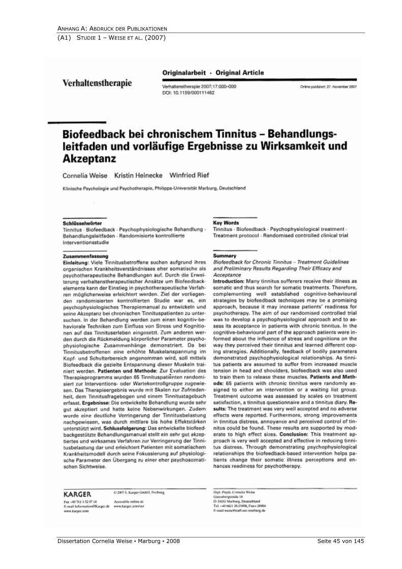
ANHANG A: ABDRUCK DER PUBLIKATIONEN
(A1) STUDIE 1 – WEISE ET AL. (2007)
Dissertation Cornelia Weise Marburg 2008 Seite 45 von 145

ANHANG A: ABDRUCK DER PUBLIKATIONEN
(A1) STUDIE 1 – WEISE ET AL. (2007)
Seite 46 von 145 Dissertation Cornelia Weise Marburg 2008
ANHANG A: ABDRUCK DER PUBLIKATIONEN
(A1) STUDIE 1 – WEISE ET AL. (2007)
Dissertation Cornelia Weise Marburg 2008 Seite 47 von 145
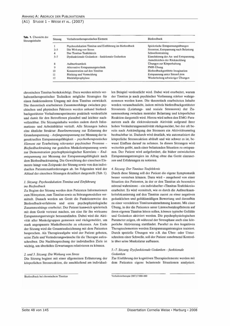
ANHANG A: ABDRUCK DER PUBLIKATIONEN
(A1) STUDIE 1 – WEISE ET AL. (2007)
Seite 48 von 145 Dissertation Cornelia Weise Marburg 2008
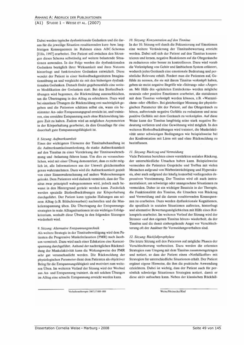
ANHANG A: ABDRUCK DER PUBLIKATIONEN
(A1) STUDIE 1 – WEISE ET AL. (2007)
Dissertation Cornelia Weise Marburg 2008 Seite 49 von 145
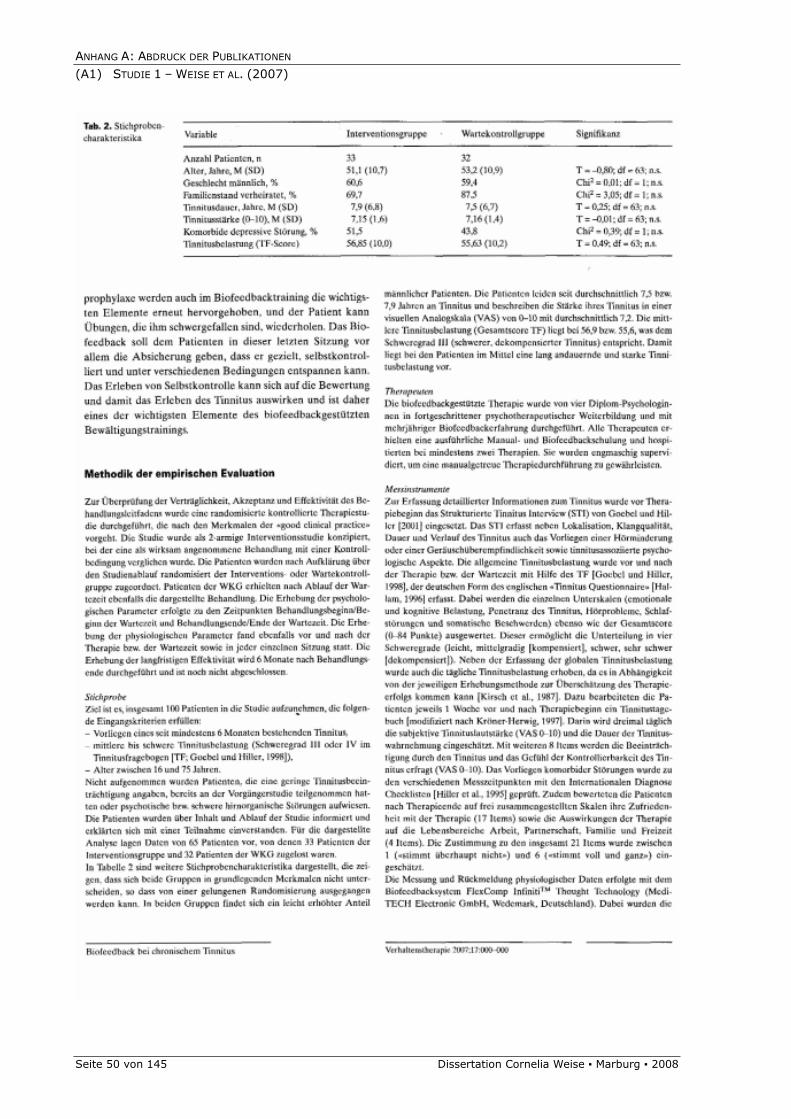
ANHANG A: ABDRUCK DER PUBLIKATIONEN
(A1) STUDIE 1 – WEISE ET AL. (2007)
Seite 50 von 145 Dissertation Cornelia Weise Marburg 2008

ANHANG A: ABDRUCK DER PUBLIKATIONEN
(A1) STUDIE 1 – WEISE ET AL. (2007)
Dissertation Cornelia Weise Marburg 2008 Seite 51 von 145
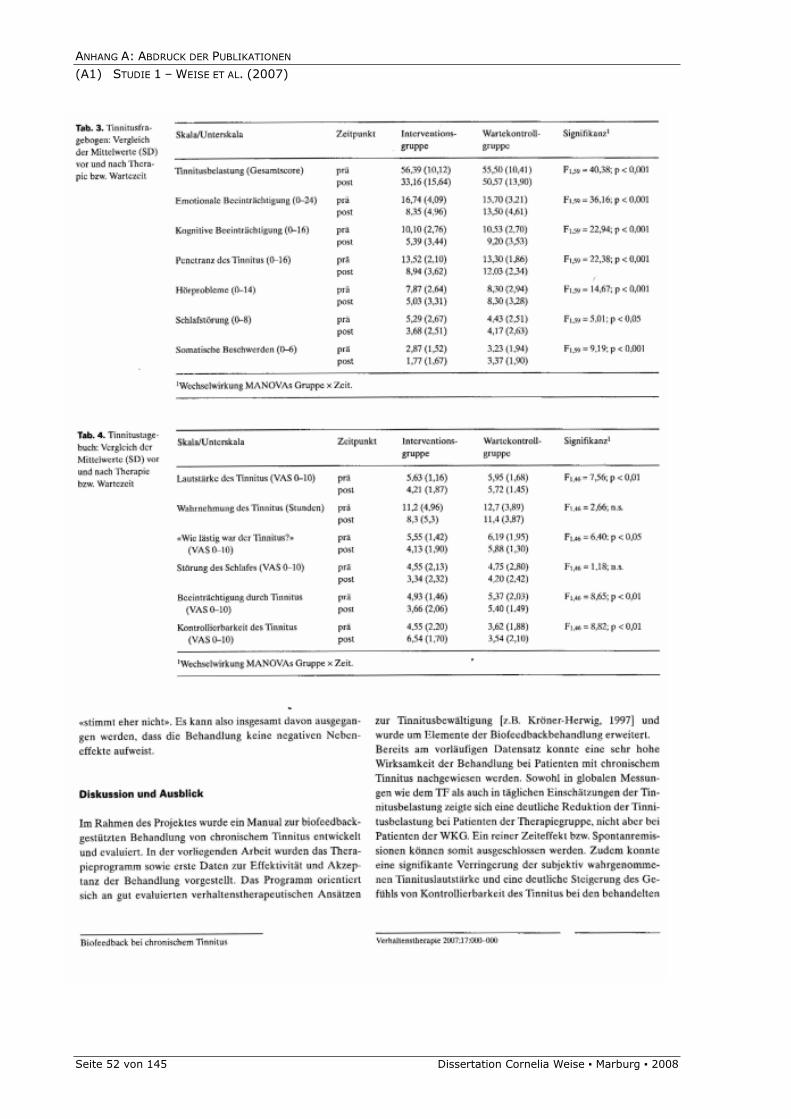
ANHANG A: ABDRUCK DER PUBLIKATIONEN
(A1) STUDIE 1 – WEISE ET AL. (2007)
Seite 52 von 145 Dissertation Cornelia Weise Marburg 2008
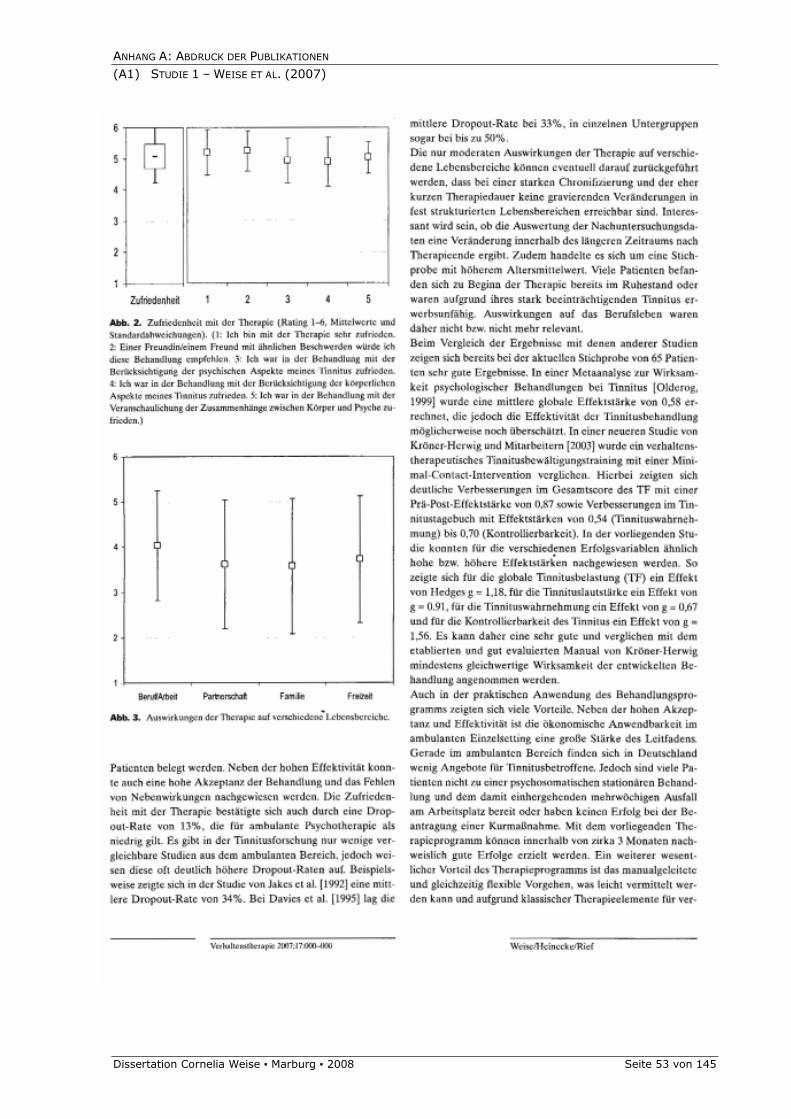
ANHANG A: ABDRUCK DER PUBLIKATIONEN
(A1) STUDIE 1 – WEISE ET AL. (2007)
Dissertation Cornelia Weise Marburg 2008 Seite 53 von 145

ANHANG A: ABDRUCK DER PUBLIKATIONEN
(A1) STUDIE 1 – WEISE ET AL. (2007)
Seite 54 von 145 Dissertation Cornelia Weise Marburg 2008
ANHANG A: ABDRUCK DER PUBLIKATIONEN
(A1) STUDIE 1 – WEISE ET AL. (2007)
Dissertation Cornelia Weise Marburg 2008 Seite 55 von 145


ANHANG A: ABDRUCK DER PUBLIKATIONEN
(A2) STUDIE 2 – WEISE ET AL. (SUBMITTED-A)
Dissertation Cornelia Weise Marburg 2008 Seite 57 von 145
Title of the manuscript: Biofeedback-based behavioural treatment for chronic tinnitus—
results of a randomised controlled trial
Running title: Biofeedback-based treatment for chronic tinnitus
Submission date: January 6th, 2008
ANHANG A: ABDRUCK DER PUBLIKATIONEN
(A2) STUDIE 2 – WEISE ET AL. (SUBMITTED-A)
Seite 58 von 145 Dissertation Cornelia Weise Marburg 2008
Abstract
Objectives: Many tinnitus sufferers suspect an organic cause for their tinnitus and therefore
look for medical rather than psychological treatments. Etiological models assume that tinnitus is asso-
ciated with negative appraisal, dysfunctional attention shift and heightened psychophysiological
arousal. Thus cognitive-behavioural interventions and biofeedback are commonly suggested treat-
ments. We developed and investigated the efficacy of a biofeedback-based cognitive-behavioural
treatment.
Methods: One hundred and thirty tinnitus patients were randomly assigned to an intervention or
a waitlist group. Treatment consisted of 12 sessions of biofeedback-based behavioural intervention
over a 3-month period. Patients in the waitlist group received treatment after the waiting period.
Results: Results showed clear improvements regarding tinnitus annoyance, diary ratings of
loudness and feelings of controllability. Furthermore, changes in control and coping cognitions as well
as in depressive symptoms were detected. Improvements were maintained over a 6-month follow-up
period where medium to large effect sizes were observed.
Conclusions: The developed treatment is highly accepted and leads to clear and stable im-
provements. Through demonstrating psychophysiological interrelationships, the treatment enables
patients to change their somatic illness perceptions to a more psychosomatic point of view.
Keywords: Tinnitus, CBT, psychophysiological treatment, RCT
Trial registration: This study is registered with ClinicalTrials.gov, number NCT00397007.

ANHANG A: ABDRUCK DER PUBLIKATIONEN
(A2) STUDIE 2 – WEISE ET AL. (SUBMITTED-A)
Dissertation Cornelia Weise Marburg 2008 Seite 59 von 145
Introduction
Tinnitus is defined as an acoustic perception such as ringing or buzzing in the ear in the ab-
sence of any apparent auditory stimulation. It is characterised as subjective if it is not audible to an
external listener and as chronic if it lasts for more than 6 months.
Tinnitus is very common in the population, with epidemiological studies showing prevalence
rates up to 14% for prolonged spontaneous tinnitus and 1–3% for severe and distressing tinnitus
(Davis & El Refaie, 2000). These data confirm results of a survey in Germany, where prevalence rates
of up to 4% for chronic distressing tinnitus were detected (Pilgramm et al., 1999). Severe tinnitus—or
decompensated tinnitus—is characterised by subjective reports of intrusiveness as well as cognitive
and emotional distress. It often leads to difficulties in communication and sleeping and disrupts every-
day activities. Furthermore, tinnitus is often accompanied by comorbid psychological disorders such
as depression, anxiety and somatoform disorders.
To date, no theoretical model has fully explained the processes of tinnitus development and
maintenance. However, the habituation model by Hallam and colleagues (1984) accounts for the fact
that neither subjective loudness nor quality of the ear noises are substantially related to the degree of
suffering and annoyance (Kirsch et al., 1989a). The habituation model assumes that the normal
response to tinnitus is habituation, while permanent tinnitus suffering results from a failure in
habituation. As possible causes of missing habituation, the authors propose different factors such as
the characteristics of the stimulus and the perceiver, the significance of the stimulus, and central state
characteristics such as the level of cortical arousal (Hallam, 1987). The assumption of a heightened
arousal level is supported by subjective reports of many tinnitus sufferers and first results of recent
studies. Rief and colleagues (Rief et al., 2004) found first hints for a heightened physiological arousal
level in tinnitus patients while listening to a tinnitus-similar sound. A study by Hébert and Lupien
(2007) evidenced a link between tinnitus, stress and changed hormone release.
As a consequence of the habituation model and the expected heightened arousal level, relaxa-
tion techniques and biofeedback have been applied in tinnitus sufferers. Quite contrary to the first
promising results, meta-analytic reviews (Andersson & Lyttkens, 1999; Andersson et al., 1995)
ANHANG A: ABDRUCK DER PUBLIKATIONEN
(A2) STUDIE 2 – WEISE ET AL. (SUBMITTED-A)
Seite 60 von 145 Dissertation Cornelia Weise Marburg 2008
showed that the more controlled the studies were, the less evidence for the efficacy of biofeedback
was found. A study by Haralambous and colleagues (1987) analysed the effects of frontal electromy-
ographic (EMG) biofeedback on tinnitus suffering and showed that, though EMG-levels significantly
decreased, tinnitus related measures did not improve. Kirsch and colleagues (Kirsch et al., 1987) also
investigated the effects of EMG biofeedback on tinnitus disturbance and differentiated between global
measures and daily diary data. Although they found significant improvements on global scales, the
daily diary data revealed little if any treatment effect. Altogether, as a sufficient pool of high quality
studies is missing, definite statements regarding the efficacy of biofeedback are not yet possible.
Many tinnitus sufferers additionally describe emotional and cognitive problems accompanying
their tinnitus, and also the habituation model assumes inadequate coping processes in the individual.
Therefore an adequate treatment has to account for the multiple comorbid complaints; a purely relaxa-
tion-focused treatment appears to be insufficient (Kirsch et al., 1989b). Thus, cognitive-behavioural
therapy (CBT) has been commonly implemented and investigated in various randomised controlled
trials (Andersson et al., 2002; Hiller & Haerkötter, 2005; Kröner-Herwig et al., 2003; Kröner-Herwig
et al., 1995). All studies found improvements regarding global tinnitus distress and annoyance. Some
of the studies additionally reported improved coping abilities, enhanced feelings of controllability and
self-efficacy (Kröner-Herwig et al., 2003; Kröner-Herwig et al., 1995), and improvements regarding
the ratings of depression, anxiety and psychosocial functioning (Andersson et al., 2002; Hiller &
Haerkötter, 2005). A recent Cochrane review (Martinez Devesa et al., 2007) underlined these findings
and showed that CBT led to improvements in global tinnitus severity, quality of life and coping abil-
ity. Similar to previous results, the review did not find clear evidence for changes in perceptual or
sensory parameters such as tinnitus loudness.
Although CBT was shown to be effective, providing psychological treatment alone may be not
sufficient for many patients who experience their tinnitus as caused and maintained by organic factors
(e.g., muscle contractions or a cervical spine syndrome, Bernhardt et al., 2004; Sanchez et al., 2002).
Therefore we decided to combine well-founded CBT techniques with a psychophysiological treatment
approach. This intervention addresses the biomedical illness beliefs of many patients and offers a link
to biopsychosocial concepts. Results of a randomised controlled pilot study (Rief et al., 2005) re-
ANHANG A: ABDRUCK DER PUBLIKATIONEN
(A2) STUDIE 2 – WEISE ET AL. (SUBMITTED-A)
Dissertation Cornelia Weise Marburg 2008 Seite 61 von 145
vealed positive and long-term stable improvements in tinnitus-specific variables, including tinnitus
distress, controllability of tinnitus, and health-related life satisfaction.
The aim of our randomised controlled trial was to improve and to enlarge the biofeedback-
based treatment approach and to evaluate the effects of this treatment on a larger sample and in com-
parison to a waitlist group. We expected a clear efficacy regarding the primary outcome variable,
tinnitus annoyance, in general as well as in diary data. Furthermore, we hypothesised that our treat-
ment would be effective in improving secondary outcome variables like depressive symptoms, coping
and control cognitions, and self-efficacy.
Method
All investigations and treatment sessions of this randomised controlled trial were conducted in
an outpatient treatment center for psychological interventions between January 2005 and November
2007. We aimed to recruit a minimum of 45 patients in each group to detect medium effects with 80%
power at 5% significance (Cohen, 1988). This was based on the results of previous studies (Rief et al.,
2005) and on the waitlist design. As outpatient studies generally show discontinuation rates between
15 and 25%, we planned to recruit at least 60 patients per group in order to compensate for drop-outs.
Sample
In order to be included in the study, patients needed to: (a) have tinnitus for more than 6 months
(chronic tinnitus); (b) have a serious or severe tinnitus annoyance; and (c) be within the age range of
16 to 75 years. Patients with a mild degree of annoyance or with tinnitus following Menière's disease,
as well as patients with psychosis or seriously disabling brain damage, were excluded. Other psychiat-
ric disorders like depression or anxiety were not excluded but accepted as comorbidities.
Patients were recruited by means of newspaper articles and an article on the webpage of the
German Tinnitus Association (Deutsche Tinnitus-Liga e. V.). To check the primary inclusion criteria
of interested patients, we investigated the duration of tinnitus and the tinnitus annoyance with the 12
questions of the Mini-Tinnitus Questionnaire (Hiller & Goebel, 2004). Two hundred and ten patients
reported tinnitus for more than 6 months with serious or severe annoyance and were eligible for the
study. These patients were invited for a detailed diagnostic interview in which further inclusion crite-
ANHANG A: ABDRUCK DER PUBLIKATIONEN
(A2) STUDIE 2 – WEISE ET AL. (SUBMITTED-A)
Seite 62 von 145 Dissertation Cornelia Weise Marburg 2008
ria were assessed. Seventy-two patients were excluded because of insufficient tinnitus annoyance, 2
reported to have no further interest in the study, and 6 failed to show up at the first appointment. The
remaining 130 patients fulfilled all inclusion criteria and were randomly assigned to a waitlist group
(WLG) or an intervention group (IG) by a permuted-block-randomisation proceeding. The random
block was generated by a research worker not involved in the study.
All patients were informed about the treatment procedure and the nature of the study and gave
written informed consent to participate in the study, which was approved by the Ethics Committee of
the German Federation of Psychological Associations (DGPs). None of the included patients was
already included in the pilot study of Rief et al. (2005).
Procedure and assessment points
Subjects of the IG were assessed three times; patients of the WLG were assessed four times.
During the first assessment, two structured diagnostic interviews regarding general psychopathology
and tinnitus-specific variables were conducted, and patients filled in several questionnaires. There-
after, patients of the intervention group received the treatment (approximately 3 months) while pa-
tients of the waitlist group waited for a comparable time period. Afterwards, patients of both groups
were reassessed with the same assessment instruments for the second time. Following this, patients of
the WLG received the treatment and were reassessed for the third time. Follow-up assessments took
place 6 months after the end of the treatment in both groups (see Figure 1 for details).
Assessment instruments
At the first assessment point we assessed general tinnitus characteristics such as duration, hear-
ing problems, medical causes, as well as associated psychological problems with the Structured Tinni-
tus Interview (Goebel & Hiller, 2001) and comorbid psychiatric disorders with the International
Diagnostic Checklists for ICD-10 (Hiller et al., 1995).
The primary outcome measures were global tinnitus annoyance and a daily rating of tinnitus
disturbance. Global tinnitus annoyance was measured by the German version of the Tinnitus Ques-
tionnaire (TQ) (Goebel & Hiller, 1998; Hallam, 1996), which is a 52-item, 3-point self-rating scale
with a total score ranging from 0 to 84. It provides information about the most common complaints on
six subscales including 'emotional distress', 'cognitive distress', 'intrusiveness', 'auditory perceptual
ANHANG A: ABDRUCK DER PUBLIKATIONEN
(A2) STUDIE 2 – WEISE ET AL. (SUBMITTED-A)
Dissertation Cornelia Weise Marburg 2008 Seite 63 von 145
difficulties', 'sleep disturbances' and 'somatic complaints'. Furthermore it allows a classification of the
degree of annoyance from mild (0-30) or moderate (31-46) to serious (47-59) and very severe (60-84).
As global data tend to overestimate treatment effects (Kirsch et al., 1987), patients additionally filled
in a tinnitus diary (modified after Kröner-Herwig, 1997) for one week at all assessment points. In this
diary, patients rated subjective loudness, sleep disturbance, impairment, distress due to tinnitus and
feelings of controllability on visual analogue scales (VAS) ranging from 0 to 10 as well as the dura-
tion of tinnitus perception per day ranging from 0 to 18 hours.
Secondary outcome measures included further self-reported measures. With the German ver-
sion of the Beck Depression Inventory (BDI, Hautzinger et al., 1995) the existence and severity of
depressive symptoms was assessed. The total score ranges from 0 to 63 and indicates mild (11 to 17),
moderate (18 to 23) or severe (24+) depression. General psychopathology was assessed with the self-
rating questionnaire Symptom Checklist (SCL-90R, Derogatis, 1983). This test contains 90 items on a
5-point scale which measure nine primary symptom dimensions. The Global Severity Index (GSI) is a
mean of all items with a higher score indicating a greater symptom severity. Furthermore the Pain-
Related Self-Statements Scale as well as the Pain-Related Control Scale (Flor et al., 1993) were
adopted for tinnitus sufferers and used in the present study. The Tinnitus-Related Self-Statements
Scale (TRSS) assesses with 18 items, which are situation-specific aspects of patients' cognitive coping
and are divided into the sections 'catastrophising' and 'coping'. The Tinnitus-Related Control Scale
(TRCS) measures general attitudes towards the tinnitus with 15 items on two subscales, 'helplessness'
and 'resourcefulness'. Furthermore, patients rated their feelings of perceived self-efficacy on the Gen-
eral Self-efficacy Scale (Schwarzer & Jerusalem, 1995). This 10-item scale assesses the ability to
cope with daily hassles and adaption after experiencing stressful life events. After the end of the
treatment, patients reported their satisfaction with the therapy and possibly occurring negative side
effects on a self-developed, 6-point, 19-item scale with higher values indicating higher satisfaction
and more adverse effects, respectively.
Intervention
The treatment consisted of 12 1-hour sessions of biofeedback-based behavioural intervention
which took place as individual therapy and were conducted by four therapists. Each session contained
ANHANG A: ABDRUCK DER PUBLIKATIONEN
(A2) STUDIE 2 – WEISE ET AL. (SUBMITTED-A)
Seite 64 von 145 Dissertation Cornelia Weise Marburg 2008
physiological and psychological treatment elements and followed a structured manual (Weise et al.,
2007). One main element in all 12 sessions was a biofeedback-based relaxation training for individual
highly tensed muscles. Different training techniques with and without feedback were implemented to
enhance self-control over bodily processes. Furthermore, biofeedback played a decisive role for de-
monstrating psychophysiological interrelationships (e.g. bodily responses on different stress tasks).
The other main treatment element was a collection of CBT techniques, which were also implemented
in every session. The main purpose of the first session was to provide information about tinnitus,
hearing and possible etiological models. Sessions 2 and 3 focused on the influence of stress on tinni-
tus suffering and maintenance. The importance of cognitions on emotions and tinnitus was explained
and demonstrated in the sessions 4 to 8. Therefore, individual stressful events and their effect on tinni-
tus were analysed, and possible alternative cognitions as well as helpful coping strategies were deve-
loped. Another important coping strategy was implemented in the sessions 8 to 10, when patients
practised directing attention towards and away from their tinnitus. Session 11 focused on the role of
avoidance behaviour in social situations. The purpose of the last session was to resume the individual-
ly important treatment elements and to provide strategies for a relapse prevention.
Statistical analyses
All computations were carried out using the SPSS statistical package (Version 12.0). Possible
baseline differences between the groups regarding the sociodemographic characteristics were tested
with chi-square and t tests. Differences regarding the primary and secondary outcome variables were
tested with multivariate analyses of variance (MANOVA).
Results of the randomised controlled trial (pre-post comparison of IG and WLG) were firstly
calculated as completer analyses. Single missing values were replaced with imputed values using the
multiple imputation procedure and the NORM program (Schafer & Graham, 2002). Following this,
results were calculated on an intention-to-treat basis, and missing data of the dropouts were replaced
according to the principle of 'last observation carried forward'.
To identify differences between the groups, several repeated-measures, Bonferroni-corrected
MANOVAs with the factors group (IG and WLG) and time (pre- and post-treatment respectively
waiting) were performed. If the main effect showed significance, univariate ANOVAs were con-
ANHANG A: ABDRUCK DER PUBLIKATIONEN
(A2) STUDIE 2 – WEISE ET AL. (SUBMITTED-A)
Dissertation Cornelia Weise Marburg 2008 Seite 65 von 145
ducted subsequently. For the following investigation of treatment efficacy in the WLG (cross-over),
also several repeated measures, Bonferroni-corrected MANOVAs with the factor time (pre- and post-
treatment) and univariate ANOVAs were conducted.
To analyse the overall treatment effect with general effect size measures as well as the mainte-
nance of the improvements, we merged the data of the two groups for the three assessment points
(pre, post, follow-up). Then we carried out several repeated measures, Bonferroni-corrected
MANOVAs with the factor time (pre, post, follow up), followed by univariate ANOVAs, if the
multivariate tests revealed significance. Additionally, further MANOVAs were used to investigate
changes from pre- to post-treatment as well as from pre-treatment to follow-up.
Primary outcomes were the global tinnitus annoyance and the daily rating of tinnitus distress.
Secondary outcomes included general psychopathology, self-efficacy, coping and control cognitions,
and satisfaction with treatment.
Effect sizes for the randomised controlled part of the study were computed using Hedges' g:
gHedges=
¯¯¯X EG -
¯¯¯X KG
(nEG-1)*s2EG
+(nKG-1)*s2KG
nEG +nKG-2
which refers to the difference between the means of the two
groups (IG and WLG) divided by the pooled standard deviation (intergroup effect size; Hedges &
Olkin, 1995). For the evaluation of the general improvement (pre-post) as well as of the maintenance
of the results for the whole group, we computed effect sizes using the formula by McGaw and Glass
(1980): g=M1 - M2
s12 + s2
2-2r*s1*s2 which accounts for pre-post or pre-follow-up correlations of the out-
come measures. All effect sizes were computed separately for completer and intention-to-treat analy-
ses.
Results
Sample characteristics and patient flow
An overview of the patient flow in the different stages of the study according to the CONSORT
statement is displayed in Figure 1. From the 130 patients that were included in the study, five patients

ANHANG A: ABDRUCK DER PUBLIKATIONEN
(A2) STUDIE 2 – WEISE ET AL. (SUBMITTED-A)
Seite 66 von 145 Dissertation Cornelia Weise Marburg 2008
dropped out before the beginning of the treatment, seven IG patients (5.6%) discontinued intervention
and seven WLG patients (5.6%) discontinued the waiting period. Another seven patients (5.6%) of the
WLG discontinued the treatment after waiting. Based on those patients who started the intervention
(n = 118) the overall attrition rate is 11.9% at post-treatment. Of the 104 patients who completed the
treatment, 89 patients returned the follow-up assessments, resulting in a return rate of 85,6%1.
Background data and baseline characteristics are shown in Table 1. Subjects suffered predomi-
nantly from severe tinnitus, and their tinnitus lasted in average for more than seven years. The chi-
square and t tests for the sample characteristics as well as the MANOVAs for the primary and secon-
dary outcome variables did not show any significant differences between the IG and the WLG at pre-
treatment, which implies effective randomisation.
Randomised controlled trial
Main outcome—tinnitus distress
Results for the primary outcome variables are shown in Table 2. For the Tinnitus Questionnaire
(TQ) and the tinnitus diary, the MANOVA showed a statistically significant interaction for time x
group (F[13,97]=5.16; p<.001) for the completer analysis. In the subsequent univariate ANOVAs,
significant interactions for the total score of the TQ and for all subscales were detected. Patients in the
intervention group reported significantly less emotional and cognitive distress, perceived their tinnitus
as less intrusive and reported less auditory perceptual difficulties, less sleep disturbance and less so-
matic complaints than at pre-treatment, while patients of the WLG did not improve significantly. Re-
garding the results of the tinnitus diary, the subsequent ANOVAs also showed statistically significant
interactions for time x group. Patients in the IG rated their tinnitus after the treatment as quieter and
less disruptive of sleep, they felt less tormented, impaired and distressed by the tinnitus, and they also
perceived higher feelings of controllability regarding the ear noises. Furthermore, patients reported
the time listening to tinnitus daily as significantly decreased in comparison to pre-treatment and to the
WLG.
1 At this time, 11 follow up assessments are still outstanding and 4 patients refused to send back the
questionnaires.
ANHANG A: ABDRUCK DER PUBLIKATIONEN
(A2) STUDIE 2 – WEISE ET AL. (SUBMITTED-A)
Dissertation Cornelia Weise Marburg 2008 Seite 67 von 145
When an intention-to-treat approach was used, both the MANOVA (F[13,111] = 4.15;
p < .001) and all subsequent univariate ANOVAs confirmed the significant interactions.
To investigate the clinical relevance of the achieved results, effect sizes were computed sepa-
rately. For the completer analysis we found large effect sizes for the total score of the TQ with
Hedges' g of 1.15 as well as for the TQ subscales emotional and cognitive distress, intrusiveness and
auditory perceptual difficulties in the range of 0.85 to 1.07 (see Table 2 for details). Regarding the
tinnitus diary large effect sizes were detected for the impairment and the feelings of controllability
(0.84 and 0.81), while medium effect sizes in the range of 0.55 to 0.77 were found for the remaining
ratings.
As expected, effect sizes for the intention-to-treat analyses showed lower, but still medium or
large, effect sizes for most variables (see Table 2 for details).
Secondary outcome measures
The MANOVA for the secondary outcome measures showed a statistically significant main in-
teraction effect for time x group (F[7,103]=10.36; p<.001). Subsequent univariate ANOVAs (Table 2)
showed significant interactions for the BDI and the Global Severity Index (GSI) of the SCL-90-R,
which means that patients of the IG reported less depressive and other psychopathological symptoms
compared to pre-assessment and to the WLG. Furthermore the ANOVAs showed a significant inter-
action for time and group for the General Self-Efficacy Scale, which suggests that patients of the IG
but not those of the WLG perceived their feelings of self-efficacy as clearly improved after the treat-
ment. Significant interactions were also found for the subscales of the Tinnitus-related Control Scale
and for those of the Tinnitus-related Self-Statements Scale. Patients in the IG reported less feelings of
helplessness, increased feelings of resourcefulness, less catastrophising self-statements, and more
helpful coping self-statements compared to pre-treatment and to the WLG.
Intention-to-treat analysis again confirmed significant main interaction for time x group
(F[7,117]= .37; p < .001) in the MANOVA and significant interactions for all variables in the univari-
ate ANOVAs.
The computed completer effect sizes showed large effect sizes between g=0.97 and g=1.17 for
the subscales of the TRCS and the TRSS. In contrast, Hedges' g for the BDI (0.13) and the GSI (0.03)
ANHANG A: ABDRUCK DER PUBLIKATIONEN
(A2) STUDIE 2 – WEISE ET AL. (SUBMITTED-A)
Seite 68 von 145 Dissertation Cornelia Weise Marburg 2008
did not evidence clinical relevance for the significant improvements. For self-efficacy at least a small
effect size (0.27) was detected. Though groups were randomised and the pre-treatment MANOVA did
not show significant differences between the groups, slight group differences existed. Therefore addi-
tionally the effect-gain was computed by subtracting the pre-effect-size from the post-effect-size. This
procedure showed an effect-gain of 0.30 and 0.33 for the BDI and the GSI respectively. Also for the
self efficacy a medium effect gain of 0.55 was detected.
The effect sizes computed for the intention-to-treat analysis showed comparable but somewhat
smaller values (see Table 2 for details).
Cross-over
After the waiting period the waitlist group also received the treatment, and 52 patients finished.
Regarding the primary outcome variables, the MANOVA showed a significant main time effect
(F[13,39] = 8.43; p < .001). In the subsequent ANOVAS we found significant time effects for the
total score of the TQ, its subscales and for all diary VAS ratings. Also, for the secondary outcome
variables, a significant main time effect in the MANOVA (F[7,45] = 17.55; p < .001) and significant
effects for all subscales in the ANOVAs were detected.
Maintenance of the results
To estimate the overall effect of the treatment, we merged the data of the two groups after all
patients of the WLG also entered treatment and were followed-up 6 months later. Results for primary
and secondary outcome variables for the merged group with mean scores, F-statistics for the pre-post
and the pre-follow-up MANOVAs as well as effect sizes are shown in Table 3.
For the primary outcome variables, the general MANOVA showed a statistically significant
main time effect (pre, post, follow-up) (F[26,63]=9.82; p<.001). The additional MANOVAs investi-
gating changes between pre- and post-treatment (F[13,76]=15.07; p<.001) and between pre-treatment
and follow-up (F[13,76]=11.66; p<.001) also showed significant main time effects. All subsequent
ANOVAs evidenced significant time effects.
Regarding the secondary outcome, the main MANOVA showed a statistically significant main
time effect (pre, post, follow-up) (F[14,75]=15.85; p<.001). This result was supported by the subse-
quent pre-post MANOVA (F[7,82]=31.17; p<.001) as well as by the pre-follow-up MANOVA

ANHANG A: ABDRUCK DER PUBLIKATIONEN
(A2) STUDIE 2 – WEISE ET AL. (SUBMITTED-A)
Dissertation Cornelia Weise Marburg 2008 Seite 69 von 145
(F[7,82]=13.60; p<.001) which both showed significant main time effects. Additionally all subse-
quent ANOVAs revealed significant time effects for the several subscales.
The intention-to-treat-analyses for the merged group included the 14 treatment dropouts (miss-
ing data replaced by pre-treatment data) as well as the 15 patients who failed to return the follow-up
questionnaires (missing data replaced by post-treatment data). For the primary outcome variables, the
general MANOVA (pre, post, follow-up) (F[26,92]=8.75; p<.001), the pre-post MANOVA
(F[13,105]=14.61; p<.001), the pre-follow-up MANOVA (F[13,105]=12.78; p<.001) as well as all
subsequent ANOVAs confirmed, with the significant time effects, the results of the completer analy-
ses. For the secondary outcome variables, significant main time effects were found for the general
MANOVA (F[14,104]=14.26; p<.001), the pre-post MANOVA (F[7,111]=28.57; p<.001) and the
pre-follow-up MANOVA (F[7,111]=15.30; p<.001). All subsequent ANOVAs showed significant
time effects.
The clinical relevance of the data was further investigated by computing effect sizes which are
shown in Table 3. Analysing the treatment completer showed pre-post effect sizes for the Tinnitus
Questionnaire and its subscales in the range of 0.62-1.41 and pre-follow-up effect sizes in the range of
0.63-1.17. For the several visual analogue scales of the tinnitus diary, we found pre-post effect sizes
between 0.56-0.90 and pre-follow-up effect sizes in the range of 0.35-0.79. Regarding the secondary
outcome variables, we found for the BDI effect sizes of 0.70 (pre-post) and 0.45 (pre-follow-up), and
for the GSI, effect sizes of 0.57 (pre-post) and 0.37 (pre-follow-up). For self-efficacy, we found me-
dium pre-post (0.58) and follow-up (0.57) effect sizes. Large effect sizes were detected for the sub-
scales of the TRCS and the TRSS in the range of 0.93-1.26 for pre-post and 0.69-0.81 for pre-follow-
up. As expected, effect sizes for the intention-to-treat analyses were somewhat smaller.
Satisfaction with treatment and adverse effects
In addition to the above presented data for the primary and secondary outcome, patients rated
their satisfaction with the treatment as well as possibly occurring adverse effects after the end of the
treatment. The internal consistency of this scale indicates a unidimensional scale with a Cronbach's
alpha of .91 and with item-total correlations in the range of .24 to .77. Descriptive analyses showed a
mean score of 5.16 (SD = 0.51, range = 1 to 6) for the satisfaction with therapy subscale, which
ANHANG A: ABDRUCK DER PUBLIKATIONEN
(A2) STUDIE 2 – WEISE ET AL. (SUBMITTED-A)
Seite 70 von 145 Dissertation Cornelia Weise Marburg 2008
means that patients were quite content with the treatment. Patients agreed to items like 'I would
recommend this treatment to a friend with a similar disorder' (mean = 5.34, SD = 0.65). With a mean
of 1.51 (SD = 0.63, range = 1 to 6) the adverse effects subscale indicated that the majority of the pa-
tients did not experience negative side effects caused by the treatment.
Discussion
We investigated the efficacy of a biofeedback-based behavioural treatment on tinnitus patients
using a randomised controlled design. Furthermore, we investigated the overall effect of the treatment
and the maintenance of treatment effects over 6 months.
The results for the group comparison showed statistically significant improvements in patients
of the intervention group for all primary outcome variables. Improvements in tinnitus annoyance were
not only found in global but also in diary data. Medium-to-large effect sizes for most variables sup-
ported the clinical relevance of the data.
Regarding the secondary outcome variables we also found significant improvements in the in-
tervention group. The computed effect sizes showed clinical relevance only for self-efficacy and the
subscales of the Tinnitus-Related Control and Self-Statements scales. This result indicated less
feelings of helplessness and catastrophising cognitions as well as improved feelings of coping ability
and resourcefulness in treated patients. For depressive and general psychopathological symptoms the
computed effect sizes revealed no clinical relevance of the achieved improvements, but the effect-gain
showed at least small effect.
Results of the merged group showed significant improvements for all primary and secondary
outcome variables, which implies that improvements were maintained over the 6-month follow-up
period. These significant time effects were underpinned by at least small but mostly medium-to-large
pre-post and pre-follow-up effect sizes for all variables. Comparing effect sizes for post-treatment and
follow-up confirmed no worsening after end of treatment for all but three variables. Therefore, we can
conclude that the improvements in both tinnitus annoyance and tinnitus-associated psychological
problems achieved by the developed treatment approach are stable.
ANHANG A: ABDRUCK DER PUBLIKATIONEN
(A2) STUDIE 2 – WEISE ET AL. (SUBMITTED-A)
Dissertation Cornelia Weise Marburg 2008 Seite 71 von 145
In conclusion, the results showed that the treatment led to significant and long-term stable im-
provements in tinnitus annoyance, cognitive and emotional distress, intrusiveness of the tinnitus, sleep
disturbance and somatic complaints. With large improvements in the Tinnitus Questionnaire from pre
(52.5) to post (33,9) and follow-up (34,7) the average degree of tinnitus annoyance in the patients
after the treatment was no longer in the serious (total score: 47 to 59) but, rather, in the moderate
range (31-46). Thus many tinnitus sufferers can be named as compensated instead of decompensated.
The effects regarding the Tinnitus Questionnaire are as good as or even better than those of previous
trials. The improvements in the perceived tinnitus loudness and the daily tinnitus perception can be
named as exceptionally positive when compared with results of previous studies, which found only
little if any improvements with only small or moderate effect sizes (Andersson et al., 2002; Hiller &
Haerkötter, 2005; Kaldo et al., 2007; Kröner-Herwig et al., 2003), particularly for diary data.
Regarding depressive and general psychopathological symptoms, contrary to our hypothesis,
the inter-group comparison failed to show clinically relevant improvements for the BDI and the GSI.
However, the investigation of the merged group showed medium pre-post but small pre-follow-up
effects for the improvement in depressive and general psychopathological symptoms. Our results are
consistent with previous findings (Hiller & Haerkötter, 2005; Kaldo et al., 2007; Kröner-Herwig et al.,
2003). Descriptive analyses of our data showed that patients reported only mild depression (both at
pre- [14.9], post- [11.3] and follow-up assessment [12.0]) and had only slightly higher-than-average
values in the GSI (T-values 62, 60 and 60), which may be one reason for the small changes. Another
reason may be our treatment approach itself, which is possibly not sufficient to treat comorbid disor-
ders and should therefore be extended if tinnitus is accompanied by serious psychopathological prob-
lems.
Further quite important improvements after the end of the treatment were shown for general
self-efficacy and tinnitus-related control and coping cognitions. Previous reviews showed the im-
portance of coping and recommended increasing coping abilities as a decisive treatment goal in tinni-
tus therapy (Andersson et al., 1995; Kirsch et al., 1989b). Scott and colleagues (Scott et al., 1985)
emphasised additionally the relevance of relaxation as an important coping technique. As we com-
bined CBT techniques to increase coping abilities with biofeedback-based relaxation, possibly this
ANHANG A: ABDRUCK DER PUBLIKATIONEN
(A2) STUDIE 2 – WEISE ET AL. (SUBMITTED-A)
Seite 72 von 145 Dissertation Cornelia Weise Marburg 2008
combination was one reason for the large improvements in coping and control cognitions. Another
reason may be the biofeedback itself, which has high face validity, leads to positive treatment expec-
tations, increases patients' feelings of self-control, and decreases feelings of helplessness.
Concerning the long-term results of self-efficacy, as well as coping and control cognitions,
slightly lower effect sizes were found. As control beliefs are relatively stable constructs (Flor et al.,
1993; Williams & Thorn, 1989) it may be difficult for patients to consistently implement the new
beliefs and strategies in everyday life after the end of treatment.
Our study leads to some important conclusions. The efficacy of the developed treatment ap-
proach was investigated based on a large sample size and the results showed clear and long-term sta-
ble improvements over a cost-effective short treatment period. Patients reported no negative side ef-
fects and rated their satisfaction with the therapy as very good. The satisfaction with the treatment is
additionally reflected in the treatment dropout rate of 11.9%, which is considered as low when com-
pared with general outpatient clinical research (Wierzbicki & Pekarik, 1993) and tinnitus treatment
studies which show dropout rates up to 50% (Davies et al., 1995; Jakes et al., 1992). Additionally, it
seems that in particular males with reluctant attitudes towards solely psychological interventions
benefit from the developed approach.
There are further reasons why a biofeedback-based behavioural treatment may presumably be
better accepted by many tinnitus sufferers than an exclusively psychological treatment approach. With
its biofeedback component, our treatment also addresses patients with somatic illness perceptions,
who are very common among tinnitus sufferers, and patients feel they are being taken seriously in
their somatic point of view. Through demonstrating psychophysiological interrelationships, the treat-
ment helps patients to bridge the gap from biomedical to psychosocial illness beliefs. This may facili-
tate the critical examination of accompanied psychological problems and may increase the efficacy of
cognitive-behavioural techniques. Studies regarding somatoform disorders could affirm these assump-
tions. For instance Wickramasekera (1989) assumed less efficacy of purely psychotherapeutic treat-
ments in pain patients but suggested adjunct therapies like biofeedback which use psychophysiologi-
cal demonstrations to clarify the influence of mental factors on bodily reactions.

ANHANG A: ABDRUCK DER PUBLIKATIONEN
(A2) STUDIE 2 – WEISE ET AL. (SUBMITTED-A)
Dissertation Cornelia Weise Marburg 2008 Seite 73 von 145
Despite the promising results of our study, there is one obvious shortcoming. The study allows
no statements concerning the additive effect of biofeedback in this integrative approach, because the
trial design contains only a waitlist and not an active treatment control group. Only with an active
control group (CBT without biofeedback), the additive effect of biofeedback would be measurable.
Therefore we can compare our results only with the pilot study of Rief and colleagues (2005) and with
trials investigating CBT without biofeedback. In comparison to those studies, our results show as high
and partially even higher effect sizes, which may indicate that the additional application of biofeed-
back can improve the treatment outcome. At the very least, it offers an important alternative if people
reject pure psychological intervention.
In consideration of previous results it seems to be recommendable to implement our promising
treatment program in a stepped care approach, as it was recently suggested (Kröner-Herwig et al.,
2003), consisting of (1) medical treatment, (2) education and (3) intense cognitive-behavioural ther-
apy extended by biofeedback. Such a stepped care approach that includes psychophysiological ther-
apy strategies may improve the general treatment outcome and may especially be very helpful for
tinnitus sufferers with somatic illness attributions.
Acknowledgments/Grants
This study was supported by the German Research Foundation (Ri 574/12-1). Particularly we
thank all our patients for participating as well as Anne Raudschus, Kristin Schwarz and Eva Petrenko
for assistance with data collection.
Notes
The trial protocol was approved by the Ethics Committee of the German Federation of Psycho-
logical Associations (DGPs) and meets the ethical standards of the Declaration of Helsinki.
This study was registered with ClinicalTrials.gov, number NCT00397007.

ANHANG A: ABDRUCK DER PUBLIKATIONEN
(A2) STUDIE 2 – WEISE ET AL. (SUBMITTED-A)
Seite 74 von 145 Dissertation Cornelia Weise Marburg 2008
References
Andersson, G., & Lyttkens, L. (1999). A meta-analytic review of psychological treatments for tinnitus.
British Journal of Audiology, 33(4), 201-210.
Andersson, G., Melin, L., Hägnebo, C., & Scott, B. (1995). A review of psychological treatment
approaches for patients suffering from Tinnitus. Annals of Behavioral Medicine, 17(4), 357-366.
Andersson, G., Stroemgren, T., Stroem, L., & Lyttkens, L. (2002). Randomized controlled trial of
Internet-based cognitive behavior therapy for distress associated with tinnitus. Psychosomatic
Medicine, 64(5), 810-816.
Bernhardt, O., Gesch, D., Schwahn, C., Bitter, K., Mundt, T., Mack, F., et al. (2004). Signs of
temporomandibular disorders in tinnitus patients and in a population-based group of volunteers:
results of the Study of Health in Pomerania. Journal of Oral Rehabilitation, 31(4), 311-319.
Cohen, J. (1988). Statistical power analysis for the behavioral sciences (2nd ed.). Hillsdale, NJ:
Erlbaum.
Davies, S., McKenna, L., & Hallam, R. S. (1995). Relaxation and cognitive therapy: A controlled trial
in chronic tinnitus. Psychology and Health, 10(2), 129-143.
Davis, A., & El Refaie, A. (2000). Epidemiology of tinnitus. In R. S. Tyler (Ed.), Tinnitus Handbook
(pp. 1-21). San Diego: Singular Publishing Grout.
Derogatis, L. R. (1983). The SCL-90 Manual: scoring administration procedures for the SCL-90-R.
Baltimore: John Hopkins University School of Medicine, Clinical Psychometrics Unit.
Flor, H., Behle, D. J., & Birbaumer, N. (1993). Assessment of pain-related cognitions in chronic pain
patients. Behaviour Research and Therapy, 31(1), 63-73.
Goebel, G., & Hiller, W. (1998). Tinnitus-Fragebogen (TF) [Tinnitus Questionnaire (TQ)]. Göttingen:
Hogrefe.
Goebel, G., & Hiller, W. (2001). STI Strukturiertes Tinnitus-Interview [Structured Tinnitus Interview].
Göttingen: Hogrefe.
Hallam, R. S. (1987). Psychological approaches to the evaluation and management of tinnitus distress.
In J. Hazell (Ed.), Tinnitus. Edinburgh: Churchill Livingstone.

ANHANG A: ABDRUCK DER PUBLIKATIONEN
(A2) STUDIE 2 – WEISE ET AL. (SUBMITTED-A)
Dissertation Cornelia Weise Marburg 2008 Seite 75 von 145
Hallam, R. S. (1996). Manual of the Tinnitus Questionnaire (TQ). London: The Psychological
Corporation.
Hallam, R. S., Rachman, S., & Hinchcliffe, R. (1984). Psychological aspects of tinnitus. In S.
Rachman (Ed.), Contributions to medical psychology (Vol. 3, pp. 31-53). Oxford: Pergamon.
Haralambous, G., Wilson, P. H., Platt-Hepworth, S., Tonkin, J. P., Hensley, V. R., & Kavanagh, D.
(1987). EMG biofeedback in the treatment of tinnitus: An experimental evaluation. Behaviour
Research and Therapy, 25(1), 49-55.
Hautzinger, M., Bailer, M., Worall, H., & Keller, F. (1995). Beck-Depressions-Inventar (BDI). [Beck-
Depression-Inventory (BDI)] (Vol. 2). Bern: Hans Huber.
Hebert, S., & Lupien, S. J. (2007). The sound of stress: blunted cortisol reactivity to psychosocial
stress in tinnitus sufferers. Neuroscience Letters, 411(2), 138-142.
Hedges, L. V., & Olkin, I. (1995). Statistical methods for meta-analysis. Orlando: Academic Press.
Hiller, W., & Goebel, G. (2004). Rapid assessment of tinnitus-related psychological distress using the
Mini-TQ. International Journal of Audiology, 43(10), 600-604.
Hiller, W., & Haerkötter, C. (2005). Does sound stimulation have additive effects on cognitive-
behavioral treatment of chronic tinnitus? Behaviour Research and Therapy, 43(5), 595-612.
Hiller, W., Zaudig, M., & Mombour, W. (1995). IDCL-Internationale Diagnosen Checklisten für ICD-
10 [IDCL-International Diagnostic Checklists for ICD-10]. Bern, Göttingen: Huber.
Jakes, S. C., Hallam, R. S., McKenna, L., & Hinchcliffe, R. (1992). Group cognitive therapy for
medical patients: An application to tinnitus. Cognitive Therapy and Research, 16(1), 67-82.
Kaldo, V., Cars, S., Rahnert, M., Larsen, H. C., & Andersson, G. (2007). Use of a self-help book with
weekly therapist contact to reduce tinnitus distress: A randomized controlled trial. Journal of
Psychosomatic Research, 63(2), 195-202.
Kirsch, C. A., Blanchard, E. B., & Parnes, S. M. (1987). A multiple-baseline evaluation of the
treatment of subjective tinnitus with relaxation training and biofeedback. Biofeedback and Self
Regulation, 12(4), 295-312.
Kirsch, C. A., Blanchard, E. B., & Parnes, S. M. (1989a). Psychological characteristics of individuals
high and low in their ability to cope with tinnitus. Psychosomatic Medicine, 51(2), 209-217.

ANHANG A: ABDRUCK DER PUBLIKATIONEN
(A2) STUDIE 2 – WEISE ET AL. (SUBMITTED-A)
Seite 76 von 145 Dissertation Cornelia Weise Marburg 2008
Kirsch, C. A., Blanchard, E. B., & Parnes, S. M. (1989b). A review of the efficacy of behavioral
techniques in the treatment of subjective tinnitus. Annals of Behavioral Medicine, 11(2), 58-65.
Kröner-Herwig, B. (Ed.). (1997). Psychologische Behandlung des chronischen Tinnitus
[Psychological treatment of chronic tinnitus]. Weinheim: Beltz PVU.
Kröner-Herwig, B., Frenzel, A., Fritsche, G., Schilkowsky, G., & Esser, G. (2003). The management
of chronic tinnitus: Comparison of an outpatient cognitive-behavioral group training to minimal-
contact interventions. Journal of Psychosomatic Research, 54(4), 381-389.
Kröner-Herwig, B., Hebing, G., van Rijn Kalkmann, U., Frenzel, A., Schilkowsky, G., & Esser, G.
(1995). The management of chronic tinnitus--comparison of a cognitive-behavioural group
training with yoga. Journal of Psychosomatic Research, 39(2), 153-165.
Martinez Devesa, P., Waddell, A., Perera, R., & Theodoulou, M. (2007). Cognitive behavioural
therapy for tinnitus. Cochrane Database of Systematic Reviews(1), Art. No.: CD005233.
McGaw, B., & Glass, G. V. (1980). Choice of the Metric for Effect Size in Meta-Analysis. American
Educational Research Journal, 17(3), 325-337.
Pilgramm, M., Rychlik, R., Lebisch, H., Siedentop, H., Goebel, G., & Kirchhoff, D. (1999). Tinnitus
in der Bundesrepublik Deutschland - eine repräsentative epidemiologische Studie [Tinnitus in
the Federal Republic of Germany - A representative epidemiological study]. HNO aktuell, 7,
261-265.
Rief, W., Sander, E., Guenther, M., & Nanke, A. (2004). Aufmerksamkeitslenkung bei Tinnitus: Eine
experimentelle psychophysiologische Untersuchung [Steering selective attention in tinnitus: An
experimental psychophysiological study]. Zeitschrift für Klinische Psychologie und
Psychotherapie, 33(3), 230-236.
Rief, W., Weise, C., Kley, N., & Martin, A. (2005). Psychophysiologic Treatment of Chronic Tinnitus:
A Randomized Clinical Trial. Psychosomatic Medicine, 67, 833-838.
Sanchez, T. G., Guerra, G. C., Lorenzi, M. C., Brandao, A. L., & Bento, R. F. (2002). The influence of
voluntary muscle contractions upon the onset and modulation of tinnitus. Audiology and Neuro-
Otology, 7(6), 370-375.
ANHANG A: ABDRUCK DER PUBLIKATIONEN
(A2) STUDIE 2 – WEISE ET AL. (SUBMITTED-A)
Dissertation Cornelia Weise Marburg 2008 Seite 77 von 145
Schafer, J. L., & Graham, J. W. (2002). Missing data: Our view of the state of the art. Psychological
Methods, 7(2), 147-177.
Schwarzer, R., & Jerusalem, M. (1995). Generalized Self-Efficacy scale. In J. Weinman, S. Wright &
M. Johnston (Eds.), Measures in health psychology: A user’s portfolio. Causal and control
beliefs (pp. 35-37). Windsor, UK: Nfer-Nelson.
Scott, B., Lindberg, P., Lyttkens, L., & Melin, L. (1985). Psychological treatment of tinnitus. An
experimental group study. Scandinavian Audiology, 14(4), 223-230.
Weise, C., Heinecke, K., & Rief, W. (2007). Biofeedback bei chronischem Tinnitus –
Behandlungsleitfaden und vorläufige Ergebnisse zu Wirksamkeit und Akzeptanz [Biofeedback
for chronic tinnitus - Treatment guidelines and preliminary results regarding their efficacy and
acceptance]. Verhaltenstherapie, 17(4), 220-230.
Wickramasekera, I. (1989). Enabling the somatizing patient to exit the somatic closet: A high-risk
model. Psychotherapy - Theory, Research, Practice, Training, 26(4), 530-544.
Wierzbicki, M., & Pekarik, G. (1993). A meta-analysis of psychotherapy dropout. Professional
Psychology: Research and Practice, 24(2), 190-195.
Williams, D. A., & Thorn, B. E. (1989). An empirical assessment of pain beliefs. Pain, 36(3), 351-
358.
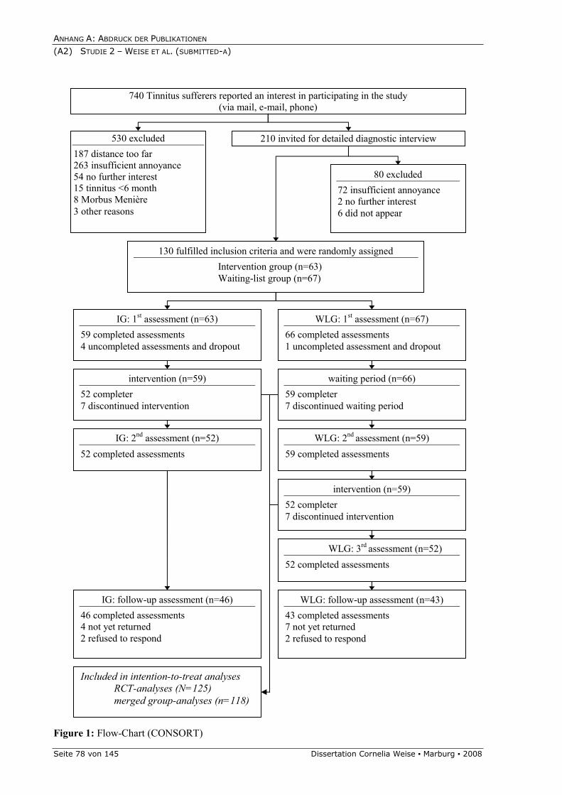
ANHANG A: ABDRUCK DER PUBLIKATIONEN
(A2) STUDIE 2 – WEISE ET AL. (SUBMITTED-A)
Seite 78 von 145 Dissertation Cornelia Weise Marburg 2008
Figure 1: Flow-Chart (CONSORT)
740 Tinnitus sufferers reported an interest in participating in the study (via mail, e-mail, phone)
530 excluded
187 distance too far 263 insufficient annoyance 54 no further interest 15 tinnitus <6 month 8 Morbus Menière 3 other reasons
210 invited for detailed diagnostic interview
80 excluded
72 insufficient annoyance 2 no further interest 6 did not appear
130 fulfilled inclusion criteria and were randomly assigned
Intervention group (n=63) Waiting-list group (n=67)
WLG: 1st assessment (n=67)
66 completed assessments 1 uncompleted assessment and dropout
IG: 1st assessment (n=63)
59 completed assessments 4 uncompleted assessments and dropout
intervention (n=59)
52 completer 7 discontinued intervention
waiting period (n=66)
59 completer 7 discontinued waiting period
IG: 2nd assessment (n=52)
52 completed assessments
WLG: 2nd assessment (n=59)
59 completed assessments
intervention (n=59)
52 completer 7 discontinued intervention
WLG: 3rd assessment (n=52)
52 completed assessments
IG: follow-up assessment (n=46)
46 completed assessments 4 not yet returned 2 refused to respond
WLG: follow-up assessment (n=43)
43 completed assessments 7 not yet returned 2 refused to respond
Included in intention-to-treat analyses
RCT-analyses (N=125)
merged group-analyses (n=118)
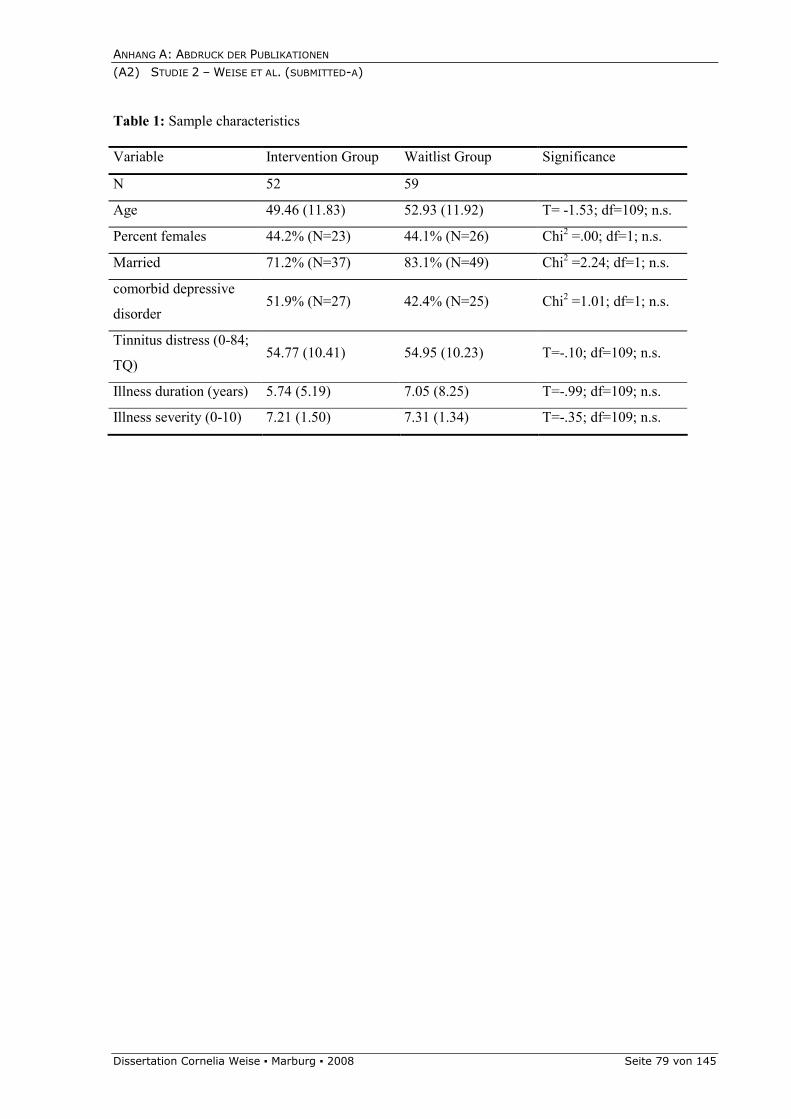
ANHANG A: ABDRUCK DER PUBLIKATIONEN
(A2) STUDIE 2 – WEISE ET AL. (SUBMITTED-A)
Dissertation Cornelia Weise Marburg 2008 Seite 79 von 145
Table 1: Sample characteristics
Variable Intervention Group Waitlist Group Significance
N 52 59
Age 49.46 (11.83) 52.93 (11.92) T= -1.53; df=109; n.s.
Percent females 44.2% (N=23) 44.1% (N=26) Chi2 =.00; df=1; n.s.
Married 71.2% (N=37) 83.1% (N=49) Chi2 =2.24; df=1; n.s.
comorbid depressive
disorder 51.9% (N=27) 42.4% (N=25) Chi2 =1.01; df=1; n.s.
Tinnitus distress (0-84;
TQ) 54.77 (10.41) 54.95 (10.23) T=-.10; df=109; n.s.
Illness duration (years) 5.74 (5.19) 7.05 (8.25) T=-.99; df=109; n.s.
Illness severity (0-10) 7.21 (1.50) 7.31 (1.34) T=-.35; df=109; n.s.

ANHANG A: ABDRUCK DER PUBLIKATIONEN
(A2) STUDIE 2 – WEISE ET AL. (SUBMITTED-A)
Seite 80 von 145 Dissertation Cornelia Weise Marburg 2008
Table 2: RCT—primary and secondary outcomes: mean (SD), F-statistics and effect sizes
variable IG (N=52) WLG (N=59)
Interaction ga g (I-T-T)b
pre 54.77 (10.41) 54.95 (10.23) TQ: total score (0-84)
post 32.52 (15.96) 49.54 (13.75) F1,109=55.40; p<.001
1.15 0.95
pre 16.00 (4.08) 15.41 (4.02) TQ: emotional distress (0-24) post 8.15 (4.90) 13.10 (5.12)
F1,109=46.22; p<.001
0.99 0.78
pre 9.88 (2.85) 10.64 (2.71) TQ: cognitive distress (0-16) post 5.45 (3.48) 9.27 (3.63)
F1,109=31.13; p<.001
1.07 0.85
pre 13.17 (2.11) 13.37 (1.82) TQ: intrusiveness (0-16) post 8.86 (3.72) 12.05 (2.55)
F1,109=32.14; p<.001
1.01 0.85
pre 7.35 (2.87) 7.64 (2.95) TQ: auditory perceptual difficulties (0-14)
post 4.59 (3.43) 7.50 (3.40) F1,109=24.16; p<.001
0.85 0.78
pre 5.25 (2.46) 4.66 (2.58) TQ: sleep disturbance (0-8) post 3.46 (2.46) 4.47 (2.62)
F1,109=13.78; p<.001
0.40 0.35
pre 3.12 (1.52) 3.22 (1.89) TQ: somatic complaints (0-6) post 2.00 (1.96) 3.13 (1.86)
F1,109=9.51; p<.01
0.59 0.50
pre 5.68 (1.32) 6.06 (1.66) Diary: loudness (VAS 0-10) post 4.36 (1.82) 5.69 (1.68)
F1,109=10.83; p<.01
0.77 0.60
pre 5.74 (1.49) 6.12 (1.79) Diary: mental torture (VAS 0-10) post 4.38 (1.89) 5.70 (1.71)
F1,109=9.87; p<.01
0.74 0.64
pre 4.62 (2.51) 4.56 (2.78) Diary: sleep disturb. (VAS 0-10) post 3.21 (2.20) 4.58 (2.71)
F1,109=9.93; p<.01
0.55 0.44
pre 5.18 (1.71) 5.37 (1.87) Diary: impairment (VAS 0-10) post 3.73 (1.76) 5.20 (1.75)
F1,109=18.71; p<.001
0.84 0.70
pre 5.02 (1.72) 4.93 (1.92) Diary: distress (VAS 0-10) post 4.18 (1.65) 5.22 (1.78)
F1,109=10.36; p<.01
0.61 0.45
pre 4.52 (2.20) 3.93 (2.23) Diary: controllability (VAS 0-10) post 6.60 (2.22) 4.61 (2.59)
F1,109=7.30; p<.01
0.82 0.82
pre 10.89 (4.30) 11.81 (4.57) Diary: time per day listening to tinnitusc post 7.81 (4.29) 10.96 (4.41)
F1,109=9.38; p<.01
0.73 0.58
pre 0.95 (0.49) 0.80 (0.52) GSI (SCL-90-R)
post 0.75 (0.56) 0.76 (0.50) F1,109=7.61; p<.01
0.03 0.07
pre 15.82 (8.06) 14.53 (8.08) BDI
post 12.29 (8.91) 13.38 (7.38) F1,109=6.98; p<.01
0.13 0.12
pre 25.97 (4.50) 27.53 (6.41) Self-efficacy
post 28.52 (3.84) 27.08 (6.27) F1,109=12.44; p<.01
0.27 0.17
pre 24.27 (6.33) 25.33 (6.62) TRCS- helplessness post 18.10 (6.37) 24.61 (6.81)
F1,109=20.22; p<.001
0.99 0.80
pre 29.06 (4.93) 27.78 (5.76) TRCS- resourcefulness post 34.37 (4.75) 28.33 (5.99)
F1,109=19.22; p<.001
1.11 0.91
pre 31.38 (7.26) 31.31 (9.52) TRSS- catastrophising post 21.41 (8.83) 29.94 (8.72)
F1,109=33.71; p<.001
0.97 0.81
pre 34.80 (6.06) 32.53 (9.23) TRSS- coping post 42.96 (5.90) 34.72 (7.88)
F1,109=23.98; p<.001
1.17 1.12
Notes: TQ, Tinnitus Questionnaire; GSI, Global Severity Index of the SCL-90-R; BDI, Beck Depres-
sion Inventory; TRCS, Tinnitus-Related Control Scale; TRSS, Tinnitus-Related Self-Statements Scale a effect size Hedges' g for completer analysis; b effect size Hedges' g for the intention-to-treat analysis; c time per day in hours
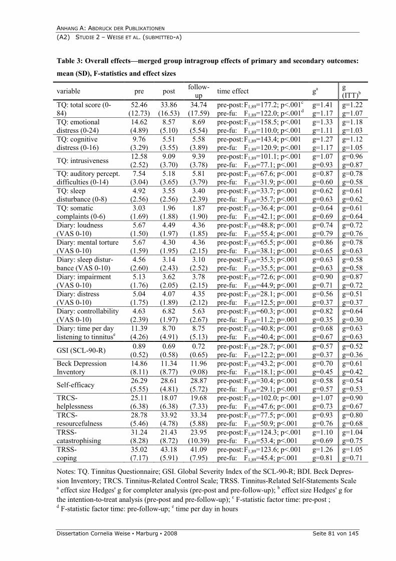
ANHANG A: ABDRUCK DER PUBLIKATIONEN
(A2) STUDIE 2 – WEISE ET AL. (SUBMITTED-A)
Dissertation Cornelia Weise Marburg 2008 Seite 81 von 145
Table 3: Overall effects—merged group intragroup effects of primary and secondary outcomes:
mean (SD), F-statistics and effect sizes
variable pre post follow-up
time effect ga g (ITT)b
TQ: total score (0-84)
52.46 (12.73)
33.86 (16.53)
34.74 (17.59)
pre-post: F1,89=177.2; p<.001c
pre-fu: F1,89=122.0; p<.001d
g=1.41 g=1.17
g=1.22 g=1.07
TQ: emotional distress (0-24)
14.62 (4.89)
8.57 (5.10)
8.69 (5.54)
pre-post: F1,89=158.5; p<.001 pre-fu: F1,89=110.0; p<.001
g=1.33 g=1.11
g=1.18 g=1.03
TQ: cognitive distress (0-16)
9.76 (3.29)
5.51 (3.55)
5.58 (3.89)
pre-post: F1,89=143.4; p<.001 pre-fu: F1,89=120.9; p<.001
g=1.27 g=1.17
g=1.12 g=1.05
TQ: intrusiveness 12.58 (2.52)
9.09 (3.70)
9.39 (3.78)
pre-post: F1,89=101.1; p<.001 pre-fu: F1,89=77.1; p<.001
g=1.07 g=0.93
g=0.96 g=0.87
TQ: auditory percept. difficulties (0-14)
7.54 (3.04)
5.18 (3.65)
5.81 (3.79)
pre-post: F1,89=67.6; p<.001 pre-fu: F1,89=31.9; p<.001
g=0.87 g=0.60
g=0.78 g=0.58
TQ: sleep disturbance (0-8)
4.92 (2.56)
3.55 (2.56)
3.40 (2.39)
pre-post: F1,89=33.7; p<.001 pre-fu: F1,89=35.7; p<.001
g=0.62 g=0.63
g=0.61 g=0.62
TQ: somatic complaints (0-6)
3.03 (1.69)
1.96 (1.88)
1.87 (1.90)
pre-post: F1,89=36.4; p<.001 pre-fu: F1,89=42.1; p<.001
g=0.64 g=0.69
g=0.61 g=0.64
Diary: loudness (VAS 0-10)
5.67 (1.50)
4.49 (1.97)
4.36 (1.85)
pre-post: F1,89=48.8; p<.001 pre-fu: F1,89=55.4; p<.001
g=0.74 g=0.79
g=0.72 g=0.76
Diary: mental torture (VAS 0-10)
5.67 (1.59)
4.30 (1.95)
4.36 (2.15)
pre-post: F1,89=65.5; p<.001 pre-fu: F1,89=38.1; p<.001
g=0.86 g=0.65
g=0.78 g=0.63
Diary: sleep distur-bance (VAS 0-10)
4.56 (2.60)
3.14 (2.43)
3.10 (2.52)
pre-post: F1,89=35.3; p<.001 pre-fu: F1,89=35.5; p<.001
g=0.63 g=0.63
g=0.58 g=0.58
Diary: impairment (VAS 0-10)
5.13 (1.76)
3.62 (2.05)
3.78 (2.15)
pre-post: F1,89=72.6; p<.001 pre-fu: F1,89=44.9; p<.001
g=0.90 g=0.71
g=0.87 g=0.72
Diary: distress (VAS 0-10)
5.04 (1.75)
4.07 (1.89)
4.35 (2.12)
pre-post: F1,89=28.1; p<.001 pre-fu: F1,89=12.5; p=.001
g=0.56 g=0.37
g=0.51 g=0.37
Diary: controllability (VAS 0-10)
4.63 (2.39)
6.82 (1.97)
5.63 (2.67)
pre-post: F1,89=60.3; p<.001 pre-fu: F1,89=11.2; p=.001
g=0.82 g=0.35
g=0.64 g=0.30
Diary: time per day listening to tinnituse
11.39 (4.26)
8.70 (4.91)
8.75 (5.13)
pre-post: F1,89=40.8; p<.001 pre-fu: F1,89=40.4; p<.001
g=0.68 g=0.67
g=0.63 g=0.63
GSI (SCL-90-R) 0.89 (0.52)
0.69 (0.58)
0.72 (0.65)
pre-post: F1,89=28.7; p<.001 pre-fu: F1,89=12.2; p=.001
g=0.57 g=0.37
g=0.52 g=0.36
Beck Depression Inventory
14.86 (8.11)
11.34 (8.77)
11.96 (9.08)
pre-post: F1,89=43.2; p<.001 pre-fu: F1,89=18.1; p<.001
g=0.70 g=0.45
g=0.61 g=0.42
Self-efficacy 26.29 (5.55)
28.61 (4.81)
28.87 (5.72)
pre-post: F1,89=30.4; p<.001 pre-fu: F1,89=29.1; p<.001
g=0.58 g=0.57
g=0.54 g=0.53
TRCS- helplessness
25.11 (6.38)
18.07 (6.38)
19.68 (7.33)
pre-post: F1,89=102.0; p<.001 pre-fu: F1,89=47.6; p<.001
g=1.07 g=0.73
g=0.90 g=0.67
TRCS-resourcefulness
28.78 (5.46)
33.92 (4.78)
33.34 (5.88)
pre-post: F1,89=77.5; p<.001 pre-fu: F1,89=50.9; p<.001
g=0.93 g=0.76
g=0.80 g=0.68
TRSS- catastrophising
31.24 (8.28)
21.43 (8.72)
23.95 (10.39)
pre-post: F1,89=124.3; p<.001 pre-fu: F1,89=53.4; p<.001
g=1.10 g=0.69
g=1.04 g=0.75
TRSS- coping
35.02 (7.17)
43.18 (5.91)
41.09 (7.95)
pre-post: F1,89=123.6; p<.001 pre-fu: F1,89=45.4; p<.001
g=1.26 g=0.81
g=1.05 g=0.71
Notes: TQ. Tinnitus Questionnaire; GSI. Global Severity Index of the SCL-90-R; BDI. Beck Depres-
sion Inventory; TRCS. Tinnitus-Related Control Scale; TRSS. Tinnitus-Related Self-Statements Scale a effect size Hedges' g for completer analysis (pre-post and pre-follow-up); b effect size Hedges' g for
the intention-to-treat analysis (pre-post and pre-follow-up); c F-statistic factor time: pre-post ; d F-statistic factor time: pre-follow-up; e time per day in hours

ANHANG A: ABDRUCK DER PUBLIKATIONEN
(A3) STUDIE 3 – WEISE ET AL. (SUBMITTED-B)
Dissertation Cornelia Weise Marburg 2008 Seite 83 von 145
Stability of physiological variables in chronic tinnitus sufferers.
Running Title: Stability of physiological variables
Cornelia Weise, Kristin Heinecke, Winfried Rief
Address of the authors:
University of Marburg
Faculty of Psychology
Department of Clinical Psychology and Psychotherapy
Gutenbergstrasse 18
35032 Marburg
Germany
phone: +49-6421-2825498
fax: +49-6421-2828904
email: [email protected]
Correspondence should be addressed to:
Cornelia Weise
Abbreviations:
EMG = Electromyography
SCL = Skin conductance level
ANHANG A: ABDRUCK DER PUBLIKATIONEN
(A3) STUDIE 3 – WEISE ET AL. (SUBMITTED-B)
Seite 84 von 145 Dissertation Cornelia Weise Marburg 2008
Abstract
The etiological tinnitus models propose that suffering can be caused and aggravated by height-
ened physiological arousal. Therefore psychophysiological treatments are applied. Stability of the
measured parameters is essential for the use of biofeedback as well as to permit the attribution of
changes to the administered treatment.
The aim of our study was to investigate the 3-month reproducibility of psychophysiological pa-
rameter in 60 tinnituspatients. Using a repeated-measures design, the activity of these parameters was
assessed twice during various stress and relaxation trials.
The results showed that the measurements of frontalis, masseter and trapezius muscles were
stable, while for the sternocleidomastoid, the skin conductance level and the skin temperature retest-
stability could not be evidenced. For all parameters, the stability of relative scores was insufficient.
In conclusion, our study has important implications for applied psychophysiology research: (1)
the measurement of EMG assessed in a clinical sample is stable over a 3-month interval; (2) in con-
trast, the retest-stability of SCL and skin temperature has to be marked as insufficient; (3) all
parameters are less stable when investigated as difference scores; and (4) because of the evident
stability in tinnitus patients, changes in EMG-parameters can be attributed to the administered
treatment.
Keywords: stability, psychophysiological parameters, surface-EMG, tinnitus
ANHANG A: ABDRUCK DER PUBLIKATIONEN
(A3) STUDIE 3 – WEISE ET AL. (SUBMITTED-B)
Dissertation Cornelia Weise Marburg 2008 Seite 85 von 145
Introduction
Tinnitus is the perception of a sound such as ringing or whistling in the ear in the absence of an
apparent acoustic stimulus. The common case of subjective tinnitus means sounds, which are not au-
dible to external listeners while objective tinnitus represents sounds that are objectively audible. Tinni-
tus is considered chronic if it lasts for more than 6 months. The distress which accompanies tinnitus
can vary substantially between individuals. A severe and so-called complex chronic tinnitus is often
accompanied by various psychiatric or psychosomatic conditions such as depression, anxiety or in-
creased physiological arousal.
The prevalence of the symptom tinnitus varies depending on its severity. A study by Davis and
El Refaie (2000) found that 10.1% of the adult UK population had tinnitus for 5 or more minutes; 5%
of the study population described a moderately to severely annoying tinnitus and 0.5% reported that
their tinnitus was severe and led to many problems in everyday life. These data confirm results of
earlier studies which reported prevalence rates of up to 4% for distressing tinnitus (Coles, 1984;
Pilgramm et al., 1999).
Up to now, the pathophysiology of tinnitus has been unclear. Tinnitus can have several medical
causes, such as cochlear lesions (e.g. after noise bursts) or diseases of the auditory nerve (Crummer &
Hassan, 2004), or it can be caused and aggravated by stress, as well as psychosocial and psychological
factors (Hebert & Lupien, 2007). Furthermore, studies have reported heightened physical tension in
cephalic and neck muscles as well as in masticatory muscles, thus implying possible sources of tinni-
tus (Bernhardt et al., 2004; Peroz, 2003; Sanchez et al., 2002). Findings of a recent study by Rief and
colleagues (2004) supported subjective reports of tinnitus sufferers who described increased muscle
tension as a reason for tinnitus development and maintenance. The authors found first hints of in-
creased muscle reactivity in tinnitus patients while listening to their own tinnitus or to an external tin-
nitus-similar sound.
In addition, the psychological model of tinnitus by Hallam and colleagues (Hallam, 1987;
Hallam et al., 1984) proposes that habituation to tinnitus noises is a normal response, while suffering
and complaint can be explained as a failure of habituation. The authors listed different variables that

ANHANG A: ABDRUCK DER PUBLIKATIONEN
(A3) STUDIE 3 – WEISE ET AL. (SUBMITTED-B)
Seite 86 von 145 Dissertation Cornelia Weise Marburg 2008
can be considered as influencing the process of habituation—for example, the characteristics and the
meaning of the stimulus, environmental conditions, and also central state characteristics such as the
level of cortical arousal. The neurophysiological tinnitus model by Jastreboff and Hazell (1993) also
emphasizes the role of heightened arousal as well as dysfunctional attention and appraisal processes.
Various studies have evidenced a link between tinnitus and stress with consistently heightened stress
levels that are accompanied by an increased cortical arousal level (Alpini & Cesarani, 2006; Hebert &
Lupien, 2007; Horner, 2003).
This increased arousal level as well as the relationship between muscle tension and tinnitus are
part of the rationale for using biofeedback-based relaxation in treating chronic tinnitus. Studies on the
efficacy of biofeedback in tinnitus differ in regard to the outcome in physiological variables (e.g.,
EMG-levels) versus psychological variables (e.g., self-ratings of tinnitus distress) (Haralambous et al.,
1987). This discrepancy between psychological and physiological measures in tinnitus may reflect an
actual phenomenon, but this divergence can also result from lower reliability of physiological vari-
ables. The aim of a psychophysiological treatment is to enable the subject to reduce the heightened
physiological arousal level by feeding the physiological processes back to the individual. The stability
of the parameters' measurements is an important requirement to achieve this aim.
To our knowledge, no studies have examined the stability of physiological data in tinnitus suf-
ferers. However, some studies have investigated the reliability of various physiological parameters in
healthy subjects and pain patients. Arena et al. (1989) and Waters et al. (1987) investigated the stabil-
ity of frontalis muscles over a 1-month and 2-week interval, respectively, and found correlation coeffi-
cients ranging from .14 to .79 and .0 to .55, respectively. Burdette and Gale (1990) found test-retest
correlations across sessions for the surface EMG of the masseteric area to be between .56 and .65,
which were clearly lower than the within-session correlations. The reproducibility of trapezius EMGs
over a period of 1 year was analysed by Elert et al. (1998). The test-retest correlation coefficients in a
patient sample with neck and shoulder problems did not exceed .35. Regarding the skin conductance
level (SCL), Schell et al. (2002) showed temporal stability over a 1-year period in schizophrenia pa-
tients (r = .43) and normals (r = .61) and El-Sheik (2007) showed long-term stability (r = .30) of SCL
ANHANG A: ABDRUCK DER PUBLIKATIONEN
(A3) STUDIE 3 – WEISE ET AL. (SUBMITTED-B)
Dissertation Cornelia Weise Marburg 2008 Seite 87 von 145
in children. With regard to skin temperature, Waters et al. (1987) found significant stability coeffi-
cients between .47 and .61 over a 2-week interval, while Arena and colleagues (Arena et al., 1983;
Arena & Hobbs, 1995) showed high reliability coefficients only if sessions were repeated within 1
week and not for longer test-retest intervals. Furthermore, the measurements were only stable when
investigated as absolute data and were particularly unreliable when examined as relative measures.
To summarise, previous studies regarding the stability of physiological responses have
investigated primarily few response systems in rather small samples of mostly healthy subjects across
short test-retest intervals. Many studies showed that the longer the time period of retesting, the lower
the stability of the physiological measure. Furthermore, the correlation coefficients were mostly
reliable if absolute scores were evaluated, while relative measures produced smaller and less reliable
coefficients.
In light of the previous research, several questions still remain unanswered: (1) Are psycho-
physiological parameters in clinical populations stable measures, and do they show reliable associa-
tions between multiple assessment points for longer test-retest intervals? (2) Do muscle activity, SCL
and temperature have trait or state characteristics, and what are consequential implications for admin-
istered treatments? (3) Are the assessed data reliable enough to permit the attribution of changes in
physiological parameters to an administered biofeedback treatment. (4) Is the reliability of psycho-
physiological measurements sufficient to assess treatment effects of a psychophysiological treatment
not only by investigating psychological but also physiological parameters and is the use of psycho-
physiological assessments in the investigation of treatment effects recommendable?
Biofeedback is a favoured treatment approach in chronic tinnitus. Two own studies evidenced
that a biofeedback-based treatment is highly effective and well accepted by the patients (Rief et al.,
2005; Weise et al., 2007). As long as no information on the stability of physiological parameters
exists, it remains difficult to evaluate whether psychophysiological treatment approaches lead to stable
modifications in the physiological arousal. Therefore, the aim of our study was to investigate the
3-month reproducibility of the activity of frontalis, masseter, trapezius and sternocleidomastoid mus-
cles as well as of the measurements of SCL and skin temperature. We hypothesised that: (1) the elec-

ANHANG A: ABDRUCK DER PUBLIKATIONEN
(A3) STUDIE 3 – WEISE ET AL. (SUBMITTED-B)
Seite 88 von 145 Dissertation Cornelia Weise Marburg 2008
tromyographic measures, assessed with a biofeedback device, would be clearly associated between
two assessments and therefore stable measures; (2) the frequently and successfully used parameters of
frontalis and masseter activity and skin temperature would show the best stability coefficients, while
the SCL would show less stability coefficients because of its high inter-individual variability; and (3)
absolute values would be continuously more reliable than change scores.
Methods
Sample
Main inclusion criteria for participation in the study were tinnitus for more than 6 months result-
ing in serious distress. Tinnitus-associated distress was defined by a minimum score in the Tinnitus
Questionnaire (TQ ≥ 40). Patients were required to be between the ages of 16 and 75 and needed to
have sufficient language skills. Patients with tinnitus as a result of a medical disease (e.g., Menière's
disease) as well as patients with psychosis, dementia or other seriously disabling cognitive impairment
were excluded. Other psychiatric comorbidities such as depression or anxiety were not excluded, but
carefully diagnosed and registered. The sample of the present study was the waitlist group of a larger
intervention trial for chronic tinnitus. The sample size was determined to allow the detection of me-
dium correlations with a statistical power of 0.8. Two hundred and ten patients responded to articles in
newspapers, and of them 132 fulfilled the inclusion criteria. Two patients refused participation. The
resulting 130 patients gave their informed consent and were randomly assigned by a permuted-block
randomisation proceeding to either the intervention group or waitlist group. Five out of the 65 patients
from the waitlist group discontinued the waiting period, resulting in 60 patients with complete data.
Details about sample characteristics are shown in Table 1.
– Please insert Table 1 here –
Procedure
The first assessment was administered in two parts: first, patients filled in different question-
naires; then a standardised physiological measurement took place to assess physiological variables.
This was carried out by one of three blinded assessors, who were not informed about the severity and
ANHANG A: ABDRUCK DER PUBLIKATIONEN
(A3) STUDIE 3 – WEISE ET AL. (SUBMITTED-B)
Dissertation Cornelia Weise Marburg 2008 Seite 89 von 145
degree of tinnitus annoyance as well as about probable individual causes of tinnitus. After this first
assessment (t1), patients waited 3 months for the beginning of the biofeedback-based therapy. After
the waiting period, patients were reassessed for the second time (t2). The physiological data were as-
sessed by the same standardised measurement. The assessors were three graduate psychology students,
who were extensively trained and supervised by biofeedback-experienced psychologists.
For the physiological measurement, several skin areas were cleansed with a propanol solution.
Electrodes were then applied over the belly of the relevant muscles (frontalis, masseter, sternocleido-
mastoid and trapezius). The skin conductance sensor was applied on two fingers of the non-dominant
hand, and the temperature sensor was fixed on the same hand to assess skin temperature. The measure-
ment was standardised and consisted of 13 phases (see Fig. 1) beginning with a baseline-period of 2
minutes in order to adapt to the experimental environment. During this period, patients were asked to
sit quietly and try not to speak. The physiological data of the baseline trial were not evaluated, as this
phase was only used for adaptation. Following this, a 5-minute relaxation phase was initiated, in which
patients were asked to try to relax. Subsequently five different 3-minute stress tasks were initiated,
which comprised tinnitus-specific stressors as well as cognitive stressors. The stressors were: (1) lis-
tening to loud noise; (2) listening to a tinnitus-similar noise; (3) listening to their own tinnitus; (4) an
arithmetic task (counting backwards by multiples of 7 starting from 700); and (5) a test for divided
attention—a subtest from the alertness-test battery 'TAP' (Zimmermann & Fimm, 1994). The sequence
of the stressors 1 to 4 was permuted, and the test for divided attention was always the last stressor
because a change of position was needed. A post-stressor 2-minute relaxation period followed each
stressor. Following the five stressors, a final 5-minute relaxation phase completed the measurement.
Afterwards, the electrodes were carefully removed and patients were debriefed.
– Please insert Figure 1 here –
Measures
The physiological data were assessed by a biofeedback device (FlexComp Infiniti™, Thought
Technology Ltd.) offering 10 high-speed channels (2048 samples/sec). For assessing the EMG, seven
pre-amplified MyoScan-Pro™ EMG sensors with an input impedance of 10 GΩ and a bandwidth of
ANHANG A: ABDRUCK DER PUBLIKATIONEN
(A3) STUDIE 3 – WEISE ET AL. (SUBMITTED-B)
Seite 90 von 145 Dissertation Cornelia Weise Marburg 2008
20-500 Hz were used. We used disposable silver/silver-chloride surface Triode™ electrodes with
standard 2 cm spacing, which ensured a constant distance between the electrodes. The skin conduc-
tance was measured by a SC-Flex/Pro Sensor™ with a signal input range of 0-30 µS and skin tem-
perature was measured by a temperature sensor with a range of 10-45 °C.
For assessing demographic and tinnitus-specific data, we used the Structured Tinnitus Interview
(STI) (Goebel & Hiller, 2001). This interview evaluates characteristics of tinnitus–like duration or
accompanied hearing problems, medical causes and psychological aspects in the development and
maintenance of tinnitus. The existence of comorbid psychiatric disorders was evaluated by using the
International Diagnostic Checklists for the ICD-10 (Hiller et al., 1995).
Patients also answered the German version of the Tinnitus Questionnaire (TQ) (Goebel &
Hiller, 1998), which is a 52-item self-rating scale, originally developed by Hallam and colleagues
(1996). The total score (0-84) provides information on the severity of tinnitus distress. The impairment
is classified as mild (0-30), moderate (31-46), serious (47-59), and severe (60-84).
Data processing
As filters for the EMG signals, we used, first, a 50 Hz notch-filter and, second, a wide-
bandwidth filter from 20 to 500 Hz. This does not exclude electrocardiographic artefacts but allows
the detection of the majority of EMG activity that lies in the range between 30 and 100 Hz, which is
important for measuring slow and fatigued muscles. For the raw EMG signal, the root mean square
was computed for the rectification and integration of the bipolar signal. This was done by the non-
sliding window algorithm with an averaging factor of 10 and a time period of 1 second. No filters
were used for SCL and skin temperature. We defined a sampling rate of 32 samples per second.
Artefact rejection and averaging of each single phase and for each single measure were per-
formed by a customised LabVIEW programme called 'BioCalc'. This program computed means and
standard deviations for each phase and each person. Outliers, which were more than three standard
deviations above the individual phase mean, were rejected.
After this automatic artefact rejection, a manual control for artefacts followed, in which upper
limits were defined. Frontalis, masseter and sternocleidomastoid values that exceeded 30 µV and tra-
ANHANG A: ABDRUCK DER PUBLIKATIONEN
(A3) STUDIE 3 – WEISE ET AL. (SUBMITTED-B)
Dissertation Cornelia Weise Marburg 2008 Seite 91 von 145
pezius values that exceeded 45 µV were replaced by missing values. For SCL, the upper limit was set
at 20 µS and for skin temperature all values lower than 20°C or higher than 40 °C were replaced.
Finally, the measurements of the bilaterally recorded muscles (masseter, trapezius and sterno-
cleidomastoid) were pooled in order to increase the reliability of the data. Relative measures (change
values) were computed by subtracting the mean of the initial 5-minute relaxation phase from the mean
of each of the following reaction periods.
Statistical analyses
All computations were carried out using the SPSS statistical package (Version 12.0). To identify
possible differences between the two assessments, repeated-measures, Bonferroni-corrected
MANOVAs with the factor time (t1 and t2) were performed for the several psychophysiological
parameters. To determine the intersession temporal stability (3 months) of the absolute values, correla-
tional analyses using Pearson's r were conducted. Due to the fact that most of the physiological vari-
ables were skewed, average scores of the absolute data had to be transformed by the logarithmic trans-
formation (y=log [x+1]) to approximate the data to normal distribution. After this transformation, all
variables, with the exception of the skin temperature, were normally distributed. For this reason, the
temporal stability of the skin temperature was assessed by computing correlations with Spearman's
Rho.
Change scores of the absolute values were also skewed and were therefore calculated using the
logarithmically transformed values. These difference scores were normally distributed, again with the
exception of skin temperature. To assess the intersession temporal stability of the change scores, Pear-
son's correlation coefficients were computed for the EMG-values and for the SCL. The stability of the
skin temperature was again investigated by computing correlations with Spearman's Rho.
As adjustment for multiple comparisons the Bonferrroni correction was used. Therefore the sig-
nificance level for the correlational analyses of absolute scores was set at p ≤ .004 (p=0.05 divided by
12 [comparisons]). For the correlational analyses of change scores the significance level was set at
p ≤ .005 (p=0.05 divided by 11 [comparisons]). Stability of a variable in one phase from the test-retest
point of view was considered when a significant correlation coefficient between the first and the sec-

ANHANG A: ABDRUCK DER PUBLIKATIONEN
(A3) STUDIE 3 – WEISE ET AL. (SUBMITTED-B)
Seite 92 von 145 Dissertation Cornelia Weise Marburg 2008
ond measurement was found. All correlational analyses were computed separately for the various
physiological variables, the various trials (initial and final 5-minute relaxation, stress tasks and post-
stress relaxations) and separately for absolute and change values. To further investigate stability, a
composite stress mean score and a composite relaxation mean score were computed by averaging the
means of the five stress trials respectively of the five post-stress relaxation trials separately for all
muscles. For the stress and relaxation mean scores, further correlational analyses were conducted.
Results
Stability of absolute values
The several MANOVAs, which were conducted primarily, did not show any significant main
time effect between the first and the second assessment for the different psychophysiological parame-
ters.
The stability coefficients for the logarithmically transformed absolute values of the EMG data
as well as means and standard deviations of the untransformed data are presented in Table 21.
– Please insert Table 2 here –
The best stability coefficients were obtained for the frontalis muscle. For all but one phases of
the standardised measurement, significant correlations (p ≤ .004) in the range of .36 to .75 were de-
tected. One correlation coefficient for the short relaxation after the fourth stressor (listening to tinni-
tus) failed the alpha-adjusted significance level. The data of the masseter muscle also showed high
stability for the two measurements, with significant correlations for 11 out of 12 phases (p ≤ .004) and
a range from .37 to .67. Only the correlation coefficient for the short relaxation after the first stressor
(listening to loud noise) failed to reach significance. The EMG of the trapezius showed somewhat less
but still satisfactory stability with seven significant correlations out of 12. The size of these significant
correlations was modest, with a range from .37 to .56, and again the significance level was at p ≤ .004
1 All analyses were also carried out on the basis of the untransformed variables. Retest-stability was assessed by
computing parametric as well as non-parametric correlations. The results of the several analyses revealed gener-
ally comparable results to those presented in the following.
ANHANG A: ABDRUCK DER PUBLIKATIONEN
(A3) STUDIE 3 – WEISE ET AL. (SUBMITTED-B)
Dissertation Cornelia Weise Marburg 2008 Seite 93 von 145
for all correlations. The correlations for the five stress trials were significant, while only two of the
five post-stressor relaxation phases reached statistical significance. Furthermore, the initial relaxation
phase failed the alpha-adjusted significance level (r=.32, p=.014) and the final relaxation phase did not
reveal a significant correlation between the two assessment points for trapezius activity. The EMG
measurements of the sternocleidomastoid muscles showed a weak stability. Only two correlations out
of 12 reached the alpha-adjusted significance level of p=.004. The correlation coefficients were gener-
ally lower, with a range from .25 to .43. Significant correlations were only detected for the stress trials
3 (listening to own tinnitus) and 5 (test for divided attention). Five of the further correlations failed the
alpha-adjusted significance level and further five phases were not at all significantly associated
between the two assessments.
– Please insert Table 3 here –
Table 3 shows the means and standard deviations of the untransformed SCL values for the first
and second measurement as well as Pearson correlations coefficients for the logarithmically trans-
formed values. For the absolute values no stability was found. Indeed six out of 12 correlations
reached significance (p ≤ .05), but failed the alpha-adjusted significance level (p ≤ .004). Altogether
correlations were weak, with a range from .26 to .29 (p ≤ .05).
– Please insert Table 4 here –
Finally, Table 4 shows the means and standard deviations of the skin temperature values and the
non-parametric correlation coefficients (Spearman's Rho). Eight out of 12 correlations were signify-
cant, but only two of them reached the alpha-adjusted significance level (p ≤ .004). The size of the
correlations was rather small, with a range from .29 to .39.
In addition to the separate analyses of the various phases, a composite stress mean score and a
relaxation mean score were computed by averaging all stress all relaxation periods separately for the
two assessment points. Pearson's correlation coefficients for the stress score and for the relaxation
score were significant (p ≤ .004) for the muscles: frontalis (.66 and .64), masseter (.68 and .63), and
trapezius (.65 and .43). Both composite scores for the muscle sternocleidomastoid failed the alpha
ANHANG A: ABDRUCK DER PUBLIKATIONEN
(A3) STUDIE 3 – WEISE ET AL. (SUBMITTED-B)
Seite 94 von 145 Dissertation Cornelia Weise Marburg 2008
adjusted significance level (.31 and .29; p<.05). For the SCL, neither the correlation coefficient for the
stress nor for the relaxation score reached significance. Regarding skin temperature, Spearman's Rho
did not reach the alpha-adjusted significance level for the relaxation score (.28; p<.05) and did not
reveal significance for the stress score.
Stability of change scores
The change scores yielded weaker results for all EMG measures as well as for the measure-
ments of SCL and skin temperature. Change scores for the EMG measures and the SCL were com-
puted by subtracting the logarithmically transformed mean of the initial relaxation phase from all sub-
sequent logarithmically transformed period means. For skin temperature, change scores were com-
puted by subtracting the untransformed value of the initial relaxation phase from each of the following
untransformed phase means.
– Please insert Table 5 here –
Table 5 shows means and standard deviations of the change scores of the EMG measures. Only
a few correlation coefficients reached significance, and most change scores were not stable. For the
frontalis muscle, only three out of 11 correlations reached the alpha-adjusted significance level (p ≤
.005): stress trials 1 (listening to loud noise), 3 (listening to the own tinnitus), and 5 (test for divided
attention) were significantly associated between the two assessment points, with correlation coeffi-
cients in the range of .40 to .42. Of the measurements of the masseter muscles, four out of 11 correla-
tions were significant but only two of them reached the alpha-adjusted significance level, namely the
correlations for stress trials 1 (listening to loud noise) and 5 (test for divided attention), with coeffi-
cients of .55 and .49 respectively. For the trapezius muscles five correlations out of 11 were signifi-
cantly associated between the assessments, but only the correlation for the stressor 4 (arithmetic)
reached the alpha-adjusted significance level (r=.36; p ≤ .005). Finally, for the change scores of the
sternocleidomastoid, none of the correlations was significant (p ≤ .005). Altogether, there was no ob-
vious pattern for the stability of change scores between the muscles; persistent stability for all different
ANHANG A: ABDRUCK DER PUBLIKATIONEN
(A3) STUDIE 3 – WEISE ET AL. (SUBMITTED-B)
Dissertation Cornelia Weise Marburg 2008 Seite 95 von 145
muscle reactions could be shown for none of the various phases. Even for the 5-minute final relaxation
phase, stability relative to the initial relaxation phase could not be detected.
Concerning the SCL change scores, none of the difference scores correlated significantly be-
tween the two assessment points (see Table 3). Table 4 shows that seven out of 11 of the computed
Spearman's Rho correlation coefficients for skin temperature were significant. However, only three of
them reached the alpha-adjusted significance level. For the stress trial 3 (arithmetic) and the subse-
quent relaxation phases as well as for the stress trial 5 (test for divided attention) stability coefficients
in the range of .38 to .48 were detected.
Discussion
To our knowledge, this is the first study that has examined the reliability of surface EMG levels,
SCL and skin temperature in chronic tinnitus patients. The results of our study are consistent with
previous research that investigated stability of those parameters in healthy subjects or in pain patients.
Considering the absolute values of the EMG measures, the frontalis and masseter muscles ap-
pear to show the most stable pattern, which is consistent with our hypothesis. For both muscles 11 out
of 12 coefficients for the stress and relaxation phases reached significance and were at a moderate to
strong level. The stability of the EMG for the trapezius can be rated as satisfactory, with seven signifi-
cant coefficients out of 12 computed correlations, which were all of moderate size. These results were
confirmed by significant correlations for the composite stress and relaxation mean scores. These com-
posite scores showed significant associations between the two assessment points for all muscles,
which were typically higher than those of single stress or relaxation periods. In contrast to these results
no stability was found for the EMG of the sternocleidomastoid, for the SCL, and for the skin tempera-
ture. Indeed half of the correlations computed for these parameters showed significant associations
between the assessment points, but most of them did not reach the Bonferroni-corrected significance
level. These results are confirmed by the composite stress and relaxation mean scores, of which both
did not reach significance.
ANHANG A: ABDRUCK DER PUBLIKATIONEN
(A3) STUDIE 3 – WEISE ET AL. (SUBMITTED-B)
Seite 96 von 145 Dissertation Cornelia Weise Marburg 2008
In conclusion, the results regarding the muscles frontalis, masseter and trapezius were consistent
to our first hypothesis, which assumed that electromyographic measures assessed with a biofeedback
device would be stable over a longer test-retest interval. The frontalis and masseter muscles, in parti-
cular, appeared to be substantially associated across the two assessment points. Contrary to our hypo-
thesis no stability was found for the sternocleidomastoid. Furthermore, our second hypothesis, that
frontalis and skin temperature would be substantially stable measures while SCL would show less
stability, was only partly confirmed by our data, because we did not find stability for the skin tempera-
ture.
The results of our study regarding the absolute values are comparable to previous research.
However, previous studies investigated the stability of EMG measures as well as SCL and hand tem-
perature mostly in healthy subjects and over short test-retest intervals. Our results for the frontalis and
masseter muscles are as good as those of previous studies regarding the extent of significant
correlation coefficients, but we were able to show stability coefficients in a clinical sample and,
moreover, for a test-retest interval of approximately 3 months, while the longest test-retest interval in
previous studies was 28 days (Arena et al., 1983; Arena et al., 1989; Arena & Hobbs, 1995; Burdette
& Gale, 1990). Our results regarding the stability of the trapezius muscle show slightly higher stability
coefficients compared to results of Elert et al. (1998), who investigated a 1-year test-retest interval.
Regarding the SCL our results are weaker than those of previous studies. Indeed the extent of our
correlations is comparable to the results of El-Sheik (2007) and Waters et al. (1987). But taking the
Bonferroni correction for multiple comparisons into account, none of the correlations in our study met
the alpha-adjusted significance level. Finally, for skin temperature over a 3-month interval our study
showed rather low stability coefficients which failed to reach significance after alpha-adjustment. This
result is somewhat comparable to previous studies, which showed high correlation coefficients for a
2- or 3-week interval but a clear decrease in stability with increasing test-retest intervals (Arena et al.,
1983). Altogether the reliability coefficients are in a small-to-moderate range. But as the above cited
studies showed, low to moderate correlation coefficients are usual in psychophysiology research
ANHANG A: ABDRUCK DER PUBLIKATIONEN
(A3) STUDIE 3 – WEISE ET AL. (SUBMITTED-B)
Dissertation Cornelia Weise Marburg 2008 Seite 97 von 145
which investigates EMG-stability. Also a study by Cram and colleagues (1990) found a median corre-
lation of only r=.64 for the reliability of surface EMG.
A quite different pattern was found regarding the stability of change scores: in accord with our
third hypothesis, difference values relative to the initial relaxation phase produced clearly smaller and
less significant stability coefficients for all muscles. For the EMG measures, a maximum of three out
of 11 correlations reached the alpha-adjusted significance level. Furthermore the coefficients did not
exceed a value of r = .56. Because of these small and few significant correlations, the stability of
change scores has to be marked as insufficient, which is consistent with our hypothesis. Treating the
responses of the SCL as difference values also did not increase reliability; in fact, the SCL was com-
pletely unreliable. Also the skin temperature scores showed a weak stability when considering the
difference values, with three out of 11 correlations reaching the Bonferroni-corrected significance
level.
By comparing absolute and relative scores, not only the loss of significant correlation coeffi-
cients is apparent but also a decrease in the extent of correlations. Furthermore, the patterns for situa-
tions and parameters revealing stable results show variability.
The substantial divergence between absolute and change scores was also found in previous stu-
dies (Arena et al., 1983; Arena et al., 1989; Arena et al., 1990). Differences were found in healthy sub-
jects as well as in pain patients. Arena et al. (1990) showed that relative measures of different condi-
tions were consistently less reliable in lower back pain patients than in normals. The authors explained
these differences by the 'inadequate homeostatic restraint' theory of Sternbach (1966, quoted from
Arena, 1990), which postulates that individuals with psychophysiological disorders show inconstant
responses in this psychophysiological modality which is presumed aberrant. Therefore they recom-
mended relying more on absolute than on relative values when employing surface EMG with pain
patients.
As many previous studies showed methodological shortcomings, we tried to control some of the
factors that may influence the reliability of surface EMG data. We kept technical factors like the inter-
electrode distance constant by using triode electrodes. Furthermore, the same equipment with equal

ANHANG A: ABDRUCK DER PUBLIKATIONEN
(A3) STUDIE 3 – WEISE ET AL. (SUBMITTED-B)
Seite 98 von 145 Dissertation Cornelia Weise Marburg 2008
settings for filters, amplifiers and data processing was used for the two assessments. Experimental
factors concerning the measurement procedure were controlled through instructing the assessors to
prepare the skin carefully, to use a measuring tape when placing the electrodes and to consider the
distance to a reference point (e.g., the cervical spine). In addition to those experimenter instructions,
patients got a standardised instruction for the course of the measurement in order to keep conditions
constant between different subjects and different experimenters.
Nevertheless, there are some shortcomings in our study, which may have reduced the stability
of the data. Because of our 3-month test-retest interval, not all environmental conditions such as inside
and outside temperature or humidity were kept constant. Also, temporal conditions regarding the
measuring time (day of the week or time of day) were not always the same for the two assessments.
Additionally, in 30% of the measurements, the experimenter changed from the first to the second as-
sessment. Another limitation of our study concerns the planned test-retest interval of 90 days, which
was not strictly adhered to. The mean interval was 93.6 days, with a range of 71 to 133 days and with
one outlier at 172 days due to a longer illness. Furthermore, although patients were instructed to sit
quietly and try not to speak during the measuring phases, not all patients followed these instructions,
which led to head or body movements. Frontalis and masseter muscles are involved in speech or facial
expressions, so associated artefacts are not completely avoidable.
In spite of the listed shortcomings, our study has some important implications for the practical
use of biofeedback and applied psychophysiology research. With our results, some of the aforemen-
tioned questions can be answered: (1) EMG-parameters, which are commonly used in applied psycho-
physiology and assessed with a biofeedback device in a practitioner related context appear to be reli-
able measures in a clinical sample. In our tinnitus sample, these parameters were stable over a 3-month
test-retest interval when investigated as absolute data. The activity of the sternocleidomastoid, the
SCL and, the skin temperature as well as treating the data as difference scores appear to be less reli-
able. (2) Electromyographic parameters seem to be trait rather than state variables, while the SCL and
skin temperature seem to have state characteristics and their stability is insufficient. Muscle relaxation
training should therefore focus on tonic rather than on phasic changes. (3) Because our study showed
ANHANG A: ABDRUCK DER PUBLIKATIONEN
(A3) STUDIE 3 – WEISE ET AL. (SUBMITTED-B)
Dissertation Cornelia Weise Marburg 2008 Seite 99 von 145
good reliability coefficients for electromyographic measures in a clinical sample, it should be justify-
able to use psychophysiological treatments and to attribute changes in those parameters to the adminis-
tered treatment or experimental manipulation. This is important information for applied psychophysio-
logy research, as it allows the interpretation of not only psychological but also physiological data. We
recommend carefully following guidelines regarding the application of electrodes, filtering of raw
data, data processing, and the correction of artefacts in order to obtain reliable data. (4) If psycho-
physiological parameters are carefully assessed, their use in psychophysiological treatments and the
subsequent evaluation and interpretation of those data, as well as of possible changes through treat-
ment, is meaningful and can be recommended. Because this is the first study which has investigated
psychophysiological stability in tinnitus sufferers, further research in other clinical populations is also
required to replicate our findings.
Acknowledgments/Grants
This study was supported by the German Research Foundation (Ri 574/12-1). Additionally we
thank all our patients for participating, as well as Anne Raudschus, Kristin Schwarz and Eva Petrenko
for assistance with data collection.
Notes
The trial protocol has been approved by the Ethics Committee of the German Federation of Psy-
chological Associations (DGPs) and meets the ethical standards of the Declaration of Helsinki. This
study was registered with ClinicalTrials.gov, number NCT00397007.

ANHANG A: ABDRUCK DER PUBLIKATIONEN
(A3) STUDIE 3 – WEISE ET AL. (SUBMITTED-B)
Seite 100 von 145 Dissertation Cornelia Weise Marburg 2008
Figures and Tables
Figure
min
baseline
stressor 1 stressor 2 stressor 3 stressor 4 stressor 5
initial
relaxation
short
relaxation
short
relaxation
short
relaxation
short
relaxation
short
relaxation
2' 5' 3' 5'2' 3' 2' 3' 2' 3' 2' 3' 2'
final
relaxation
min
baseline
stressor 1 stressor 2 stressor 3 stressor 4 stressor 5
initial
relaxation
short
relaxation
short
relaxation
short
relaxation
short
relaxation
short
relaxation
2' 5' 3' 5'2' 3' 2' 3' 2' 3' 2' 3' 2'2' 5' 3' 5'2' 3' 2' 3' 2' 3' 2' 3' 2'
final
relaxation
Figure 2: Procedure of the standardised measurement
Notes: Stressor 1, listening to loud noise; Stressor 2, listening to a tinnitus-similar noise; Stressor 3,
listening to the own tinnitus; Stressor 4, arithmetic; Stressor 5, test for divided attention.
Tables
Table 4: Sample characteristics
Variable
N 60
Age 52.8 (11.95)
Percent females 43.3% (N=26)
Married 81.7% (N=49)
Illness duration (years) 7.48 (8.24)
Illness severity (0-10) 7.33 (1.3)
Comorbid depressive disorder 40% (N=24)
Tinnitus distress (0-84; TQ) 54.85 (10.26)

ANHANG A: ABDRUCK DER PUBLIKATIONEN
(A3) STUDIE 3 – WEISE ET AL. (SUBMITTED-B)
Dissertation Cornelia Weise Marburg 2008 Seite 101 von 145
Table 5: Absolute values (untransformed) of electromyographic activity: Means and standard deviations in µV for the first (t1) and second (t2) measurements
and Pearson's correlation coefficients (r) for the logarithmically transformed values2
Frontalis Masseter Trapezius Sternocleidomastoid
t1 t2 r t1 t2 r t1 t2 r t1 t2 r
Initial relax. 4.75 (2.5) 4.58 (2.2) .50*** 2.12 (1.0) 2.34 (1.5) .40*** 4.06 (3.3) 4.38 (4.1) .32* 4.97 (2.5) 5.01 (3.2) .15
Stressor 1a 6.59 (4.4) 6.23 (3.0) .73*** 3.49 (2.5) 3.29 (3.0) .67*** 5.00 (5.1) 4.80 (4.5) .37*** 5.76 (3.6) 5.59 (3.4) .15
Short relax. 5.72 (4.1) 5.39 (3.1) .59*** 2.53 (1.7) 2.53 (2.5) .25 4.27 (4.7) 3.71 (2.4) .21 4.97 (2.8) 5.38 (4.3) .21
Stressor 2b 6.48 (3.8) 6.59 (3.4) .63*** 2.83 (1.7) 3.30 (3.3) .49*** 4.32 (3.7) 5.33 (6.0) .42*** 5.44 (3.2) 5.82 (3.6) .24
Short relax. 5.40 (2.9) 5.91 (3.8) .57*** 2.46 (2.2) 2.50 (2.2) .41*** 3.72 (3.3) 4.14 (4.9) .13 5.07 (3.1) 5.39 (3.5) .17
Stressor 3c 5.83 (2.8) 6.37 (4.2) .39*** 3.48 (2.4) 2.80 (1.8) .55*** 5.16 (3.9) 4.63 (2.6) .45*** 6.33 (3.3) 5.95 (3.2) .43***
Short relax. 5.43 (4.1) 5.33 (3.7) .36** 2.63 (1.4) 2.77 (1.7) .58*** 4.41 (3.7) 5.01 (6.0) .46*** 5.65 (3.5) 5.65 (3.2) .30*
Stressor 4d 5.24 (3.5) 5.03 (2.8) .64*** 2.36 (2.4) 2.16 (1.3) .52*** 5.61 (6.0) 5.72 (6.1) .56*** 5.52 (3.5) 5.56 (3.3) .30*
Short relax. 5.28 (2.9) 5.53 (4.1) .62*** 2.30 (1.8) 1.98 (1.0) .50*** 3.84 (3.1) 4.16 (4.7) .57*** 5.31 (3.3) 5.06 (3.2) .25*
Stressor 5e 7.55 (3.6) 7.25 (3.8) .58*** 3.37 (2.3) 3.11 (2.1) .55*** 8.97 (6.4) 10.04 (6.2) .52*** 6.93 (3.8) 6.83 (3.9) .41***
Short relax. 6.15 (3.4) 5.72 (3.6) .49*** 2.49 (1.2) 2.69 (1.9) .39*** 4.97 (5.4) 4.89 (3.7) .25 5.55 (3.6) 5.61 (4.0) .32*
Final relax. 4.99 (2.9) 4.88 (3.2) .53*** 2.26 (1.8) 2.52 (1.7) .37*** 3.82 (3.3) 4.59 (4.4) .18 4.53 (2.3) 5.51 (4.0) .31*
Notes: a listening to loud noise b listening to a tinnitus-similar noise c arithmetic d listening to own tinnitus e test for divided attention * p≤.05 ** p≤.01 *** p≤.004 (alpha-adjusted significance level) 2 Means and standard deviations shown in the tables are always the untransformed values, as this method of presentation allows for a self-evident view of the data. However,
correlations were computed by using the logarithmically transformed values.
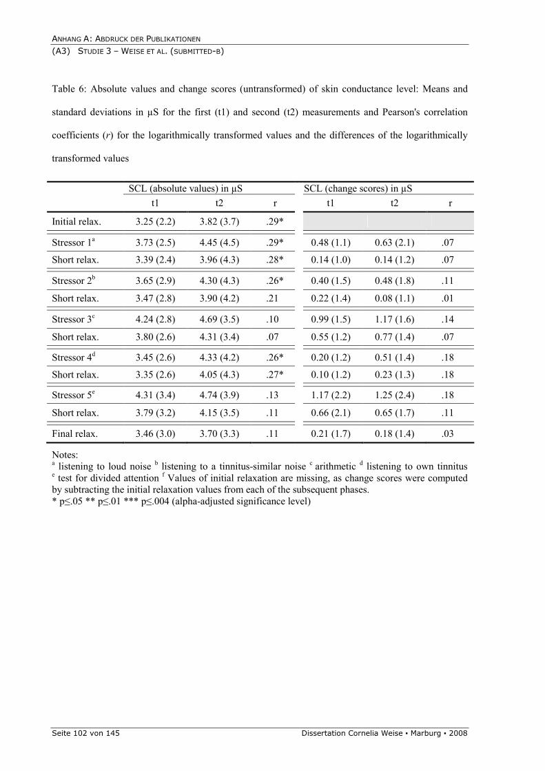
ANHANG A: ABDRUCK DER PUBLIKATIONEN
(A3) STUDIE 3 – WEISE ET AL. (SUBMITTED-B)
Seite 102 von 145 Dissertation Cornelia Weise Marburg 2008
Table 6: Absolute values and change scores (untransformed) of skin conductance level: Means and
standard deviations in µS for the first (t1) and second (t2) measurements and Pearson's correlation
coefficients (r) for the logarithmically transformed values and the differences of the logarithmically
transformed values
SCL (absolute values) in µS SCL (change scores) in µS
t1 t2 r t1 t2 r
Initial relax. 3.25 (2.2) 3.82 (3.7) .29*
Stressor 1a 3.73 (2.5) 4.45 (4.5) .29* 0.48 (1.1) 0.63 (2.1) .07
Short relax. 3.39 (2.4) 3.96 (4.3) .28* 0.14 (1.0) 0.14 (1.2) .07
Stressor 2b 3.65 (2.9) 4.30 (4.3) .26* 0.40 (1.5) 0.48 (1.8) .11
Short relax. 3.47 (2.8) 3.90 (4.2) .21 0.22 (1.4) 0.08 (1.1) .01
Stressor 3c 4.24 (2.8) 4.69 (3.5) .10 0.99 (1.5) 1.17 (1.6) .14
Short relax. 3.80 (2.6) 4.31 (3.4) .07 0.55 (1.2) 0.77 (1.4) .07
Stressor 4d 3.45 (2.6) 4.33 (4.2) .26* 0.20 (1.2) 0.51 (1.4) .18
Short relax. 3.35 (2.6) 4.05 (4.3) .27* 0.10 (1.2) 0.23 (1.3) .18
Stressor 5e 4.31 (3.4) 4.74 (3.9) .13 1.17 (2.2) 1.25 (2.4) .18
Short relax. 3.79 (3.2) 4.15 (3.5) .11 0.66 (2.1) 0.65 (1.7) .11
Final relax. 3.46 (3.0) 3.70 (3.3) .11 0.21 (1.7) 0.18 (1.4) .03
Notes: a listening to loud noise b listening to a tinnitus-similar noise c arithmetic d listening to own tinnitus e test for divided attention f Values of initial relaxation are missing, as change scores were computed by subtracting the initial relaxation values from each of the subsequent phases. * p≤.05 ** p≤.01 *** p≤.004 (alpha-adjusted significance level)

ANHANG A: ABDRUCK DER PUBLIKATIONEN
(A3) STUDIE 3 – WEISE ET AL. (SUBMITTED-B)
Dissertation Cornelia Weise Marburg 2008 Seite 103 von 145
Table 7: Absolute values and change scores (untransformed) of skin temperature: Means and standard
deviations in °C for the first (t1) and second (t2) measurements and Spearman's correlation coeffi-
cients (Rho) for the untransformed values
Temp (absolute values) in °C Temp (change scores) in °C
t1 t1 Rho t1 t2 Rho
Initial relax. 34.00 (3.1) 33.99 (3.7) 0.39***
Stressor 1a 34.06 (3.1) 34.44 (2.5) 0.29* 0.06 (0.9) 0.45 (2.0) .19
Short relax. 34.12 (3.1) 34.47 (2.4) 0.33* 0.12 (1.0) 0.48 (2.2) .28*
Stressor 2b 34.14 (3.0) 34.49 (2.5) 0.32* 0.14 (0.6) 0.50 (2.1) .29*
Short relax. 34.25 (3.0) 34.58 (2.4) 0.33* 0.25 (0.7) 0.59 (2.3) .34*
Stressor 3c 33.91 (2.8) 34.22 (2.6) 0.22 -0.08 (1.0) 0.23 (1.7) .44***
Short relax. 34.07 (2.8) 34.33 (2.5) 0.22 0.07 (1.4) 0.34 (1.9) .38***
Stressor 4d 34.01 (3.1) 34.30 (2.9) 0.38*** 0.02 (1.0) 0.30 (2.2) .13
Short relax. 34.16 (3.1) 34.41 (2.8) 0.32* 0.17 (1.0) 0.41 (2.4) .18
Stressor 5e 33.21 (2.9) 33.39 (3.0) 0.21 -0.95 (2.0) -0.61 (3.3) .48***
Short relax. 33.18 (3.0) 33.30 (3.2) 0.26 -0.98 (2.1) -0.66 (3.3) .38**
Final relax. 33.28 (3.3) 33.76 (2.8) 0.36** -0.72 (2.0) -0.24 (2.8) .36
Notes: a listening to loud noise b listening to a tinnitus-similar noise c arithmetic d listening to own tinnitus e test for divided attention f Values of initial relaxation are missing, as change scores were computed by subtracting the initial relaxation values from each of the following phases. * p≤.05 ** p≤.01 *** p≤.004 (alpha-adjusted significance level)
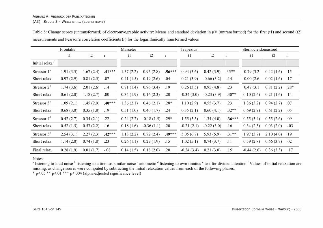
ANHANG A: ABDRUCK DER PUBLIKATIONEN
(A3) STUDIE 3 – WEISE ET AL. (SUBMITTED-B)
Seite 104 von 145 Dissertation Cornelia Weise Marburg 2008
Table 8: Change scores (untransformed) of electromyographic activity: Means and standard deviation in µV (untransformed) for the first (t1) and second (t2)
measurements and Pearson's correlation coefficients (r) for the logarithmically transformed values
Frontalis Masseter Trapezius Sternocleidomastoid
t1 t2 r t1 t2 r t1 t2 r t1 t2 r
Initial relax.f
Stressor 1a 1.91 (3.5) 1.67 (2.4) .41*** 1.37 (2.2) 0.95 (2.8) .56*** 0.94 (3.6) 0.42 (3.9) .35** 0.79 (3.2 0.42 (1.6) .15
Short relax. 0.97 (2.9) 0.81 (2.5) .07 0.41 (1.5) 0.19 (2.6) .04 0.21 (3.9) -0.66 (3.2) .14 0.00 (2.6 0.02 (1.6) .17
Stressor 2b 1.74 (3.6) 2.01 (2.6) .14 0.71 (1.4) 0.96 (3.4) .19 0.26 (3.5) 0.95 (4.8) .23 0.47 (3.1 0.81 (2.2) .28*
Short relax. 0.61 (2.0) 1.18 (2.7) .00 0.34 (1.9) 0.16 (2.3) .20 -0.34 (3.0) -0.23 (3.9) .30** 0.10 (2.6) 0.21 (1.6) .14
Stressor 3c 1.09 (2.1) 1.45 (2.9) .40*** 1.36 (2.1) 0.46 (2.1) .28* 1.10 (2.9) 0.55 (3.7) .23 1.36 (3.2) 0.94 (2.7) .07
Short relax. 0.68 (3.0) 0.35 (1.8) .19 0.51 (1.0) 0.40 (1.7) .24 0.35 (2.1) 0.60 (4.1) .32** 0.69 (2.9) 0.61 (2.2) .05
Stressor 4d 0.42 (2.7) 0.34 (2.1) .22 0.24 (2.2) -0.18 (1.5) .29* 1.55 (5.5) 1.34 (4.0) .36*** 0.55 (3.4) 0.55 (2.6) .09
Short relax. 0.52 (1.5) 0.57 (2.2) .16 0.18 (1.6) -0.36 (1.1) .20 -0.21 (2.1) -0.22 (3.0) .16 0.34 (2.3) 0.03 (2.0) -.03
Stressor 5e 2.54 (3.1) 2.27 (2.3) .42*** 1.13 (2.2) 0.72 (2.4) .49*** 5.05 (6.7) 5.93 (5.9) .31** 1.97 (3.7) 2.10 (4.0) .19
Short relax. 1.14 (2.0) 0.74 (1.8) .23 0.26 (1.1) 0.29 (1.9) .15 1.02 (5.1) 0.74 (3.7) .11 0.59 (2.8) 0.66 (3.7) .02
Final relax. 0.28 (1.9) 0.01 (1.7) -.08 0.14 (1.5) 0.18 (2.0) .20 -0.24 (3.4) 0.21 (3.0) .15 -0.44 (2.6) 0.36 (3.3) .17
Notes: a listening to loud noise b listening to a tinnitus-similar noise c arithmetic d listening to own tinnitus e test for divided attention f Values of initial relaxation are missing, as change scores were computed by subtracting the initial relaxation values from each of the following phases. * p≤.05 ** p≤.01 *** p≤.004 (alpha-adjusted significance level)
ANHANG A: ABDRUCK DER PUBLIKATIONEN
(A3) STUDIE 3 – WEISE ET AL. (SUBMITTED-B)
Dissertation Cornelia Weise Marburg 2008 Seite 104 a von 145
References
Alpini, D., & Cesarani, A. (2006). Tinnitus as an alarm bell: stress reaction tinnitus model.
ORL Journal of Oto-Rhino-Laryngology and its related specialities, 68(1), 31-36.
Arena, J. G., Blanchard, E. B., Andrasik, F., Cotch, P. A., & Myers, P. E. (1983). Reliability
of psychophysiological assessment. Behaviour Research and Therapy, 21(4), 447-460.
Arena, J. G., Goldberg, S. J., Saul, D. L., & Hobbs, S. H. (1989). Temporal stability of
psychophysiological response profiles: Analysis of individual response stereotypy and
stimulus response specificity. Behavior Therapy, 20(4), 609-618.
Arena, J. G., & Hobbs, S. H. (1995). Reliability of psychophysiological responding as a
function of trait anxiety. Biofeedback and Self Regulation, 20(1), 19-37.
Arena, J. G., Sherman, R. A., Bruno, G. M., & Young, T. R. (1990). Temporal stability of
paraspinal electromyographic recordings in low back pain and non-pain subjects.
International Journal of Psychophysiology, 9(1), 31-37.
Bernhardt, O., Gesch, D., Schwahn, C., Bitter, K., Mundt, T., Mack, F., Kocher, T., Meyer,
G., Hensel, E., & John, U. (2004). Signs of temporomandibular disorders in tinnitus
patients and in a population-based group of volunteers: results of the Study of Health in
Pomerania. Journal of Oral Rehabilitation, 31(4), 311-319.
Burdette, B. H., & Gale, E. N. (1990). Reliability of surface electromyography of the
masseteric and anterior temporal areas. Archives of Oral Biology, 35(9), 747-751.
Coles, R. R. (1984). Epidemiology of tinnitus: prevalence. Journal of Laryngology and
Otology, Supplement, 9, 7-15.
Cram, J. R., Lloyd, J., & Cahn, T. S. (1990). The reliability of EMG muscle scanning.
International Journal of Psychosomatics, 37, 68-72.
Crummer, R. W., & Hassan, G. A. (2004). Diagnostic approach to tinnitus. American Family
Physician, 69(1), 120-126.
Davis, A., & El Refaie, A. (2000). Epidemiology of tinnitus. In R. S. Tyler (Ed.), Tinnitus
Handbook (pp. 1-21). San Diego: Singular Publishing Grout.
El Sheikh, M. (2007). Children's skin conductance level and reactivity: Are these measures
stable over time and across tasks? Developmental Psychobiology, 49(2), 180-186.
Elert, J., Karlsson, S., & Gerdle, B. (1998). One-year reproducibility and stability of the
signal amplitude ratio and other variables of the electromyogram: test-retest of a
shoulder forward flexion test in female workers with neck and shoulder problems.
Clinical Physiology, 18(6), 529-538.
Goebel, G., & Hiller, W. (1998). Tinnitus-Fragebogen (TF) [Tinnitus Questionnaire (TQ)].
Göttingen: Hogrefe.
Goebel, G., & Hiller, W. (2001). STI Strukturiertes Tinnitus-Interview [Structured Tinnitus
Interview]. Göttingen: Hogrefe.
Hallam, R. S. (1987). Psychological approaches to the evaluation and management of tinnitus
distress. In J. Hazell (Ed.), Tinnitus. Edinburgh: Churchill Livingstone.
Hallam, R. S. (1996). Manual of the Tinnitus Questionnaire (TQ). London: The Psychological
Corporation.
ANHANG A: ABDRUCK DER PUBLIKATIONEN
(A3) STUDIE 3 – WEISE ET AL. (SUBMITTED-B)
Seite 104 b von 145 Dissertation Cornelia Weise Marburg 2008
Hallam, R. S., Rachman, S., & Hinchcliffe, R. (1984). Psychological aspects of tinnitus. In S.
Rachman (Ed.), Contributions to medical psychology (Vol. 3, pp. 31-53). Oxford:
Pergamon.
Haralambous, G., Wilson, P. H., Platt-Hepworth, S., Tonkin, J. P., Hensley, V. R., &
Kavanagh, D. (1987). EMG biofeedback in the treatment of tinnitus: An experimental
evaluation. Behaviour Research and Therapy, 25(1), 49-55.
Hebert, S., & Lupien, S. J. (2007). The sound of stress: blunted cortisol reactivity to
psychosocial stress in tinnitus sufferers. Neuroscience Letters, 411(2), 138-142.
Hiller, W., Zaudig, M., & Mombour, W. (1995). IDCL - Internationale Diagnosen
Checklisten für ICD-10. Bern,Göttingen: Huber.
Horner, K. C. (2003). The emotional ear in stress. Neuroscience and Biobehavioral Reviews,
27(5), 437-446.
Jastreboff, P. J., & Hazell, J. W. P. (1993). A neurophysiological approach to tinnitus: clinical
implications. British Journal of Audiology, 27, 7-17.
Peroz, I. (2003). Funktionsstörungen des Kauorgans bei Tinnituspatienten im Vergleich zu
einer Kontrollgruppe [Dysfunctions of the stomatognathic system in tinnitus patients
compared to controls]. HNO, 51(7), 544-549.
Pilgramm, M., Rychlik, R., Lebisch, H., Siedentop, H., Goebel, G., & Kirchhoff, D. (1999).
Tinnitus in der Bundesrepublik Deutschland - eine repräsentative epidemiologische
Studie [Tinnitus in the Federal Republic of Germany - A representative epidemiological
study]. HNO aktuell, 7, 261-265.
Rief, W., Sander, E., Guenther, M., & Nanke, A. (2004). Aufmerksamkeitslenkung bei
Tinnitus: Eine experimentelle psychophysiologische Untersuchung [Steering selective
attention in tinnitus: An experimental psychophysiological study]. Zeitschrift fuer
Klinische Psychologie und Psychotherapie, 33(3), 230-236.
Rief, W., Weise, C., Kley, N., & Martin, A. (2005). Psychophysiologic Treatment of Chronic
Tinnitus: A Randomized Clinical Trial. Psychosomatic Medicine, 67, 833-838.
Sanchez, T. G., Guerra, G. C., Lorenzi, M. C., Brandao, A. L., & Bento, R. F. (2002). The
influence of voluntary muscle contractions upon the onset and modulation of tinnitus.
Audiology and Neuro-Otology, 7(6), 370-375.
Schell, A. M., Dawson, M. E., Nuechterlein, K. H., Subotnik, K. L., & Ventura, J. (2002).
The temporal stability of electrodermal variables over a one-year period in patients with
recent-onset schizophrenia and in normal subjects. Psychophysiology, 39(2), 124-132.
Waters, W. F., Williamson, D. A., Bernard, B. A., Blouin, D. C., & Faulstich, M. E. (1987).
Test-retest reliability of psychophysiological assessment. Behaviour Research and
Therapy, 25(3), 213-221.
Weise, C., Heinecke, K., & Rief, W. (2007). Biofeedback bei chronischem Tinnitus –
Behandlungsleitfaden und vorläufige Ergebnisse zu Wirksamkeit und Akzeptanz
[Biofeedback for chronic tinnitus - Treatment guidelines and preliminary results
regarding their efficacy and acceptance]. Verhaltenstherapie, 17(4), 220-230.
Zimmermann, P., & Fimm, B. (1994). Testbatterie zur Aufmerksamkeitsprüfung [Test battery
for attentional performance]. Würselen: Psytest.
ANHANG A: ABDRUCK DER PUBLIKATIONEN
(A4) STUDIE 4 – HEINECKE ET AL. (ACCEPTED)
Dissertation Cornelia Weise Marburg 2008 Seite 105 von 145
Physiological and psychological stress reactivity in chronic tinnitus
Kristin Heinecke, Cornelia Weise, Kristin Schwarz, and Winfried Rief
Philipps - University of Marburg
Section Clinical Psychology and Psychotherapy
Gutenbergstr. 18
35032 Marburg
Germany
Corresponding author:
Kristin Heinecke; Philipps-University of Marburg, Section Clinical Psychology and Psychotherapy,
Gutenbergstr. 18, 35032 Marburg, Germany;
e-mail: [email protected]
ANHANG A: ABDRUCK DER PUBLIKATIONEN
(A4) STUDIE 4 – HEINECKE ET AL. (ACCEPTED)
Seite 106 von 145 Dissertation Cornelia Weise Marburg 2008
Abstract
Several models of tinnitus maintenance emphasize the importance of cognitive, emotional and
psychophysiological processes. These factors contribute to distress in patients with decompensated
tinnitus symptoms. We investigated whether tinnitus patients show increased physiological levels of
arousal, more intense stress reactivity patterns and exaggerated psychological strain compared to
healthy controls.
Seventy tinnitus patients and 55 healthy controls underwent various stress tests. Muscular reac-
tivity and peripheral arousal as well as strain ratings were assessed.
Tinnitus patients reported significantly more strain during stress tests compared to healthy con-
trols. Few physiological reactivity patterns differed significantly between the two groups. The physio-
logical data thus only partly supported a hyperreactivity hypothesis. Strain reports and physiological
data were only marginally correlated.
Tinnitus patients show maladaptive appraisal processes during stress exposure, yet physiologi-
cal reactivity is only slightly affected. Treatment programs for patients with decompensated tinnitus
symptoms should account for appraisal processes and coping mechanisms in stressful situations.
ANHANG A: ABDRUCK DER PUBLIKATIONEN
(A4) STUDIE 4 – HEINECKE ET AL. (ACCEPTED)
Dissertation Cornelia Weise Marburg 2008 Seite 107 von 145
Introduction
Tinnitus noises are very prevalent: about one third of all adults report experiencing tinnitus at
some time, and 10-15% are affected chronically (Heller, 2003). About 6% to 25% of these report a
substantial amount of psychological distress (Heller, 2003; Lynn et al., 2003; Axelsson & Ringdahl,
1989; Coles, 1984) and impairments in daily functioning, such as sleep disturbances (Andersson et al.,
1999; Folmer et al., 2000) and concentration deficits (Stobik et al, 2003). If tinnitus is associated with
psychological problems, the term “decompensated” (or “complex”) tinnitus is used (Duckro et al.,
1984). On the basis of the above-mentioned estimates, the prevalence of decompensated chronic tinni-
tus is about 1-2.4 % in the adult population (Axelsson & Ringdahl, 1989, Coles, 1984).
Mechanisms of tinnitus onset and maintenance are not fully understood. However, the role of
emotional, cognitive and psychophysiological factors in maintaining distress has been emphasized in
current tinnitus models. Hallam (1984) postulates that habituation is complicated by negative appraisal
of tinnitus symptoms, and by a lack of attention shift. Jastreboff and colleagues’ (1993) subsequent
neurophysiological model emphasizes processes that facilitate the detection of tinnitus signals from
spontaneous neuronal activity. Non-audiological brain areas like the limbic system play a crucial role
in the maintenance of tinnitus symptoms. In both models, habituation is attenuated by rumination and
worry about tinnitus and a strong negative emotional reaction to the associated sounds. Several studies
found empirical evidence for the importance of appraisal processes and irrational beliefs in maintain-
ing tinnitus-related distress (Erlandsson & Hallberg, 2000; Erlandsson et al, 1992; Hallberg &
Erlandsson, 1993; Langenbach et al., 2005, Rienhoff et al., 2002). Stress is supposed to play an impor-
tant part in the development of tinnitus: first, it might act as a potential trigger for sudden hearing loss
and onset of tinnitus (Schmitt et al., 2000). Second, it might unfavorably influence habituation via
hyperarousal processes (Hallam, 1984). Third, tinnitus symptoms themselves act as a stressor resulting
in higher physiological arousal and psychological distress. Considerations on a stress reaction tinnitus
model have been put forward by Alpini and Cesarani (2006), resulting in an adaptation of Selyes’
stress model (1976) to tinnitus symptoms. Empirical evidence for a stress reactivity hypothesis is

ANHANG A: ABDRUCK DER PUBLIKATIONEN
(A4) STUDIE 4 – HEINECKE ET AL. (ACCEPTED)
Seite 108 von 145 Dissertation Cornelia Weise Marburg 2008
discussed below, and psychological and physiological findings concerning a stress hyperreactivity hy-
pothesis in chronic tinnitus sufferers will be presented.
Psychological findings
Indirect evidence concerning the stress hypothesis is based on the high psychiatric comorbidity
of tinnitus: about 10-60% of chronic tinnitus patients suffer from a depressive disorder and 28-45%
show clinically relevant anxiety symptoms (Andersson et al., 2002; Reynolds et al., 2003). Assuming
that tinnitus patients have a greater amount of stress reactivity, maladjustment to daily stress situations
could be a consequence. It has been demonstrated that decompensated tinnitus patients use more mal-
adaptive coping strategies compared to controls (Budd & Pugh, 1995, 1996; D´Amelio et al., 2004,
Kirsch et al., 1989, Schmitt et al., 2000). Another argument is the high efficacy of psychotherapy in
the treatment of tinnitus sufferers (Dobie, 1999; Andersson & Lyttkens, 1999; Olderog, 1999). Most of
these treatment programs address coping strategies, which influence stress reactivity.
Physiological findings
Concerning the physiological factors, maladaptive stress reactivity in chronic tinnitus patients
should lead to a hyperreactivity in the autonomous nervous system. Some evidence for this can be
found in studies addressing the role of abnormal physiological stress reactions in the onset and main-
tenance of tinnitus symptoms (Horner, 2003; Sahley et al, 2001, Datzov, 1999). Studies testing the
effect of relaxation training emphasize psychophysiological factors in chronic tinnitus patients. Here,
changes in endocrine and immunological parameters as well as peripheral indicators (e.g. muscle ten-
sion) and perceived stress were demonstrated (Weber et al., 2002, Rief et al., 2005). Few studies have
tested the hypothesis of higher stress reactivity using experimental stress induction. For a maladaptive
stress reactivity pattern in tinnitus patients, results are partly inconsistent. It has been shown that tinni-
tus patients respond with a different cortisol reaction pattern towards stress induction compared to
controls (Hébert et al., 2004, Hébert et al., 2007). Contrary to prediction, however, stress ratings and
baseline cortisol levels were rather similar. Another indicator of stress reactivity are changes in elec-
tromyography (EMG) levels (Lundberg, 1994). Sanchez et al. (2002) demonstrated the importance of
muscle contraction in tinnitus modulation. Confirming the hyperreactivity hypothesis, tinnitus patients
ANHANG A: ABDRUCK DER PUBLIKATIONEN
(A4) STUDIE 4 – HEINECKE ET AL. (ACCEPTED)
Dissertation Cornelia Weise Marburg 2008 Seite 109 von 145
reported a generally higher level of muscle tension (in face, jaw and shoulders) than healthy controls
(Peroz, 2003; Andersson, 1999). Unfortunately, physiological parameters of stress reactivity like EMG
change scores or skin conductance level were not assessed in these studies. A further tinnitus study by
Rief et al. (2004) reported correlations between psychophysiological changes and strain ratings during
tasks with different levels of attentional focus.
Although the linkage between stress and tinnitus is commonly accepted and part of many psy-
chological interventions, only few studies have so far addressed the issue of experimental stress induc-
tion in chronic tinnitus patients, and none of these included all essential characteristics such as psycho-
logical and physiological assessment, a control group, and the use of several stressors.
Hypotheses
In consideration of the available evidence, we hypothesized that tinnitus patients would expe-
rience more subjective strain during a stress test compared to controls, due to the tinnitus patients’
higher responsiveness to stressful events. In addition, tinnitus patients are supposed to show more
autonomous arousal than controls, resulting in higher physiological arousal levels in baseline assess-
ments. We predicted that tinnitus patients have more difficulty relaxing and therefore expected higher
physiological levels during relaxation phases. This might be the case for relaxation ability in general
and for regeneration periods shortly after the stressor phases. We further expected that tinnitus patients
would demonstrate higher stress reactivity than controls, resulting in an increase of physiological
arousal during the stress tests.
Methods
Participants
From May 2005 to February 2007, tinnitus patients were recruited via advertisements in a local
newspaper to participate in an outpatient treatment program at the University of Marburg (Weise et al,
in press). The program consisted of 15 sessions of cognitive behavioral therapy especially developed
for chronic tinnitus sufferers and included stress tests as part of its diagnostic process. 210 tinnitus
patients were invited for diagnostic interview. Eligibility criteria for participation were: chronic de-

ANHANG A: ABDRUCK DER PUBLIKATIONEN
(A4) STUDIE 4 – HEINECKE ET AL. (ACCEPTED)
Seite 110 von 145 Dissertation Cornelia Weise Marburg 2008
compensated tinnitus symptoms with a duration of at least 6 months, a moderate to strong tinnitus
annoyance with scores greater than 46 in a tinnitus questionnaire (Goebel & Hiller, 1998) and an age
ranging from 16 to 75 years. Participants had to understand German well enough to be able to follow
instructions and to complete questionnaires. Exclusion criteria were tinnitus symptoms caused by a
known underlying disease, psychotic episodes, dementia or substance abuse. 132 of 210 patients ful-
filled the inclusion criteria, 2 of these refused to participate. Seventy-one tinnitus sufferers have so far
completed the stress tests, the other participants placed on a waiting list. One data set was not usable
because of technical problems, resulting in 70 suitable data sets.
Sixty-eight controls were recruited from the local community via newspaper advertisements and
were paid €20 for participation. Inclusion and exclusion criteria for controls were the same as for the
tinnitus patients; additionally no audiological diagnosis and chronic pain should exist. Controls were
matched by age and sex. Thirteen persons had to be excluded because of chronic pain symptoms
(headache or lower back pain). Fifty-five control subjects were included for analysis.
Procedure
The stress tests were conducted by three graduate psychology students, who had been trained to
instruct the participants. After subjects consented to participate, they were seated in an adjustable
straight-back chair. Then, sensors for physiological assessment were attached. Participants had to pass
a baseline phase lasting for two and a half minutes, in which they were instructed not to speak and to
restrict movements to a minimum. Afterwards the first relaxation period of five minutes took place.
Participants were asked to sit comfortable and to relax as much as possible by attending to their
muscles tension. Participants were then exposed to five stressor phases with a duration of 3 minutes
each, presented in counterbalanced order, except the attention task stated below. For the exposition to
different sounds, participants were seated in front of two loudspeaker (Philips PC Sound System) with
an output power of 1.5 watt (RMS) each. The distance between ear and loudspeaker was approxi-
mately 1.15 metres. The stressors included tinnitus-specific stimuli and performance tests: “noise”
(mixed sounds composed of traffic sounds, ringing of a phone and artificial noises; noise level was 70
and 79 dB(A) with a mean of about 75 dB(A) measured at ear), “tinnitus-similar-noise” (tinnitus pa-
ANHANG A: ABDRUCK DER PUBLIKATIONEN
(A4) STUDIE 4 – HEINECKE ET AL. (ACCEPTED)
Dissertation Cornelia Weise Marburg 2008 Seite 111 von 145
tients had to select an audio-signal that best matched their own tinnitus from 12 examples; for controls
audio-signals were presented in randomised order; the noise level was 70 to 78 dB(A) with a mean of
approximately 74dB(A)), “arithmetics” (subtracting 700-7-7 along with an audio signal), and
“listening to own tinnitus” (tinnitus patients had to concentrate on tinnitus sounds, while wearing ear-
phones typically used for noise protection; controls had to listen to their heartbeat or other bodily
sounds).
At the end of the four randomised stressor phases, a test of divided attention was conducted at
last place (TAP, Series of tests for attention; Zimmermann & Fimm, 1993). The TAP is a computer--
based program testing attentional performance. In the subtest “divided attention” participants have to
react to visual and audiological stimuli simultaneously.
After each of the five stressor phases, there was a regeneration phase lasting 2 minutes. After
each of these post-stress phases, subjects were asked to rate the question: “During the stressor phase,
how much strain did you feel?” on an 11-point-Likert-scale ranging from “0= no strain at all” to
“10=extreme strain”. Finally, there was a second relaxation phase lasting 5 minutes. Questionnaires
were given at the end of the session. The entire procedure lasted approximately 75-85 minutes. An
overview of the stress test is shown in Figure 1.
Assessment Instruments
Physiological assessment (EMG and peripheral data)
All physiological data were registered by Flexcomp Infinity™ by Thought Technology Ltd., a
ten-channel computer-operated encoder. The software used was BioGraph Infinitiy™ (version2.5.3).
Muscle activity was assessed for forehead (frontalis region), jaws (masseter, bilateral),shoulder (trape-
zius, bilateral), and neck (sternocleidomastoideus, bilateral). According to recommendations by Cram
(1990), the electrodes were placed parallel to muscle fibres to maximize sensitivity and selectivity.
The EMG sensors used were SA9503M MyoScan EMG, a preamplified sensor with input impedance
greater than 10 GΩ and an active range from 10 to 500 Hz, according to Tassinary et al. (2000). We
used dry T3402M Triodes as electrodes (2 cm distance). The raw EMG signal was converted to root
mean square (RMS) using the non-sliding-window algorithm with an averaging factor of 10 and a time
ANHANG A: ABDRUCK DER PUBLIKATIONEN
(A4) STUDIE 4 – HEINECKE ET AL. (ACCEPTED)
Seite 112 von 145 Dissertation Cornelia Weise Marburg 2008
period of 1 second. The sampling rate for physiological data was 32 samples per second. To avoid data
loss, only a 50-Hz notch filter was implemented. The electrocardiogram artefact (especially in shoul-
der EMG) was tolerated to ensure measurement of slow muscle activity. Additionally, skin conduc-
tance level (SCL) was recorded by SA9309M Skin Conductance Flex/Pro with a signal range from 0
to 30 microsiemens. Finger temperature was measured by SA9309M sensor, signal ranging from 12.5
to 40.5 degrees Celsius. SCL and finger temperature signal was not filtered. Skin was cleaned with
antiseptic before attaching the triodes.
Self-report measures
Beck Depression Inventory (BDI)
We assessed depressive symptoms with the Beck depression inventory (Beck et al., 1961; Ger-
man version: Hautzinger et al., 1995). The German version shows good internal consistency (Cron-
bachs alpha=.73-.95), retest reliability (r=.68-.75 over a period of two weeks) and validity concerning
self-assessment questionnaires (r=.76).
Symptom checklist revised (SCL-90-R)
The SCL-90-R (Derogatis, 1986; German version: Franke et al., 2002) is a 90 item question-
naire for measuring psychological distress within a week. Reliability of the German version is found to
be high (Cronbachs Alpha ranging between .75 and .97; retest-reliability between .69 and .92). Con-
current validity coefficients ranged from .45 to .72. For analysis, the global severity index (GSI) is
used.
Tinnitus Questionnaire (TQ)
Tinnitus annoyance was assessed using the tinnitus questionnaire (TQ, Hallam et al., 1996,
German version: Goebel & Hiller, 1994). The TQ is a standard questionnaire in tinnitus research
showing good reliability in terms of retest-reliability (r=.94) and internal consistency (Cronbachs
Alpha=.94). Validity coefficients are moderate to high for psychological distress (r=.5 up to >.7) and
high for tinnitus annoyance (r=.69 to r=.74). For correlation analysis the total score was used.
Data reduction and correction
ANHANG A: ABDRUCK DER PUBLIKATIONEN
(A4) STUDIE 4 – HEINECKE ET AL. (ACCEPTED)
Dissertation Cornelia Weise Marburg 2008 Seite 113 von 145
Physiological data was imported into Biocalc, a specially adapted computer program designed
to read physiological data. The program computed means and standard deviations for every phase per
person. Biocalc rejected data when outliers exceeded three standard deviations of individual trial
means, defining them as artifacts. Finally, data was imported into SPSS for further statistical analysis.
Additionally, artifacts were corrected manually according to the following rules: single values were
replaced by missing when the numerical value was greater than 30 microvolts for frontalis, masseter
and sternocleidomastoideus region, respectively 45 microvolts for the trapezius muscles, and 20
microsiemens for skin conductance level, indicating an error value. To reduce the number of variables
and enhance stability, bilateral measurements of the masseter, trapezius and sternocleidomastoideus
muscles were averaged.
Despite the instruction not to move during baseline assessment, we identified a great number of
artifacts in this phase. We therefore decided to use the first relaxation phase as baseline equivalent.
Statistical analyses
For statistical analysis SPSS 12.0 for Windows was used. Statistical significance was set at an
alpha level of p< 0.05. Demographic variables (age and sex) and depressive symptoms were tested
using independent t tests for differences in means and chi-square tests for categorical variables. Stress
induction was tested using multiple analyses of variance (MANOVA) with repeated measures, with a
2 (group) x 13 (treatment) design. The two groups were tinnitus and non-tinnitus. The 13 treatment
conditions were: baseline, relaxation at the beginning, the five stressor episodes with five regeneration
periods and the second relaxation phase. We decided to use the frontalis region and SCL as validation
of whether stress had been induced, because they act as typical indicators of physiological stress reac-
tions (Passchier, J. & v Helm-Hylkema, 1981). Manipulation was considered successful when the
MANOVA showed a significant effect for treatment. Further contrast analyses indicated which
stressor trials differed from the first relaxation phase. If a significant difference was found, this single
treatment phase stressor could be assumed to have acted as a stressor producing a substantial change in
physiological measures. In order to compare the tonic level of the relaxation phases between the tinni-
tus patients and the controls we also used a MANOVA. Differences between groups were calculated
ANHANG A: ABDRUCK DER PUBLIKATIONEN
(A4) STUDIE 4 – HEINECKE ET AL. (ACCEPTED)
Seite 114 von 145 Dissertation Cornelia Weise Marburg 2008
for all muscles (four muscle regions) and in a separate analysis for peripheral data (two measures:
temperature and SCL). To test potential differences in reactivity during single stressor phases, a 2
(group factor) x 4 (muscles) MANOVA for each stressor phase was conducted. Additionally, a 2
(group factor) x 2 (SCL and temperature) MANOVA was calculated. Therefore, EMG change scores
were computed (mean stressor-mean baseline). Subsequently, univariate analyses of every single
physiological region (muscles or peripheral data) were conducted, with adjustment using the Bon-
ferroni procedure, even if multivariate analyses failed to reach significance level. We then explored
the data looking for potential group differences in terms of descriptive statistics, as high variability in
the physiological data of some of the parameters could hinder the detection of effects. The same ap-
proach was used for the post-stressor regeneration phases belonging to each stressor phase. Differ-
ences in variability of group means were tested by the Levine test. Additionally, MANOVA tests were
carried out for individual standard variations, resulting from averaging each physiological parameter
within each treatment phase. The MANOVA is considered sufficiently robust if the sample size is
adequate and approximately equal for both groups (Glass et al., 1972). For testing covariation between
physiological and psychological ratings correlation analyses with Pearson correlations were computed
(alpha adjustments with Bonferroni procedure).
Results
Matching
Table I shows the characteristics of the tinnitus and control group concerning age and sex. No
significant differences in age (t = 0.708, df = 123, p = .480) or sex (chi square = 0.010, p = .919) were
found. Thus, matching was successful. Significant differences in BDI were found, with more depress-
sive symptoms in the group of tinnitus sufferers (mean tinnitus = 15.8 vs. mean controls = 6.0,
t = 6.72, df = 115, p < .001). Additionally, both groups differed significantly in general distress (SCL-
R: GSI). These results indicate that tinnitus patients suffered from more psychological complaints than
controls (T=6.04 **p<.001).
Stress induction: Manipulation check
ANHANG A: ABDRUCK DER PUBLIKATIONEN
(A4) STUDIE 4 – HEINECKE ET AL. (ACCEPTED)
Dissertation Cornelia Weise Marburg 2008 Seite 115 von 145
Figure 2 shows means and standard error of the mean (SEM) for each stressor and relaxation
phase for both groups. As described above, we used physiological data of the frontalis region and SCL
to test stress induction. For both measures a significant effect of treatment could be demonstrated
(Wilks-Lambda = 0.418, F(12/98) = 11.38, p < .001 for SCL; and Wilks-Lambda = 0.418, F(12/98) =
8.88, p < .001 for frontalis region). There was no significant group effect, either for SCL (F(1/109) =
0.320, p = .573) or for the frontalis region (F(1/112) = 0.088, p= .768). Moreover, no significant
differences in group x treatment interaction was found for SCL (Wilks-Lambda = 0.893, F(12/98) =
0.975, p = .478) or frontalis (Wilks-Lambda = 0.855, F(12/101) = 1.424, p = .167). These results indi-
cate that each stressor was equally effective for both groups. Pairwise contrasts showed a significant
difference for each stressor phase compared to baseline (= first relaxation phase). These results con-
firm a successful stress induction. One exception, however, was stressor four (listening to own tinnitus
noise). Concerning the measurement of the frontalis region, no significant difference to baseline could
be demonstrated (mean difference =.169; 95% confidence interval: -0.996 to 0.659). The stressor “lis-
tening to own tinnitus” seems to have been an ineffective stressor for both groups.
Psychological stress reactivity
As can be seen in Figure 3, tinnitus sufferers report a greater amount of subjective strain after
each stress phase than controls (Wilks-Lambda = 0.454, F(5/104) = 24.996 p < .001). Univariate
analysis shows differences for “noise” (mean tinnitus (SEM) = 6.4 (0.3), mean controls (SEM) = 4.4
(0.3); (F(1/108) = 18.822, p < .001), “tinnitus-similar noise” (mean tinnitus (SEM) = 6.7 (0.3), mean
controls (SEM) = 4.8 (0.3); F(1/108) = 19.625, p < .001), “subtract” (mean tinnitus (SEM) = 6.6 (0.3),
mean controls (SEM) = 4.6 (0.4); F(1/108) = 16.329, p < .001) and “listening to own tinnitus” (mean
tinnitus (SEM) = 5.4 (0.3), mean controls (SEM) = 1.6 (0.3); F(1/108) = 90.062 p < .001). For “di-
vided attention”, group differences failed to reach significance level (mean tinnitus (SEM) = 5.3 (0.3),
mean controls (SEM) = 4.7 (0.3);F(1/108) = 2.633, p = .108).
Tonic level: differences in general arousal
In order to test potential differences in the tonic physiological level between tinnitus patients
and controls, MANOVA for baseline phase and relaxation phases (at the beginning and at the end of
ANHANG A: ABDRUCK DER PUBLIKATIONEN
(A4) STUDIE 4 – HEINECKE ET AL. (ACCEPTED)
Seite 116 von 145 Dissertation Cornelia Weise Marburg 2008
the stress test) were examined. No significant differences were found between the group of tinnitus
sufferers and controls in the baseline phase, either for peripheral results (SCL, F(1/122) = 0.548, p =
.461; temperature, F(1/120) = 0.444, p = .506) or for muscle activity (frontalis, F(1/121) = 0.497, p =
.482; masseter, F(1/122) = 0.059, p = .809; trapezius, F(1/122) = 1.055, p = .306; sternocleidomas-
toideus, F(1/122) = 0.120, p = .730). Similar results were found for the total relaxation phase (average
of both relaxation phases at the beginning and at the end of the stress test). Again the two groups did
not differ with regard to either SCL (F(1/122) = 0.022, p = .884) or temperature (F(1/121) = .380, p =
.539) as well as for muscles (frontalis, F(1/122) = 0.133, p = .716; masseter, F(1/122) = 1.101, p =
.296; trapezius, F(1/123) = .035, p = .851; sternocleidomastoideus, F(1/123) = 0.376, p = .541).
Physiological stress reactivity
To test potential group differences in reactivity during single stressor phases, a MANOVA for
each stressor phase was conducted.
Noise. Multivariate analyses revealed no significant group effect referring to muscle reactivity (Wilks-
Lambda = 0.951, F(4/118) = 1.536, p = .196) or peripheral data (Wilks-Lambda = 0.995, F(2/114) =
0.290, p = .748). The subsequent univariate analysis indicated a significant difference in the reactivity
of the frontalis muscles (mean (SD) tinnitus = 6.2 (3.2), mean (SD) controls = 5.3 (2.6); F(1/122) =
4.255, p = .041), revealing that tinnitus patients show increased reactivity in frontalis muscles tension
during this stressor phase.
Tinnitus-similar noise. There were no significant differences in muscle reactivity (Wilks-Lambda =
0.975, F(4/118) = 0.768, p = .548) or peripheral data (Wilks-Lambda =0.991, F (2/112) = 0.488, p =
.615) between groups referring to the stressor “listening to a tinnitus-similar noise”. Univariate analy-
sis differences failed to reach statistical significance.
Arithmetics with time pressure. There was a marginally significant multivariate group effect for
muscle reactivity in this performance task (Wilks-Lambda = 0.925, F(4/117) = 2.365 p = .057) with
tinnitus patients showing higher activity. Considering the univariate analyses, higher reactivity of the
tinnitus group is represented in masseter region (mean (SD) tinnitus = 4.1 (3.5), mean (SD) controls =
2.9 (2.4); F(1/122) = 6.680, p = .011) and marginally in the frontalis region (mean (SD) tinnitus = 6.0
ANHANG A: ABDRUCK DER PUBLIKATIONEN
(A4) STUDIE 4 – HEINECKE ET AL. (ACCEPTED)
Dissertation Cornelia Weise Marburg 2008 Seite 117 von 145
(2.5), mean (SD) controls = 5.4 (2.4); F(1/122) = 3.497, p = .064). Again, no significant differences
were found for peripheral data (Wilks-Lambda=.999, F(2/113) = 0.069, p = .933).
Listening to own tinnitus. No significant group differences were found (muscles, Wilks-Lambda =
0.945, F(4/115) = 1.686, p = .158; peripheral data, Wilks-Lambda = .998 F(2/113) = 0.106, p = .900).
Exploratory subsequent analyses showed that in the trapezius region, tinnitus patients had higher reac-
tivity while listening to their own tinnitus (mean (SD) tinnitus = 5.1 (5.4), mean (SD) controls = 3.6
(2.1); F(1/118) = 4.300, p = .040).
Test of divided attention. Contrary to the hypothesis, a significant group effect was found, indicating
that controls had higher muscle reactivity (Wilks-Lambda = 0.907, F(4/101) = 2.587, p = .041). This
effect was due to the trapezius region (mean (SD) tinnitus = 8.0 (5.9), mean (SD) controls = 11.8 (9.0);
F(1/104) = 6.639, p = .011). Because of movement artifacts during this test, especially for the shoulder
region (participants had to press a button), these results were not explored further. No differences
between the groups occurred for peripheral data (Wilks-Lambda = 0.993, F(2/112) = 0.404 p = .669).
We also tested for differences in variability between groups. Overall, the results suggest that
neither individual standard deviations per phase, nor standard deviations of the group means differed
substantially.
Reactivity in relaxation
Another aspect of stress reactivity is the ability to relax after stressor episodes. We thus
analyzed the short post-stress regeneration phases for potential group differences. First, change values
were calculated as subtraction between regeneration and baseline phases. Overall, no significant group
difference between tinnitus patients and controls could be found in regeneration phase. In a second
step, we investigated potential differences in the speed of relaxation by calculating change scores of
the first and second minute of the regeneration phase. Again, both groups showed very similar reaction
patterns. This indicates that neither the extent nor the speed of relaxation differed between the groups.
Medication as a covariate
Considering that tinnitus patients and controls differ in medication intake, multivariate analyses
were repeated with medication as a covariate. Results were similar to the results reported above, with
ANHANG A: ABDRUCK DER PUBLIKATIONEN
(A4) STUDIE 4 – HEINECKE ET AL. (ACCEPTED)
Seite 118 von 145 Dissertation Cornelia Weise Marburg 2008
one exception: group differences in the subtests “listening to own tinnitus” and “arithmetics” reached
significance, while the subtest “divided attention task” did not. For the subtest “noise”, the difference
for the frontalis region became marginally significant. The other results remained unaffected.
Correlation analysis
Strain rating and physiological data. None of the correlations between self-reported strain during the
stressor phases and physiological arousal values reached significance level after applying alpha ad-
justment. This result indicates that the physiological and psychological stress reactions were different
reaction patterns.
Actual strain rating and long-lasting distress. For the group of tinnitus patients, correlations between
state ratings and measures of psychological strain in general (BDI, TQ and SCL-90-R) were analyzed.
The BDI sum score and the subjective ratings were moderately correlated for the stressors “tinnitus-
similar noise”, “arithmetics” and “listening to their own tinnitus” ranging between r = .301 and r =
.544, (p < .05/15). Only non-significant correlations were found for the tinnitus questionnaire and sub-
jective stress ratings. None of the strain ratings were substantially correlated with GSI scores of the
SCL-90-R.
Symptoms of psychological distress and physiological stress reactivity. Correlation analyses for each
stressor phase were used to explore relationships between depressive symptoms and physiological re-
activity. Only one out of 20 possible correlations between BDI scores and muscle reactivity values
was significantly correlated (stressor “listening to own tinnitus” and masseter, r = .319, p <.05/20). For
peripheral data (SCL and temperature) no significant correlations were found. Regarding tinnitus an-
noyance and GSI scores, none of the physiological reactivity parameters were significantly associated.
Muscular intercorrelation. Muscular intercorrelations relating to the different stressor phases were
tested in order to determine whether the stressors induced a general muscular stress reactivity pattern.
Correlations were quite small, ranging from r=-.189 to .380. After alpha adjustment only 4 out of 45
scores reached significance level. In summary, stress reactivity seems not to be represented in a gen-
eral simultaneous muscular activation.
ANHANG A: ABDRUCK DER PUBLIKATIONEN
(A4) STUDIE 4 – HEINECKE ET AL. (ACCEPTED)
Dissertation Cornelia Weise Marburg 2008 Seite 119 von 145
Further analysis
To test potential subgroups of tinnitus patients, we divided them into a depressed and a non-
depressed group (BDI cut-off score: 17). Analysis revealed that not only the depressed but also the
non-depressed group showed an increased strain rating compared to controls. The tinnitus groups did
not differ in strain rating except for stressor “listening to own tinnitus”.
Another subgroup analysis was calculated with a hearing loss and a non-hearing loss group. Dif-
ferences in both groups concerning physiological and psychological stress reactivity failed to reach
statistical significance.
Discussion
In the present study, we assumedthat tinnitus patients experience more psychological and
physiological strain during various stress tests. In accordance with this expectation, tinnitus patients
rated each stressor phases with both tinnitus specific stimuli and performance tasks as more stressful
than controls. Our results indicate a greater amount of psychological stress in tinnitus patients. This is
in contrast to Hébert et al`s (2007) study, in which no differences in strain ratings were found. As an-
ticipated, patients showed more depressive symptoms and more distress generally.
Our second hypothesis presumed that the general arousal level of tinnitus patients is higher
compared to controls. The present data do not support this hypothesis. However, the results are in line
with other empirical studies concerning immunological and endocrine findings (e.g. Herbert, 2007)
but contradict the assumption of greater autonomous activation in general. Surprisingly, tinnitus pa-
tients and controls showed very similar general relaxation abilities and regeneration patterns after
stressor phases. One explanation could be that tinnitus patients are more trained in relaxation because
of past experiences with psychological treatment. Almost two third of the participants had been in
some kind of psychological therapy program, while less than one third of controls had received previ-
ous treatment. As stress induction was successful in both groups, the prerequisite for detecting poten-
tial differences in stress reactivity was met. Interestingly, “listening to own tinnitus” was rated as
highly stressful by the tinnitus group, but physiological reactivity was similar to the relaxation phase.

ANHANG A: ABDRUCK DER PUBLIKATIONEN
(A4) STUDIE 4 – HEINECKE ET AL. (ACCEPTED)
Seite 120 von 145 Dissertation Cornelia Weise Marburg 2008
This result underlines the importance of appraisal processes as postulated in models of tinnitus annoy-
ance (Hallam, 1984; Jastreboff et al., 1993).
Considering the third hypothesis, slight group differences in physiological stress reactivity were
found, but there seemed to be no general or consistent muscular hyperreactivity or abnormal reaction
pattern for peripheral measures in the tinnitus group. These results are partly in line with the studies of
immunological and endocrine findings mentioned above, showing some, but not very clear, differ-
ences between tinnitus patients and healthy controls.
A divergence between subjective strain ratings and objective physiological data emerged. This
assumption was further confirmed by the lack of significant correlations between strain ratings and
physiological reactivity. One crucial factor with regard to the ability to cope with stressful situations
seems to be the presence of depressive symptoms. They were moderately correlated with strain ratings
and slightly correlated with physiological reactivity. A similar result - confirming the association of
depression with extended physiological and emotional reactivity to daily life stress - has been found in
patients with affective disorders (Myin-Germeys et al., 2003). Considering that more than 50% of the
tinnitus patients participating in the present study suffered from affective disorder symptoms, treat-
ment should focus on these symptoms. Surprisingly, tinnitus specific annoyance and distress as a
whole seem to play a less important part in stress reactivity.
Taken all results into account, subjective data support the stress-reactivity hypothesis proposed
in models of tinnitus annoyance (Hallam, 1984; Jastreboff et al., 1993). Appraisal processes can lead
to psychological strain and intensify or cause depressive symptoms. Maladaptive and catastrophizing
thoughts should be considered and worked on - not only in relation to tinnitus specific symptoms and
annoyance, but also in the appraisal of stress situations. The present study only partly confirms the
hypothesis of autonomous hyperreactivity in chronic tinnitus sufferers.
Strengths and limitations of the study
A major strength of our study is the sample size, including tinnitus sufferers and healthy con-
trols matched for relevant variables. The number of participants for both groups was sufficient to iden-
tify even moderate differences (Cohen, 1988). We efficiently controlled for artifacts, and we counter-
ANHANG A: ABDRUCK DER PUBLIKATIONEN
(A4) STUDIE 4 – HEINECKE ET AL. (ACCEPTED)
Dissertation Cornelia Weise Marburg 2008 Seite 121 von 145
balanced multiple stressors. The stressors were ecologically valid and composed of tinnitus-specific
material as well as performance stressors. Effective stress induction was demonstrated, showing per-
sonal emotional relevance as required (Ohrbach et al., 1998).
Some limitations of the study should be taken into account: retest reliability of surface EMG is
only moderate, ranging from .41 to .94 with a median correlation of .64 (Cram et al., 1990). Therefore,
error variance and instability complicate the detection of group differences. Another critical point
might be the type of data reduction used, particularly averaging over phases. Changes that might occur
temporarily during one phase could not be shown. The small intercorrelations of EMG change scores
indicate that there seems to be no generalized muscular reaction pattern. Therefore another approach
could analyse more individual reactions of specific muscle regions for each subject, comparable with
symptom-specific reactions in chronic pain patients (Flor et al., 1992).
Despite these shortcomings, we summarized that if there were substantial differences in
autonomous reactivity, we should have been able to demonstrate them. Physiological parameters in
tinnitus sufferers seem to be less relevant peripherally and more important centrally. As demonstrated
by neurophysiological research, tinnitus patients show abnormalities relating EEG negativity during
acoustic stimuli (Weisz et al., 2004) and abnormal spontaneous neuronal activity (Weisz et al., 2005).
These processes should be further investigated.
Practical implications
It might be useful to inform tinnitus patients about the divergence between subjective strain ra-
tings and autonomous stress reactions. This might give reassurance, by confirming that their physio-
logical pattern is normal and non-pathological. Our results were in line with findings on chronic pain
patients, who overestimate muscle tension (Flor et al., 1999). 86% of our participants reported having
difficulty relaxing, and 66% reported feeling at least partially overburdened with daily stress situa-
tions. Physiological reactivity and relaxation ability via biofeedback treatment could be demonstrated
to sufferers in order to provide objective evidence. Demonstrating psychophysiological reactions
might result in insights into the overestimation processes in stress situations. Also, tinnitus patients
often report that their impairment is mainly caused by tinnitus symptoms. Over 70% of our patients

ANHANG A: ABDRUCK DER PUBLIKATIONEN
(A4) STUDIE 4 – HEINECKE ET AL. (ACCEPTED)
Seite 122 von 145 Dissertation Cornelia Weise Marburg 2008
partly agreed to the item: “Almost all of my problems are caused by tinnitus symptoms”. Contradic-
tory to these expectations, tinnitus annoyance did not correlate with physiological or psychological
stress reactions. In our psychological interventions, we focus on these catastrophizing thoughts with
the aim to adjust to symptoms and enhance quality of life irrespective of the existence of tinnitus
symptoms (Rief et al., 2005; Weise et al., in press).

ANHANG A: ABDRUCK DER PUBLIKATIONEN
(A4) STUDIE 4 – HEINECKE ET AL. (ACCEPTED)
Dissertation Cornelia Weise Marburg 2008 Seite 123 von 145
References
Alpini, D., and Cesari, A. (2006). Tinnitus as an Alarm Bell: Stress Reaction Tinnitus Model. ORL 68:
31-37.
Andersson, G., and Lyttkens, L. (1999). A meta-analytic review of psychological treatments for
tinnitus. British Journal of Audiology 33(4): 201-10.
Andersson, G., Lyttkens L., and Larsen, H.C. (1999). Distinguishing levels of tinnitus distress.
Clinical Otolaryngology & Allied Sciences 24: 404-410.
Andersson, G. (2002). Psychological aspects of tinnitus and the application of cognitive – behavioral
therapy. Clinical Psychology Review 22: 977-990.
Axelsson, A. & Ringdahl, A. (1989). Tinnitus-a study of its prevalence and characteristics. British
Journal of Audiology, 23 (1). 53-62.
Beck, A., and Steer, R. (1987). Beck depression inventory-manual. The psychological Corporation,
San Antonio.
Budd, R. J., and Pugh, R. (1995). The relationship between locus of control, tinnitus severity, and
emotional distress in a group of tinnitus sufferers. Journal of Psychosomatic Research 39(8):
1015-1018.
Budd, R. J., and Pugh R. (1996). Tinnitus coping style and its relationship to tinnitus severity and
emotional distress. Journal of Psychosomatic Research 41(4): 327-335.
Cohen, J. (1988). Statistical power analysis for the behavioral sciences. Hillsdale, Erlbaum.
Cram, J. R., Lloyd J., and Cahn, T. S. (1990). The reliability of EMG muscles scanning. International
Journal of Psychosomatic 37(1-4): 68-72.
Datzov, E., S. Danev, Haralanov, H., Naidenova, V., Sachanska, T., and Savov, A (1999). Tinnitus,
heart rate variability, and some biochemical indicators. International Tinnitus Journal 5(1): 20-
23.
Derogatis, L.R. (1983). SCL-90 : Administration, scoring and procedures manual for the revised
version. Baltimore: Clinical Psychometric Research.
Dobbie, R. (1999). A review of randomised clinical trials in tinnitus. Laryngoscope 109(8): 1202-
1211.
ANHANG A: ABDRUCK DER PUBLIKATIONEN
(A4) STUDIE 4 – HEINECKE ET AL. (ACCEPTED)
Seite 124 von 145 Dissertation Cornelia Weise Marburg 2008
Ducro, P. N., C. A. Pollard, Bray, H.D., and Scheiter, L. (1984). Comprehensive behavioral
management of complex tinnitus. Biofeedback and Selfregulation 9: 459-469.
D´Amelio, R., C. Archonti, Scholz, S., Falkai, P., Plinkert, P.K., and Delb, W. (2004). [Psychological
distress associated with acute tinnitus] Akuter Tinnitus - Psychische Korrelate und Ausmaß der
Belastung bei Patienten. HNO 52: 299-603.
Erlandsson, S. I., L. R. Hallberg, and Axelsson, A. (1992). Psychological and audiological correlates
of perceived tinnitus severity. Audiology 31(3): 168-79.
Erlandsson, S. I., and L. R. Hallberg (2000). Prediction of quality of life in patients with tinnitus.
British Journal of Audiology 34(1): 11-20.
Flor, H., Birbaumer N., Schugens, M. M., and Lutzenberger, W. (1992). Symptom-specific
psychophysiological responses in chronic pain patients. Psychophysiology 29(4): 452-460.
Flor, H., Fürst M., and Birbaumer, N. (1999). Deficient Discrimination of EMG Levels and
Overestimation of Perceived Tension in Chronic Pain Patients. Applied Psychophysiology and
Biofeedback 24(1): 55-66.
Folmer, R. L., and Griest S. E. (2000). Tinnitus and Insomnia. American Journal of Otolaryngology
21(5): 287-293.
Franke, G.H. (2002). SCL-90-R- Die Symptom-Checkliste von L.R. Derogatis-Manual (2nd 1ed.)
Beltz Test GmbH, Göttingen.
Glass, G. V., Peckham P. D., and Sanders, J. R. (1972). Consequences of failure to meet assumptions
underlying the fixed effects analysis of variance and covariance. Review of
Educational Research 42: 237-288.
Goebel, G., and Hiller W. (1998). Tinnitus-Fragebogen (TF)-Ein Instrument zur Erfassung von
Belastungen und Schweregrad bei Tinnitus (Manual). Göttingen, Hogrefe.
Hallam, R., S. Rachman, and Hinchcliffe, R. (1984). Psychological aspects of tinnitus. Contributions
to Medical Psychology. S. Rachman. Oxford, Pergamon.
Hallam, R. (1996). Manual of the Tinnitus Questionnaire (TQ). The Psychological Corporation,
London.
Hallberg, L. R., and S. I. Erlandsson (1993). Tinnitus characteristics in tinnitus complainers and
noncomplainers. British Journal of Audiology 27(1): 19-27.
ANHANG A: ABDRUCK DER PUBLIKATIONEN
(A4) STUDIE 4 – HEINECKE ET AL. (ACCEPTED)
Dissertation Cornelia Weise Marburg 2008 Seite 125 von 145
Hautzinger, M., Bailer M., Worall, H., and Keller, F. (1995). Beck-Depressions-Inventar (BDI).
Testhandbuch. Hans Huber, Bern.
Heller, A. J. (2003). Classification and epidemiology of tinnitus. Otolaryngologic Clinics of North
America 369: 239-248.
Hébert, S., Paiement P., and Lupien S. (2004). A physiological correlate for the intolerance to both
internal and external sounds. Hearing Research 190: 1-9.
Hébert, S., and Lupien S. (2007). The sound of stress: Blunted cortisol reactivity to psychosocial stress
in tinnitus sufferers. Neuroscience Letters 411: 138-142.
Horner, K. (2003). The emotional ear in stress. Neuroscience and Biobehavioral Reviews 27: 437-446.
Jastreboff, P. J., and Hazell J. W. (1993). A neurophysiological approach to tinnitus: clinical
implications. British Journal of Audiology 27: 7-17.
Kirsch, C. A., Blanchard E. B., and Parnes, S. M. (1989). Psychological characteristics of individuals
high and low in their ability to cope with tinnitus. Psychsomatic Medicine 21: 209-217.
Langenbach, M., Olderog M., Michel, O., Albus, C., and Köhle, K. (2005). Psychosocial and
personality predictors of tinnitus-related stress. General Hospital Psychiatry, 27: 73-77.
Lundberg, U., Kadefors R., Melin, B., Palmerud, G., Hassmen, P., Engstrom, M., and Elfsberg, I.
(1994). Psychophysiological stress and EMG activity of the trapezius muscle. International
Journal of Behavioral Medicine 1(4): 354-370.
Lynn, S. G., Bauch C. D., Williams, D. E., Beatty, C. W., Mellon, M. W., and Weaver, A. L. (2003).
Psychological profile of tinnitus patients using SCl-90-R and Tinnitus Handicap Inventory.
Otology & Neurotology 24(6): 878-881.
Myin-Germeys, I., Peeters F., Havermanns, R., Nicolson, N. A., deVries, M. W., Delespaul, P., and
van Os, J. (2003). Emotional reactivity to daily life stress in psychosis and affective disorder: an
experience sampling study. Acta Psychiatrica Scandinavica 107: 124-131.
Ohrbach, R., Blascovic J., Gale, J. N., McCall, W. D., and Dworkin, S. F. (1998). Psychophysiological
assessment of stress in chronic pain: Comparisons of stressful stimuli and of response system.
Journal of Dental Research 77(10): 1840-1850.
ANHANG A: ABDRUCK DER PUBLIKATIONEN
(A4) STUDIE 4 – HEINECKE ET AL. (ACCEPTED)
Seite 126 von 145 Dissertation Cornelia Weise Marburg 2008
Olderog, M. (1999). [A meta-analysis of the effects of psychological interventions in chronic decom-
pensated tinnitus] Metaanalyse zur Wirksamkeit psychologisch fundierter Behandlungskonzepte
des chronischen dekompensierten Tinnitus. Zeitschrift für Medizinische Psychologie 1: 5-18.
Passchier, J. and v. Helm-Hylkema, H. (1981). The effect of stress imagery on arousal and its
implications for biofeedback of the frontalis muscles. Biofeedback and self-regulation 6(3): 295-
303.
Peroz, I. (2003). [Dysfunctions of the stomatognathic system in tinnitus patients compared to controls]
Funktionsstörungen des Kauorgans bei Tinnituspatienten im Vergleich zu einer Kontrollgruppe.
HNO 51: 544-549.
Reynolds, P., Gardner D., and Lee, R. (2004). Tinnitus and psychological morbidity: a cross-sectional
study to investigate psychological morbidity in tinnitus patients and its relationship with severity
of symptoms and illness perception. Clinical Otolaryngology & Allied Sciences 29: 628-634.
Rief, W., Sander E., Günther, M., and Nanke, A. (2004). [Steering selective attention in tinnitus: an
experimental psychophysiological study.] Aufmerksamkeitslenkung bei Tinnitus: Eine
experimentelle psychophysiologische Untersuchung. Zeitschrift für Klinische Psychologie und
Psychotherapie 33(3): 230-236.
Rief, W., Weise C., Kley, N., and Martin, A. (2005). Psychophysiologic treatment of chronic tinnitus:
A randomised clinical trial. Psychosomatic Medicine 67: 833-838.
Rienhoff, N. K., Thimm L., Pöllmann, H., Nelting, M., and Hesse, G. (2002). [Irrational beliefs and
chronic complex tinnitus] Irrationale Einstellungen bei chronisch komplexem Tinnitus.
Zeitschrift für Klinische Psychologie und Psychotherapie 31(1): 47-52.
Sahley, T. L. and Nodar R. H. (2001). A biochemical model of peripheral tinnitus. Hearing Research
152: 43-54.
Sanchez, T. G., Guerra, G. C. Y., Lorenzi, M. C., Brandao, A. L., and Bento, R. F. (2001). The
influence of voluntary muscle contradictions upon the onset and modulation of tinnitus.
Audiology & Neurotology 7: 370-375.
Selye, H. (1976). The stress of life. Rev. ed. McGraw-Hill, New York.
Schmitt, C., Patak, M., and Kröner-Herwig, B. (2000). Stress and the onset of sudden hearing loss and
tinnitus. International Tinnitus Journal 6(1): 41-49.
ANHANG A: ABDRUCK DER PUBLIKATIONEN
(A4) STUDIE 4 – HEINECKE ET AL. (ACCEPTED)
Dissertation Cornelia Weise Marburg 2008 Seite 127 von 145
Schmitz, N., Hartkamp, N., Kiuse, J., Franke, G. H., Reister, G., and Tress, W. (2000). The Symptom
Check-List-90-R (SCL-90-R): A German validation study. Quality of Life Research, 9:185-193.
Stobik, C., Weber R. K., Münte, T.F., and Frommer, J. (2003). [Psychosomatic stress factors in
compensated and decompensated tinnitus] Psychosomatische Belastungsfaktoren bei
kompensiertem und dekompensierten Tinnitus. Psychotherapie, Psychosomatik, Medizinische
Psychologie 53: 344-352.
Tassinary, L. G., and Cacioppo, J. T. (2000). Handbook of Psychophysiology. Cambridge, University
Press.
Weber, C., Arck P., Mazurek, B., and Klapp, B. F. (2002). Impact of relaxation training on
psychometric and immunologic parameters in tinnitus sufferers. Journal of Psychosomatic
Research 52: 29-33.
Weise, C., Heinecke, K., and Rief, W. (in press). [Biofeedback for chronic tinnitus – Treatment
guidelines and preliminary results regarding their efficacy and acceptance] Biofeedback bei
chronischem Tinnitus - Behandlungsleitfaden und vorläufige Ergebnisse zur Wirksamkeit und
Akzeptanz. Verhaltenstherapie.
Weisz, N., S. Voss, Berg, P., and Elbert, T. (2004). Abnormal auditory mismatch response in tinnitus
sufferers with high-frequency hearing loss is associated with subjective distress level. BMC
Neuroscience 4(5): 8.
Weisz, N., Moratti, S., Meinzer, M., Dohrmann, K., and Elbert, T. (2005). Tinnitus perception and
distress is related to abnormal spontaneous brain activity as measured by
magnetoencephalography. PLoS Med 2(6): 153.
Zimmermann, P. and Fimm, B. (1993). [A test battery for attentional performance]Testbatterie zur
Erfassung von Aufmerksamkeitsstörungen. Version 1.02. Freiburg.
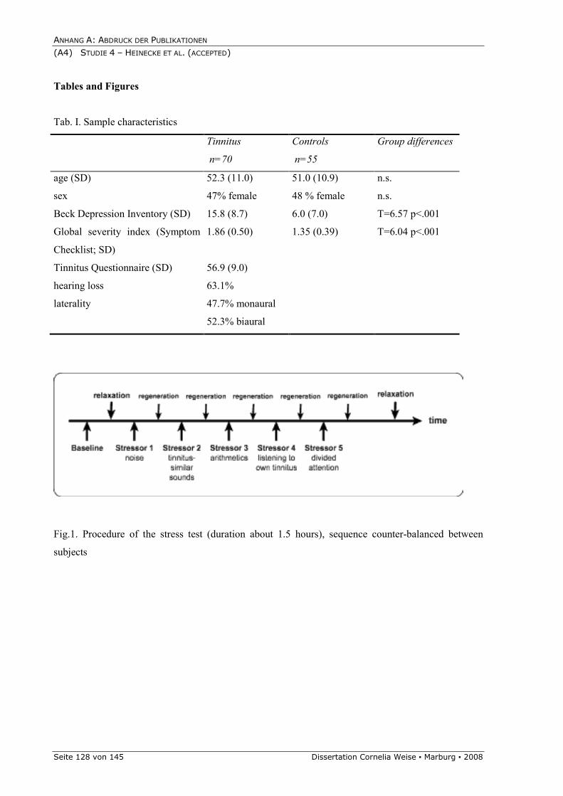
ANHANG A: ABDRUCK DER PUBLIKATIONEN
(A4) STUDIE 4 – HEINECKE ET AL. (ACCEPTED)
Seite 128 von 145 Dissertation Cornelia Weise Marburg 2008
Tables and Figures
Tab. I. Sample characteristics
Tinnitus
n=70
Controls
n=55
Group differences
age (SD) 52.3 (11.0) 51.0 (10.9) n.s.
sex 47% female 48 % female n.s.
Beck Depression Inventory (SD) 15.8 (8.7) 6.0 (7.0) T=6.57 p<.001
Global severity index (Symptom
Checklist; SD)
1.86 (0.50) 1.35 (0.39) T=6.04 p<.001
Tinnitus Questionnaire (SD) 56.9 (9.0)
hearing loss 63.1%
laterality 47.7% monaural
52.3% biaural
Fig.1. Procedure of the stress test (duration about 1.5 hours), sequence counter-balanced between
subjects

ANHANG A: ABDRUCK DER PUBLIKATIONEN
(A4) STUDIE 4 – HEINECKE ET AL. (ACCEPTED)
Dissertation Cornelia Weise Marburg 2008 Seite 129 von 145
Stress induction: skin conductance level (SCL)
2
6
base
line
noise
tinn
itus-simila
r noise
arith
metics
own tin
nitus
divide
d attention
SC
L (m
icro
sie
mens)
tinnitusn=60
controlsn=54
Stress induction: Frontalis EMG
4
8
base
line
noise
tinn
itus-simila
r noise
arith
metics
own tin
nitus
divide
d attention
EM
G (m
icro
volt)
tinnitusn=60
controlsn=54
Fig. 2. Stress induction: changes in physiological arousal (skin conductance level and frontalis EMG)
during the stress tests (mean and standard error of the mean)
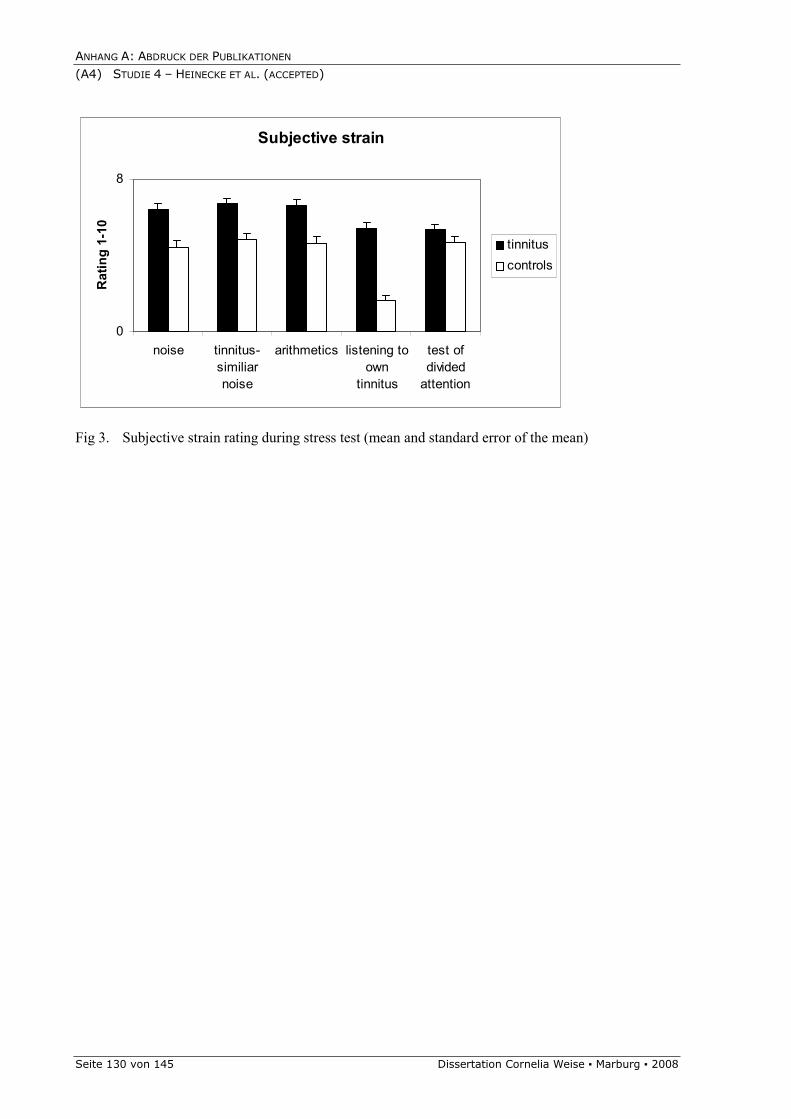
ANHANG A: ABDRUCK DER PUBLIKATIONEN
(A4) STUDIE 4 – HEINECKE ET AL. (ACCEPTED)
Seite 130 von 145 Dissertation Cornelia Weise Marburg 2008
Subjective strain
0
8
noise tinnitus-
similiar
noise
arithmetics listening to
own
tinnitus
test of
divided
attention
Rating 1-10
tinnitus
controls
Fig 3. Subjective strain rating during stress test (mean and standard error of the mean)
ANHANG B: MATERIALIEN UND AUSGEWÄHLTE MESSINSTRUMENTE (B1) EINVERSTÄNDNISERKLÄRUNG
Dissertation Cornelia Weise Marburg 2008 Seite 131 von 145
Die Inhalte und der Ablauf der Studie „Biofeedbacktherapie bei chronischem Tinnitus“, die in
der Arbeitsgruppe Klinische Psychologie und Psychotherapie am Fachbereich Psychologie der
Philipps-Universität Marburg durchgeführt wird, wurden mir von der mir zugeordneten
Therapeutin erklärt.
Meine Daten werden anonymisiert und streng vertraulich behandelt. Sie sind nur den
Forschern/innen dieses Projektes zugängig.
Ich habe alles gelesen und verstanden und erkläre mich bereit an der oben genannten
Studie und den damit einhergehenden Befragungen (vor Therapiebeginn, nach Therapie-
ende, Nachuntersuchung 6 Monate nach Therapieende) teilzunehmen. Ich kann meine Teil-
nahme an der Studie jederzeit widerrufen.
Ich bin bereit, an eventuellen weiteren Nachbefragungen zur Biofeedbacktherapie teilzunehmen.
ja nein
Ich bin damit einverstanden, dass Sie mich kontaktieren, wenn am Institut andere Studien zu Tinnitus durchgeführt werden.
ja nein
_____________________________
Name des Teilnehmers/der Teilnehmerin
_____________________________ _______________________________
Ort und Datum Unterschrift
Einverständniserklärung
Biofeedbacktherapie des chronischen Tinnitus Psychotherapie-Ambulanz Marburg
AG Klinische Psychologie & Psychotherapie
Code: _________________________ Datum: _________________________
ANHANG B: MATERIALIEN UND AUSGEWÄHLTE MESSINSTRUMENTE (B2) INFORMATIONSBLATT ZUR STUDIE
Seite 132 von 145 Dissertation Cornelia Weise Marburg 2008
Im Rahmen einer wissenschaftlichen Studie wird am Fachbereich Psychologie der Phi-
lipps-Universität Marburg eine Biofeedbacktherapie für Patienten mit chronischem Tinnitus
durchgeführt.
Chronische Ohrgeräusche gehören zu den häufigsten Symptomen, die in der Hals--
Nasen-Ohren-Arzt-Praxis angegeben werden. Der Tinnitus wird von Betroffenen als Rau-
schen, Zischen oder Brummen im Kopf wahrgenommen. In Deutschland leiden 2,9 Millionen
Menschen unter chronischem Tinnitus, 1,5 Millionen Menschen fühlen sich durch die Ohrge-
räusche mittelschwer bis zur Unerträglichkeit hin beeinträchtigt (dekompensierter Tinnitus).
Als chronisch bezeichnet man den Tinnitus, wenn er länger als 6 Monate andauert. Dekom-
pensiert bedeutet, dass die Betroffenen sich dadurch wesentlich in ihrer Lebensqualität ein-
geschränkt fühlen und den Tinnitus als Belastung empfinden. Die Beschwerden reichen von
Konzentrations- und Schlafstörungen bis zu depressiver Verstimmung und Angst. Oft berich-
ten Betroffene, dass sie sich den Ohrgeräuschen hilflos ausgeliefert fühlen und sich schwer
auf andere Dinge konzentrieren können.
Hinzu kommt, dass ein chronischer Tinnitus meist nicht durch gängige medizinische
Verfahren behoben werden kann. Anders als beim akuten Tinnitus steht beim chronischen
Tinnitus nicht die Heilung im Vordergrund, sondern die Bewältigung der tinnitusbedingten
Belastung.
In der am Fachbereich Psychologie der Philipps-Universität Marburg durchgeführten
Studie wird die Wirksamkeit einer biofeedbackgestützten Bewältigungstherapie untersucht.
Durch diese Behandlung soll die Beeinträchtigung gelindert werden und der Umgang mit
Tinnitus erleichtert werden.
Biofeedback verhilft dem Menschen, sich seiner Vorgänge im Körper bewusst zu wer-
den und diese zu optimieren. Dazu werden zunächst relevante körperliche Prozesse gemes-
sen, die normalerweise unbewusst ablaufen. Die gemessenen Prozesse werden in optische
Signale umgewandelt und dem Patienten über einen Bildschirm zurückgemeldet. Kleinste
Veränderungen der gemessenen körperlichen Aktivität werden so sichtbar. Durch diese
Rückmeldung kann der Patient lernen, die körperliche Aktivität gezielt zu beeinflussen und
erlangt dadurch zunehmend Selbstkontrolle über die gemessenen körperlichen Funktionen.
Informationen zur Studie
Biofeedbacktherapie des chronischen Tinnitus Psychotherapie-Ambulanz Marburg
AG Klinische Psychologie & Psychotherapie
ANHANG B: MATERIALIEN UND AUSGEWÄHLTE MESSINSTRUMENTE (B2) INFORMATIONSBLATT ZUR STUDIE
Dissertation Cornelia Weise Marburg 2008 Seite 133 von 145
Die körperlichen Prozesse beim Erleben des Tinnitus hängen stark mit dem erfahrenen
Stress und der verstärkten Aufmerksamkeit auf den Tinnitus zusammen. Durch Stress
verspannen sich einerseits die Muskeln in den Bereichen Kopf, Schulter und Nacken. Beim
Biofeedback wird deswegen trainiert, gezielt diese Muskeln zu entspannen. Andererseits wird
durch Stress der Tinnitus selbst verstärkt. Die Aufmerksamkeit wird stärker auf das Ohrge-
räusch gerichtet, wodurch dann wiederum die empfundene Belastung und der Stress steigt.
Mit Entspannung, die rechtzeitig und wirksam eingesetzt wird, kann man diesen Kon-
sequenzen von Stress entgegenwirken. Im entspannten Zustand ist die Aktivität der Muskeln
gering, wohingegen sie bei Stress und der folgenden Anspannung hoch ist.
Durch das Biofeedback wird die gezielte Entspannung, also die Senkung der Muskelak-
tivität, trainiert. Diese Fähigkeit kann im Alltag eingesetzt werden, wenn die Belastungen und
der Stress durch den Tinnitus stark sind oder wenn in stressigen Situationen der Tinnitus sich
zu verstärken droht.
Ziel der Studie ist es, durch gesteigerte Selbstkontrolle und den verbesserten Umgang
mit Stress die Lebensqualität, die durch den Tinnitus beeinträchtigt wurde, wieder zu erhö-
hen.
Biofeedback ist ein Verfahren, bei dem die Betroffenen eine aktive Rolle übernehmen
und selbst eine Technik erlernen, die auch im normalen Tagesablauf einsetzbar ist. Es hat
keinerlei negative Nebenwirkungen. Es wird keinerlei invasiver Eingriff vorgenommen. Die
Elektroden zur Ableitung der Muskelaktivität werden nur äußerlich auf die spezifischen Mus-
kelregionen der Stirn, des Kiefers und der Schulter angebracht.
Die Studie beinhaltet die Teilnahme an zwei diagnostischen Untersuchungen (à ca. 60
Minuten), 12 Therapieeinheiten (à 50 Minuten) sowie einer Abschlusssitzung (ca. 120 Minu-
ten). Zu Beginn der Therapie sind zwei Sitzungen pro Woche, im weiteren Verlauf ist eine
Sitzung pro Woche geplant. Die gesamte Behandlung wird sich über eine Zeitspanne von ca.
12 Wochen erstrecken.
Die Teilnahme an der Studie ist freiwillig und kostenfrei. Sie können jederzeit von der
Therapie zurücktreten. Die im Rahmen der Studie bzw. der Therapie erhobenen Daten wer-
den anonymisiert und streng vertraulich behandelt.
ANHANG B: MATERIALIEN UND AUSGEWÄHLTE MESSINSTRUMENTE (B3) TINNITUSTAGEBUCH (ADAPTIERT NACH KRÖNER-HERWIG, 1997)
Seite 134 von 145 Dissertation Cornelia Weise Marburg 2008
Anleitung zum Ausfüllen des Tagebuchs
Wann ausfüllen? Bitte nehmen Sie sich dreimal täglich Zeit zum Ausfüllen Ihres Tagesprotokolls. Es dauert nur wenige Minuten. • Beginnen Sie mit dem Ausfüllen mittags, z.B. vor dem Essen. Beurteilen Sie zu diesem
Zeitpunkt bitte den zurückliegenden Morgen hinsichtlich der durchschnittlichen Tinnitus-lautheit. Tragen Sie auch ein, wie viele Stunden Sie ihren Tinnitus in dieser Zeit wahrge-nommen haben.
• Am Abend, z.B. kurz vor dem Abendessen, beurteilen Sie bitte entsprechend den zu-rückliegenden Nachmittag.
• Kurz vor dem Schlafengehen machen Sie dann Eintragungen zum zurückliegenden Abend und beantworten bitte noch die restlichen Fragen 2-7.
Wie ausfüllen? Setzen Sie bei Ihren Angaben ein Kreuz in den Kasten der Skala, der Ihren Empfindungen am besten entspricht: Wenn Sie z.B. bei ‚durchschnittlicher Tinnituslautheit’ das Käst-chen, unter dem die Zahl 5 steht, ankreuzen, heißt das, Sie haben in den vergangenen sechs Stunden durchschnittlich einen mittellauten Tinnitus erlebt. Haben Sie in dieser Zeit keinen Tinnitus erlebt, so kreuzen Sie das Kästchen mit der Zahl 0 an. Bei der Frage 1 tragen Sie bitte außer der Tinnituslautheit noch ein, wie viele Stunden des vergangenen Zeitraumes Sie Ihren Tinnitus wahrgenommen haben und addieren diese am Ende des Tages im Kästchen ‚Gesamtstunden’ zusammen. Bitte kreuzen Sie immer jede Skala an und beantworten Sie jede Frage auch dann, wenn Sie keinen Tinnitus hatten. Bitte geben Sie auf jedem Bogen das jeweilige Datum an. Ihre Angaben werden selbstverständlich vertraulich behandelt!
Herzlichen Dank für Ihre Sorgfalt beim Ausfüllen der Bogen!
Tinnitustagebuch
adaptiert nach Kröner-Herwig, 1997
Biofeedbacktherapie des chronischen Tinnitus Psychotherapie-Ambulanz Marburg
AG Klinische Psychologie & Psychotherapie
Code: _________________________________ Bearbeitungsdatum: ______________________
Messzeitpunkt: t0 –– t1 –– t2 –– fu
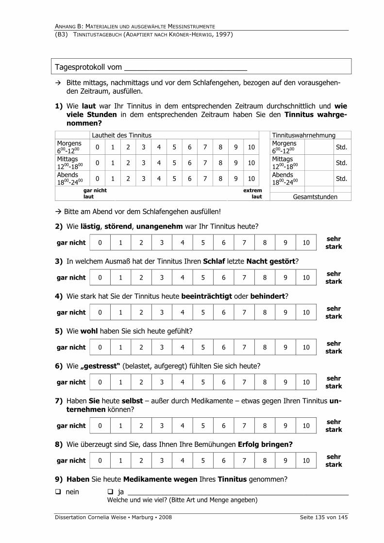
ANHANG B: MATERIALIEN UND AUSGEWÄHLTE MESSINSTRUMENTE (B3) TINNITUSTAGEBUCH (ADAPTIERT NACH KRÖNER-HERWIG, 1997)
Dissertation Cornelia Weise Marburg 2008 Seite 135 von 145
Tagesprotokoll vom _____________________________
Bitte mittags, nachmittags und vor dem Schlafengehen, bezogen auf den vorausgehen-den Zeitraum, ausfüllen.
1) Wie laut war Ihr Tinnitus in dem entsprechenden Zeitraum durchschnittlich und wie viele Stunden in dem entsprechenden Zeitraum haben Sie den Tinnitus wahrge-nommen?
Lautheit des Tinnitus Tinnituswahrnehmung Morgens 600-1200 0 1 2 3 4 5 6 7 8 9 10
Morgens 600-1200 Std.
Mittags 1200-1800 0 1 2 3 4 5 6 7 8 9 10
Mittags 1200-1800 Std.
Abends 1800-2400 0 1 2 3 4 5 6 7 8 9 10
Abends 1800-2400 Std.
gar nicht laut
extrem
laut
Gesamtstunden
Bitte am Abend vor dem Schlafengehen ausfüllen!
2) Wie lästig, störend, unangenehm war Ihr Tinnitus heute?
gar nicht 0 1 2 3 4 5 6 7 8 9 10 sehr stark
3) In welchem Ausmaß hat der Tinnitus Ihren Schlaf letzte Nacht gestört?
gar nicht 0 1 2 3 4 5 6 7 8 9 10 sehr stark
4) Wie stark hat Sie der Tinnitus heute beeinträchtigt oder behindert?
gar nicht 0 1 2 3 4 5 6 7 8 9 10 sehr stark
5) Wie wohl haben Sie sich heute gefühlt?
gar nicht 0 1 2 3 4 5 6 7 8 9 10 sehr stark
6) Wie „gestresst“ (belastet, aufgeregt) fühlten Sie sich heute?
gar nicht 0 1 2 3 4 5 6 7 8 9 10 sehr stark
7) Haben Sie heute selbst – außer durch Medikamente – etwas gegen Ihren Tinnitus un-ternehmen können?
gar nicht 0 1 2 3 4 5 6 7 8 9 10 sehr stark
8) Wie überzeugt sind Sie, dass Ihnen Ihre Bemühungen Erfolg bringen?
gar nicht 0 1 2 3 4 5 6 7 8 9 10 sehr stark
9) Haben Sie heute Medikamente wegen Ihres Tinnitus genommen?
nein ja _________________________________________________________ Welche und wie viel? (Bitte Art und Menge angeben)
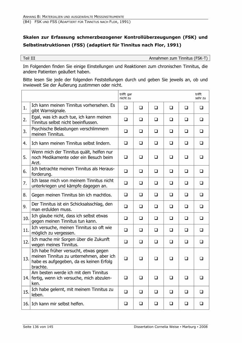
ANHANG B: MATERIALIEN UND AUSGEWÄHLTE MESSINSTRUMENTE (B4) FSK UND FSS (ADAPTIERT FÜR TINNITUS NACH FLOR, 1991)
Seite 136 von 145 Dissertation Cornelia Weise Marburg 2008
Skalen zur Erfassung schmerzbezogener Kontrollüberzeugungen (FSK) und
Selbstinstruktionen (FSS) (adaptiert für Tinnitus nach Flor, 1991)
Teil III Annahmen zum Tinnitus (FSK-T)
Im Folgenden finden Sie einige Einstellungen und Reaktionen zum chronischen Tinnitus, die andere Patienten geäußert haben.
Bitte lesen Sie jede der folgenden Feststellungen durch und geben Sie jeweils an, ob und inwieweit Sie der Äußerung zustimmen oder nicht.
trifft gar nicht zu
trifft sehr zu
1. Ich kann meinen Tinnitus vorhersehen. Es gibt Warnsignale.
2. Egal, was ich auch tue, ich kann meinen Tinnitus selbst nicht beeinflussen.
3. Psychische Belastungen verschlimmern meinen Tinnitus.
4. Ich kann meinen Tinnitus selbst lindern.
5. Wenn mich der Tinnitus quält, helfen nur noch Medikamente oder ein Besuch beim Arzt.
6. Ich betrachte meinen Tinnitus als Heraus-forderung.
7. Ich lasse mich von meinem Tinnitus nicht unterkriegen und kämpfe dagegen an.
8. Gegen meinen Tinnitus bin ich machtlos.
9. Der Tinnitus ist ein Schicksalsschlag, den man erdulden muss.
10. Ich glaube nicht, dass ich selbst etwas gegen meinen Tinnitus tun kann.
11. Ich versuche, meinen Tinnitus so oft wie möglich zu vergessen.
12. Ich mache mir Sorgen über die Zukunft wegen meines Tinnitus.
13.
Ich habe früher versucht, etwas gegen meinen Tinnitus zu unternehmen, aber ich habe es aufgegeben, da es keinen Erfolg brachte.
14. Am besten werde ich mit dem Tinnitus fertig, wenn ich versuche, mich abzulen-ken.
15. Ich habe gelernt, mit meinem Tinnitus zu leben.
16. Ich kann mir selbst helfen.
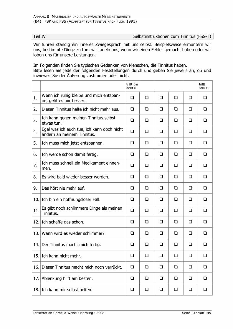
ANHANG B: MATERIALIEN UND AUSGEWÄHLTE MESSINSTRUMENTE (B4) FSK UND FSS (ADAPTIERT FÜR TINNITUS NACH FLOR, 1991)
Dissertation Cornelia Weise Marburg 2008 Seite 137 von 145
Teil IV Selbstinstruktionen zum Tinnitus (FSS-T)
Wir führen ständig ein inneres Zwiegespräch mit uns selbst. Beispielsweise ermuntern wir uns, bestimmte Dinge zu tun; wir tadeln uns, wenn wir einen Fehler gemacht haben oder wir loben uns für unsere Leistungen. Im Folgenden finden Sie typischen Gedanken von Menschen, die Tinnitus haben. Bitte lesen Sie jede der folgenden Feststellungen durch und geben Sie jeweils an, ob und inwieweit Sie der Äußerung zustimmen oder nicht.
trifft gar nicht zu
trifft sehr zu
1. Wenn ich ruhig bleibe und mich entspan-ne, geht es mir besser.
2. Diesen Tinnitus halte ich nicht mehr aus.
3. Ich kann gegen meinen Tinnitus selbst etwas tun.
4. Egal was ich auch tue, ich kann doch nicht ändern an meinem Tinnitus.
5. Ich muss mich jetzt entspannen.
6. Ich werde schon damit fertig.
7. Ich muss schnell ein Medikament einneh-men.
8. Es wird bald wieder besser werden.
9. Das hört nie mehr auf.
10. Ich bin ein hoffnungsloser Fall.
11. Es gibt noch schlimmere Dinge als meinen Tinnitus.
12. Ich schaffe das schon.
13. Wann wird es wieder schlimmer?
14. Der Tinnitus macht mich fertig.
15. Ich kann nicht mehr.
16. Dieser Tinnitus macht mich noch verrückt.
17. Ablenkung hilft am besten.
18. Ich kann mir selbst helfen.

ANHANG B: MATERIALIEN UND AUSGEWÄHLTE MESSINSTRUMENTE (B5) SKALA ZUR ERFASSUNG DER THERAPIEZUFRIEDENHEIT
Seite 138 von 145 Dissertation Cornelia Weise Marburg 2008
Skala zur Erfassung der Therapiezufriedenheit
Teil XI Therapieergebnis
Zum Abschluss nun noch einige Fragen zu Ihren persönlichen Therapieergebnissen. In wel-chem Ausmaß hat sich bei Ihnen eine Veränderung der tinnitusbezogenen Beeinträchtigung eingestellt?
Bitte geben Sie an, wie sehr Sie den einzelnen Aussagen zustimmen oder diese ablehnen.
stimmt über-haupt nicht
stimmt nicht
stimmt eher nicht
stimmt eher
stimmt
stimmt voll und ganz
1. Ich habe das Verfahren Biofeedback als sinnvoll zur Behandlung meiner Beschwer-den erlebt.
2. Ich kann mir gut vorstellen die Biofeed-backstrategien auch weiterhin im Alltag einzusetzen.
3. Ich bin mit der Therapie sehr zufrieden.
4. Ich habe mich während der Therapie in der Beziehung zur Therapeutin wohl ge-fühlt.
5. Mit dem in der Therapie Erreichten bin ich sehr zufrieden.
6. Ich habe in der Therapie gelernt, sorgsa-mer mit mir umzugehen.
7. Die Biofeedbacktherapie hat meinen Er-wartungen entsprochen.
8. Die Therapeutin hat die einzelnen Be-standteile der Therapie gut erläutert.
9. Einer Freundin/einem Freund mit ähnli-chen Beschwerden würde ich diese Be-handlung empfehlen.
10. Ich fühlte mich während der Behandlung von der Therapeutin verstanden.
11. Der Inhalt der einzelnen Sitzungen war zu umfangreich.
12. Mein persönlicher Aufwand zur Teilnahme an der Therapie hat sich für mich gelohnt.
13. Die Therapeutin war wenig am Erfolg der Therapie interessiert.
14. Ich glaube ein anderes therapeutisches Verfahren wäre für mich besser geeignet.
15. Ich war in der Behandlung mit der Berück-sichtigung der psychischen Aspekte mei-nes Tinnitus zufrieden.
16. Ich war in der Behandlung mit der Berück-sichtigung der körperlichen Aspekte meines Tinnitus zufrieden.

ANHANG B: MATERIALIEN UND AUSGEWÄHLTE MESSINSTRUMENTE (B5) SKALA ZUR ERFASSUNG DER THERAPIEZUFRIEDENHEIT
Dissertation Cornelia Weise Marburg 2008 Seite 139 von 145
stimmt über-haupt nicht
stimmt nicht
stimmt eher nicht
stimmt eher
stimmt
stimmt voll und ganz
17. Ich war in der Behandlung mit der Veran-schaulichung der Zusammenhänge zwi-schen Körper und Psyche zufrieden.
18. Durch die Behandlung habe ich jetzt mehr Beschwerden, als ich vorher hatte. Wenn ja, welche? ___________________
Was denken Sie, inwieweit die Tinnitusbehandlung Auswirkungen auf einige ihrer Lebens-bereiche hat?
überhaupt keine Aus-wirkung
starke Aus-
wirkung
Beruf/Arbeit
Partnerschaft
Familie
Freizeit

ANHANG B: MATERIALIEN UND AUSGEWÄHLTE MESSINSTRUMENTE (B6) INSTRUKTIONEN FÜR DIE PSYCHOPHYSIOLOGISCHE MESSUNG
Seite 140 von 145 Dissertation Cornelia Weise Marburg 2008
Instruktionen für die psychophysiologische Messung:
Wie Sie bereits wissen, soll in dieser Sitzung nun die physiologische Diagnostik stattfinden. Das heißt, wir messen Ihre Muskelanspannung unter verschiedene Bedingungen.
Um für alle Patienten die gleichen Bedingungen zu schaffen, wird diese Sitzung standardisiert durchgeführt. Wundern Sie sich also bitte nicht, dass wir die jeweiligen Aufgabenstellungen vorlesen und nicht frei formulieren.
Wir beginnen mit einer Messung der Grundanspannung, danach folgt eine erste Entspan-nungsphase. Im Anschluss daran wird die Muskelanspannung unter verschiedenen Bedingun-gen gemessen. Nach jeder dieser Bedingungen gibt es eine kurze Entspannungsphase. Ganz am Ende der Messung gibt es dann noch eine Abschlussentspannungsphase statt.
Instruktion zur BASELINE Als erstes werden wir die Grundanspannung messen. Dazu bitte ich Sie, möglichst ruhig zu
sitzen und nicht zu sprechen, da Bewegungen die Messgenauigkeit beeinträchtigen. Diese Pha-se wird ca. 3 Minuten dauern.
Instruktion zur ANFANGS- UND ENDENTSPANNUNG
Im Folgenden wird eine 5-minütige Entspannungsphase stattfinden. Bitte versuchen Sie dabei, so gut wie es Ihnen möglich ist, zu entspannen. Sie können dabei die Augen schließen, wenn dies angenehm für sie ist. Ich sage Ihnen, wenn die Zeit vorüber ist.
Instruktion zu den POST-STRESSOR-ENTSPANNUNGEN
Versuchen Sie jetzt bitte wieder zu entspannen und die Muskeln locker zu lassen. Ich sage Ihnen, wenn die Zeit vorüber ist.
Instruktion zum Stressor: NACHMODELLIERTER TINNITUS Im Folgenden hören Sie ein unangenehmes Geräusch, dass Ihrem Tinnitus wahrscheinlich sehr
ähnlich ist. Bitte hören Sie genau hin. Ich werde Ihnen im Nachhinein einige Fragen zu diesem Geräusch stellen. Bitte lassen Sie nun dieses unangenehme Geräusch auf sich wirken.
Instruktion zum Stressor: TINNITUS Ich möchte Sie nun bitten, sich ganz bewusst auf Ihren Tinnitus zu konzentrieren. Bitte setzen
Sie dazu diese Lärmschutzkopfhörer auf und hören Sie ganz genau auf Ihren Tinnitus. Lenken Sie sich dabei nicht ab, sondern bleiben Sie mit Ihrer Konzentration ganz bei Ihrem Tinnitus.
Instruktion zum Stressor: LÄRM In der folgenden Bedingung spiele ich Ihnen ein lautes Geräusch vor. Bitte hören Sie ganz
genau hin. Ich werde Ihnen im Nachhinein einige Fragen zu diesem Geräusch stellen. Lassen Sie nun den unangenehmen Lärm auf sich wirken.
Instruktion zum Stressor: RECHNEN
Ich werde nun mit Ihnen einen Test durchführen, bei dem es um Schnelligkeit geht. Ihre Auf-gabe ist, innerlich (also nicht laut) von 700 in 7er Schritten rückwärts zu zählen, d.h. immer 700 – 7 – 7 usw. zu rechnen. Parallel dazu wird Ihnen ein Takt über den Lautsprecher vorge-spielt. Dieser Takt gibt die Geschwindigkeit an, in der sie zählen müssen. Beim ersten Takt-schlag sollten Sie also 700-7 berechnet haben usw. Irgendwann werde ich Sie unterbrechen und fragen, bei welcher Zahl Sie angekommen sind. Haben Sie die Aufgabe verstanden?
Instruktion zum Stressor: TAP
Ich werde nun mit Ihnen einen Aufmerksamkeitstest durchführen. Damit wollen wir untersu-chen, ob es bei Tinnituspatienten Auffälligkeiten in der Aufmerksamkeit gibt. Bitte bemühen Sie sich, die Aufgabe so gut wie möglich und so schnell wie möglich zu bearbeiten. Bitte lesen Sie nun die Instruktionen auf dem Bildschirm durch. Es wird dann einen Probedurchgang ge-ben, bei dem Sie sich mit der Übung vertraut machen können.
ANHANG C: EIDESSTATTLICHE ERKLÄRUNG
Dissertation Cornelia Weise Marburg 2008 Seite 141 von 145
Eidesstattliche Erklärung
Ich versichere, dass ich meine Dissertation mit dem Titel
Biofeedback als Chance für die Tinnitusbehandlung – Entwicklung eines bio-
feedbackgestützten Therapieansatzes und Evaluation unter Berücksichtigung
psychophysiologischer Annahmen
selbständig und ohne unerlaubte Hilfe angefertigt habe.
Ich habe mich dabei keiner anderen als der von mir ausdrücklich bezeichneten Quel-
len und Hilfen bedient.
Die Dissertation wurde in der jetzigen oder einer ähnlichen Form noch bei keiner an-
deren Hochschule eingereicht und hat noch keinen sonstigen Prüfungszwecken ge-
dient.
Marburg, den 08.01.2008 Cornelia Weise

ANHANG D: CURRICULUM VITAE UND PUBLIKATIONSVERZEICHNIS
Dissertation Cornelia Weise Marburg 2008 Seite 143 von 145
Curriculum Vitae
Persönliche Angaben
Name: Cornelia Weise
Geburtsdatum: 05.07.1977
Schulische und universitäre Ausbildung
1996 Abitur (Note: sehr gut)
1996-1997 Praktikum in der Werkbund Werkstatt Nürnberg
1997-2004 Studium der Psychologie an der Philipps-Universität-Marburg 04/2004 Abschluss mit Diplom (Note: sehr gut)
Thema der Diplomarbeit:
Evaluation von psychophysiologischen Effekten durch eine Biofeed-backtherapie bei Patienten mit chronischem Tinnitus
2001 6-monatiges Praktikum in der Christoph-Dornier-Stiftung für Klini-sche Psychologie, Institut Berlin
2004-2007 Ausbildung zur psychologischen Psychotherapeutin am Institut für Psychotherapieausbildung Marburg (IPAM)
06/2005 Zwischenprüfung
Wissenschaftliches Engagement
2000 - 2001 Wissenschaftliche Hilfskraft in der AG Sozialpsychologie (Prof. Dr. U. Wagner)
2002 - 2003 Wissenschaftliche Hilfskraft in der AG Klinische Psychologie und Psy-chotherapie (Prof. Dr. W. Rief, PD Dr. Alexandra Martin)
2004-2005 Promotionsstipendium der Universität Marburg
Thema der Promotion:
Entwicklung und Evaluation eines biofeedbackgestützten Bewälti-gungstrainings für Patienten mit chronischem, dekompensiertem Tinnitus
seit Mai 2005 Wissenschaftliche Mitarbeiterin in der AG Klinische Psychologie und Psychotherapie (Prof. Dr. W. Rief)
ANHANG D: CURRICULUM VITAE UND PUBLIKATIONSVERZEICHNIS
Seite 144 von 145 Dissertation Cornelia Weise Marburg 2008
Publikationsverzeichnis
Publikationen
Weise, C., Heinecke, K., & Rief, W. (2007). Biofeedback bei chronischem Tinnitus – Behandlungsleitfaden und vorläufige Ergebnisse zu Wirksamkeit und Ak-zeptanz. Verhaltenstherapie, 17(4), 220-230.
Weise, C., Heinecke, K. & Rief, W. (submitted). Biofeedback-based behavioural treat-ment for chronic tinnitus – results of a randomized controlled trial.
Weise, C., Heinecke, K. & Rief, W. (submitted). Stability of physiological variables in chronic tinnitus sufferers.
Heinecke, K., Weise, C., Schwarz, K. & Rief, W. (accepted). Physiological and psy-chological stress reactivity in chronic tinnitus. Journal of Behavioral Medicine.
Rief, W., Weise, C., Kley, N., & Martin, A. (2005). Psychophysiologic treatment of chronic tinnitus: A randomized clinical trial. Psychosomatic Medicine, 67, 833-838.
Buchbeiträge
Kroymann, R., Weise, C., Zittlau, H., & Goebel, G. (2006). Chronischer Tinnitus. In W. Rief & N. Birbaumer (Hrsg.), Biofeedback (2. Aufl., S. 130-152). Stuttgart: Schattauer.
Weise, C. (in press). Tinnitus. In A. Martin & W. Rief (Hrsg.), Die Wirksamkeit von Biofeedback. Bern: Huber.
Abstracts
Weise, C., Heinecke, K. & Rief, W. (2007). Biofeedbackgestützte kognitiv-behaviorale Intervention bei chronischem Tinnitus – Entwicklung und Evaluation eines Behandlungsleitfadens. Verhaltenstherapie, 17 (Suppl.1), 5.
Heinecke, K., Weise, C., & Rief, W. (2007). Stressreaktivität bei Patienten mit chro-nischem dekompensiertem Tinnitus. Verhaltenstherapie, 17 (Suppl.1), 5.
Weise, C., Heinecke, K., & Rief, W. (2006). Psychophysiological treatment of chronic tinnitus. International Journal of Behavioral Medicine, 13 (Suppl.), 78.
Heinecke, K., Weise, C., & Rief, W. (2006). Attention deficits in distressed tinnitus sufferers - is there an objective impairment? International Journal of Behavioral Medicine, 13 (Suppl.), 126.
Weise, C., Martin, A., & Rief, W. (2005). Psychophysiologische Behandlung des chro-nischen Tinnitus - Ergebnisse einer randomisierten, kontrollierten Studie. Verhaltenstherapie, 15(Suppl.1).
Schmitt, B., Weise, C., Kley, N. & Rief, W. (2004). Biofeedback Treatment of Tinni-tus: A randomised clinical study. International Journal of Behavioral Medicine, 11 (Suppl.), 253.

ANHANG D: CURRICULUM VITAE UND PUBLIKATIONSVERZEICHNIS
Dissertation Cornelia Weise Marburg 2008 Seite 145 von 145
Kongressbeiträge
09/2007 Wissenschaftlicher Vortrag und Workshop beim Tinnitus Update Lecture Course (TULC), Bristol, UK
Weise, C.: Psychophysiological treatment of chronic tinnitus – Results of a randomized controlled trial
Weise, C.: Biofeedback in the treatment of chronic tinnitus
05/2007 Posterpräsentation auf dem 25. Symposium der Fachgruppe Klinische Psychologie der Deutschen Gesellschaft für Psychologie, Tübingen
Weise, C., Heinecke, K. & Rief, W.: Stabilität psychophysiologischer Reaktivitätsmaße bei Tinnitusbetroffenen
03/2007 Wissenschaftlicher Vortrag auf dem 11. Kongress der Deutschen Gesell-schaft für Verhaltensmedizin und Verhaltensmodifikation (DGVM/ DÄVT), München
Weise, C.: Biofeedbackgestützte Intervention bei chronischem Tinnitus
- Entwicklung und Evaluation eines Behandlungsleitfadens
02/2007 Workshop auf der 7. Jahrestagung der Deutschen Gesellschaft für Bio-feedback (DGBfb), Rauischholzhausen
Weise, C. & Heinecke, K.: Biofeedbackgestützte Behandlung des chroni-schen Tinnitus
12/2006 Wissenschaftlicher Vortrag beim 9th International Congress of Behav-ioral Medicine, Bangkok, Thailand
Weise, C.: Psychophysiological treatment of chronic tinnitus – results of two randomized controlled trials
03/2005 Wissenschaftlicher Vortrag auf dem 10. Kongress der Deutschen Gesell-schaft für Verhaltensmedizin und Verhaltensmodifikation (DGVM/ DÄVT), München
Weise, C.: Psychophysiologische Behandlung des chronischen Tinnitus – Ergebnisse einer randomisierten kontrollierten Studie
09/2004 Posterpräsentation auf der 5. Jahrestagung der Deutschen Gesellschaft für Biofeedback (DGBFB), Dresden
Weise, C.: Psychophysiologische Effekte einer Biofeedbacktherapie bei chronischem Tinnitus