Die genetische Varianz des Porzinen Parvovirus und die ...¼.pdf · einzelsträngiges DNA Virus mit...
78
Aus dem Institut für Tierhygiene und Öffentliches Veterinärwesen der Veterinärmedizinischen Fakultät der Universität Leipzig Die genetische Varianz des Porzinen Parvovirus und die Wirksamkeit einer neuen experimentellen Vakzine Inaugural-Dissertation zur Erlangung des Grades eines Doctor medicinae veterinariae (Dr. med. vet.) durch die Veterinärmedizinische Fakultät der Universität Leipzig eingereicht von Tessa Foerster aus Stendal Leipzig, 2016
Transcript of Die genetische Varianz des Porzinen Parvovirus und die ...¼.pdf · einzelsträngiges DNA Virus mit...

Aus dem
Institut für Tierhygiene und Öffentliches Veterinärwesen
der Veterinärmedizinischen Fakultät
der Universität Leipzig
Die genetische Varianz des Porzinen Parvovirus
und die Wirksamkeit einer neuen experimentellen
Vakzine
Inaugural-Dissertation
zur Erlangung des Grades eines
Doctor medicinae veterinariae (Dr. med. vet.)
durch die Veterinärmedizinische Fakultät
der Universität Leipzig
eingereicht von
Tessa Foerster
aus Stendal
Leipzig, 2016
Mit Genehmigung der Veterinärmedizinischen Fakultät der Universität Leipzig
Dekan: Prof. Dr. Manfred Coenen
Betreuer: Prof. Dr. Uwe Truyen
Gutachter: Prof. Dr. Uwe Truyen, Institut für Tierhygiene und
Öffentliches Veterinärwesen, Universität Leipzig,
Leipzig
Prof. Dr. Klaus Osterrieder, Institut für Virologie, Freie
Universität Berlin, Berlin
Tag der Verteidigung: 30.08.2016


V
Inhalt
Abkürzungsverzeichnis VI
Vorwort VII
Kapitel 1 – Einleitung 1
Kapitel 2 – Literaturrecherche 3
Klassifikation und Entdeckung 3
Genom 4
Morphologie 5
Neue Phänotypen 6
Neue Parvoviren 7
Epidemiologie 7
Pathogenese 8
Klinik 9
Diagnose 10
Immunität 12
Bekämpfung 13
Kapitel 3 – An inactivated whole virus porcine parvovirus vaccine protects pigs against
disease but does not prevent virus shedding even after homologous virus challenge 14
Kapitel 4 – Population dynamics and in vitro antibody pressure of porcine parvovirus
(PPV) indicate a decrease of variablility 24
Kapitel 5 – Analysis of porcine parvoviruses in tonsilis and hearts from healthy pigs
revealed high prevalence and genetic diversity in Germany 33
Kapitel 6 – Diskussion 50
Kapitel 7 – Schlussfolgerung 55
Kapitel 8 – Zusammenfassung 56
Kapitel 9 – Summary 58
Literaturverzeichnis 60
Abbildungsverzeichnis 68
Tabellenverzeichnis 69
Danksagung 70
ABKÜRZUNGSVERZEICHNIS
VI
ABKÜRZUNGSVERZEICHNIS
AB - Antibody
CO2 - Kohlenstoffdioxid
CPV - Canine Parvovirus
CPV-2 - Canine Parvovirus 2
DMEM - Dulbecco's Modified Eagle Medium
DNA - Desoxyribonucleic acid
dNTP - Desoxyribonucleotide triphosphate
EDTA - Ethylenediaminetetraacetic acid
ELISA - Enzyme-linked-immunosorbent Assay
FBS - Fetal bovine serum
FPV - feline Parvovirus
GTR - General time reversible
HI - hemagglutination inhibition test
ICNV - International Committee of Nomenclature of Viruses
ICTV - International Committee on Taxonomy of Viruses
LB - Luria Bertani
MCMC - Markov chain Monte Carlo
MEV - Nerz-Enteritis Virus
MVM - Minute Virus of Mice
NCBI - National Center for Biotechnology Information
NS - Non-structural protein
NS1 - Non-structural protein 1
NS2 - Non-structural protein 2
NS3 - Non-structural protein 3
Nuc - Nucleotide
ORF - Open reading frame
PBS - Phosphate-buffered saline solution
PCR - Polymerase chain reaction
PK15 - Pig kidney
PPV - Porcine parvovirus 2
PPV3 - Porcine parvovirus 3
PPV4 - Porcine parvovirus 4
PRRS - Porcine reproductive und respiratory syndrome
RNA - Ribonucleic acid
RPMI - Roswell Park Memorial Institute Medium
SMEDI - Stillbirth, mummification, embryonic death and infertility

INHALTSVERZEICHNIS
VII
SNT - Serumneutralisationstest
SPEV - Swine embryo kidney
STE - Swine testicular epitheloid
TCID - Tissue culture infective dose
VP - Viral protein
VP1 - Viral protein 1
VP2 - Viral protein 2
VP3 - Viral protein 3

VORWORT
VIII
VORWORT
Diese Dissertation umfasst als kumulative Studie folgende Veröffentlichungen:
Titel: An inactivated whole-virus porcine parvovirus vaccine protects pigs against
disease but does not prevent virus shedding even after homologous virus challenge
Autoren: Tessa Foerster, André Felipe Streck, Stephanie Speck, Hans-Joachim Selbitz, T.
Lindner, Uwe Truyen
zur Veröffentlichung angenommen bei: Journal of General Virology, 2016
Titel: Population dynamics and in vitro antibody pressure of porcine parvovirus (PPV)
indicate a decrease of variability
Autoren: André Felipe Streck, Timo Homeier, Tessa Foerster, Uwe Truyen
Veröffentlicht in: Journal of General Virology, 2013, 94 (9): 2050-2055
Titel: Analysis of porcine parvoviruses in tonsils and hearts from healthy pigs reveals
high prevalence and genetic diversity in Germany
Autoren: André Felipe Streck, Timo Homeier, Tessa Foerster, Stefan Fischer, Uwe Truyen
Veröffentlicht in: Archives of Virology, 2013, 158 (6):.1173–1180
KAPITEL 1 - EINLEITUNG
1
KAPITEL 1 - EINLEITUNG
Das porzine Parvovirus (PPV), 2013 vom International Committee on Taxonomy of Viruses
(ICTV) in ungulate Protoparvovirus 1 umbenannt, ist ein unbehülltes, einzelsträngiges
DNA Virus und gehört innerhalb der Familie Parvoviridae zur Subfamilie Parvovirinae. Es ist
weltweit in allen Bereichen der Schweinehaltung endemisch und verursacht große
wirtschaftliche Verluste in den Betrieben (TRUYEN und STRECK 2012).
Anders als die verwandten caninen und felinen Parvoviren (seit 2013 carnivore
Protoparvovirus 1) ist es nicht durch zum Teil tödlich verlaufende Durchfallerkrankungen,
sondern durch Fruchtbarkeitsstörungen wie Abort, Mumifikation und Unfruchtbarkeit, auch
bekannt als SMEDI – Syndrom (Stillbirth = Totgeburt, Mummification =Mumifikation, Embryonic
Death = embryonaler Tod und Infertility = Unfruchtbarkeit), gekennzeichnet. Die Schwere des
Verlaufs hängt dabei wesentlich vom Zeitpunkt sowie von dem, für die Infektion
verantwortlichen Isolats ab. Als besonders gefährdet gelten ungeimpfte Jungsauen, die
innerhalb der ersten 70 Tage der Trächtigkeit in Kontakt mit dem Virus treten.
Das Virus verfügt über eine ausgesprochen hohe Tenazität gegenüber äußeren Einflüssen. Es
ist hitzestabil, unempfindlich gegenüber pH-Werten zwischen 3-9 sowie äther- und
chloroformresistent (CARTWRIGHT und HUCK 1967, MAYR et al. 1968, JOHNSON und
COLLINGS 1969, BACHMANN 1970, MORIMOTO 1972). Einmal im Bestand bleibt es somit
über Monate infektiös. Es stehen für die Bekämpfung nur wenige Mittel zur Verfügung. Eine
entscheidende Möglichkeit ist die Einhaltung eines strikten Impfregimes, wobei Impfstoffe zum
Einsatz kommen, die seit etwa 3 Jahrzehnten auf den gleichen inaktivierten Virus-Isolaten
beruhen.
In den letzten zehn Jahren wurden zunehmend neue Isolate entdeckt, die sich, wie das
hochvirulente Isolat Kresse und das wenig virulente Isolat NADL2, nur in wenigen Aminosäuren
unterscheiden. Zum Teil weisen sie aber gravierende Unterschiede in ihrer Pathogenität auf.
Daraus ergeben sich neben dem dringenden Rat zur Beobachtung der aktuellen Entwicklung
mehrere Fragen hinsichtlich der zukünftigen Handhabung des Virus (SOARES et al. 2003,
ZIMMERMANN et al. 2006).
So sollte geklärt werden:
• wie verbreitet sind diese neuen Isolate
• was könnte ihre Entwicklung begünstigt haben
• wie effizient ist der Schutz, den herkömmliche Impfstoffe gegen die neuen Isolate bieten
• kann eines der Isolate eine Grundlage für einen neuen, effizienteren Impfstoff liefern
KAPITEL 1 - EINLEITUNG
2
Diese Dissertation umfasst insgesamt drei Veröffentlichungen, welche versuchen, die gestellten
Fragen zu beantworten. Im ersten Artikel wird die Wirksamkeit eines neuen Impfstoffes auf
Grundlage des hochvirulenten, vorherrschenden Isolat 27a untersucht. Im zweiten Manuskript
wird mit Hilfe von in vitro- und in silico- Modellen die Populationsdynamik demonstriert. Die
dritte Veröffentlichung widmet sich der Beschreibung der neuen Parvotypen (PPV2, PPV3 und
PPV4), welche aus Herzen und Tonsillen von deutschen, klinisch gesunden Schlachtschweinen
isoliert werden konnten.
Da die Veröffentlichungen teilweise bereits vor der Änderung der Taxonomie erschienen, wird
aus Gründen der Einheitlichkeit im weiteren Verlauf dieser Arbeit die alte Taxonomie
verwendet.

KAPITEL 2 - LITERATURRECHERCHE
3
KAPITEL 2 - LITERATURRECHERCHE
Klassifikation und Entdeckung
1959 kam es im Zusammenhang mit einem bei neugeborenen Hamstern
krankheitsverursachenden Virus, dem sogenannten „Hamster-osteolytic virus“, erstmals zur
Erwähnung eines Parvovirus (KILHAM und OLIVER 1959). Jedoch wurde erst 17 Jahre später
einer der Hamster-osteolytic Vertreter, das Kilham Rat Virus, als erstes entdecktes Parvovirus
identifiziert (SIEGL 1976). In der Zwischenzeit wurden in mehr als fünf verschiedenen Tierarten
und vielen Ländern Parvoviren gefunden (HALLAUER und KRONAUER 1960, BURGER et al.
1963, HAMPTON. 1964, CARTWRIGHT und HUCK 1967, ROSE et al. 1969).
MAYR und MAHNEL beschrieben 1964 ein unbekanntes Virus, welches ihre, für die Anzucht
von hog cholera Virus genutzten, primären und sekundären Schweinenieren- und
Schweinehodenzellkulturen kontaminiert hatte. Die Partikel wiesen Ähnlichkeiten mit dem Maul-
und Klauenseuche Virus auf und wurden mit dem Kilham Rat-Virus verglichen. Aufgrund seiner
Größe von 22-23 nm und seines Desoxyribonukleinsäuregehaltes wurde das Virus ursprünglich
als Picornavirus bezeichnet (MAYOR und MELNICK 1966). Nachdem es jedoch aus
Schweinenierenzellen isoliert wurde und man festgestellte, dass es sich um ein
einzelsträngiges DNA Virus mit der Fähigkeit zur Replikation handelt, wurde dem International
Committee on the Nomenclature of Viruses (ICNV) der Name Parvovirus vorgeschlagen
(ANDREWS 1970).
Heute heißt das Parvovirus laut dem International Committee on Taxonomy of Viruses
Protoparvovirus und zählt zur Familie der Parvoviridae, welche in die Unterfamilien
Densovirinae und Parvovirinae gegliedert ist. Die Densovirinae setzen sich zusammen aus den
Gattungen Ambidensovirus, Brevidensovirus, Hepandensovirus, Iteradensovirus und
Penstyldensovirus. Sie infizieren ausschließlich Arthropoden und besitzen keine klinische
Relevanz. Die Unterfamilie Parvovirinae umfasst die Gattungen Amdoparvovirus, Aveparvovirus
Bocaparvovirus, Copiparvovirus, Dependoparvovirus, Erythroparvovirus Protoparvovirus und
Tetraparvovirus, die dazugehörigen Viren besitzen eine große Bandbreite an Wirten. So
beispielsweise die Viren der Gattung Dependoparvovirus, welche auf ein Helfervirus (Adeno-,
Pox- oder Herpesvirus) angewiesen sind um sich zu replizieren. Weiterhin die Gattung
Erythroparvovirus, deren wichtigster Vertreter der Erreger der Ringelröteln, das
humanpathogene primate erythroparvovirus 1 (früher Parvovirus B19) ist. Die Viren dieser
Gattung infizieren hauptsächlich Affen und Menschen, wohingegen die Vieren der Gattung
Protoparvovirus fast ausschließlich Tiere infizieren. Die wichtigsten Vertreter dieser Gattung
sind das carnivore protoparvovirus (früher: feline Parvovirus (FPV) und canine Parvovirus
KAPITEL 2 - LITERATURRECHERCHE
4
(CPV)), primate protoparvovirus1, rodent protoparvovirus (früher Minute Virus of Mice) und das
ungulate protoparvovirus 1 (früher porzine Parvovirus(PPV)).
Letztgenanntes konnte als Ursache für die 1964 von MAYR und MAHNE beschriebene
Kontamination identifiziert werden.
Zur Zeit der Entdeckung des PPV traten gehäuft Reproduktionsstörungen in Schweineherden
auf. Eine Beteiligung des Virus war anfänglich aber umstritten. Es wurde postuliert, dass diese
Störungen durch Umwelteinflüsse, fehlerhafte Ernährung, genetisch oder toxikologisch bedingte
Ursachen ausgelöst seien (LAWSON 1961, RASBECH 1969). Erstmals beschrieben
CARTHWRIGHT und HUCK 1967 einen Zusammenhang zwischen dem Auftreten von PPV bei
Schweinen und der in Schweineherden beobachteten Unfruchtbarkeit, Aborten und
Umrauschen. Zahlreiche Infektionsversuche konnten schließlich belegen, dass PPV bei
Schweinen zu Aborten und Mumifikationen der Ferkel führt (JOHNSON und COLLINGS 1969,
REDMANN et al. 1974, CUTLIP und MENGENGELING 1975, FUJISAKI et al. 1975, JOHNSON
et al. 1976, JOO et al. 1976, MENGELING und CUTLIP 1976).
Inzwischen ist PPV weltweit als Hauptverursacher des sogenannten SMEDI Syndroms in der
Schweineindustrie bekannt (THOMPSON und PROZESKY 1994).
Genom
Ein 4500 bis 5500 Basen langer linearer Desoxyribonukleinsäure-Einzelstrang mit negativer
Polarität bildet die Grundlage des Parvovirusgenoms (MOLITOR et al. 1983, BERGERON et al.
1993). Es befinden sich zwei Haarnadelstrukturen mit je 120-200 Basen in den terminalen
Sequenzen des Virus. Diese Strukturen gelten als Primer während der DNA Replikation und
spielen eine wichtige Rolle für die Virusintegrität.
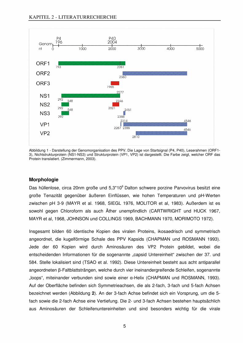
Des Weiteren sind zwei große und ein kleiner sogenannter offener Leserahmen („open reading
frames“ (ORFs)) über das Genom verteilt (Abbildung 1). Dabei ist der kleine ORF 3 vollständig
in dem am 5‘ Ende gelegenen großen ORF1 verankert. Der zweite große ORF2 befindet sich
am 3‘ Ende des Genoms. Alle drei überlappen sich gegenseitig.
Die ORF1 und ORF3 enthalten die Informationen für die Nichtstrukturproteine (NS1, NS2 und
NS3) sowie die ersten zehn Aminosäuren des Strukturproteins VP1. Die Informationen für die
Strukturproteine (VP1, VP2 und VP3) werden auf dem ORF 2 kodiert (BERGERON et al. 1996).
Durch alternatives Spleißen ist es möglich, verschiedene Leseraster abzulesen und damit die
gesamte Erbinformation auf dem kleinen Genom unterzubringen (BERNS 1990).
KAPITEL 2 - LITERATURRECHERCHE
5
Abbildung 1 - Darstellung der Genomorganisation des PPV. Die Lage von Startsignal (P4, P40), Leserahmen (ORF1-3), Nichtstrukturprotein (NS1-NS3) und Strukturprotein (VP1, VP2) ist dargestellt. Die Farbe zeigt, welcher ORF das Protein translatiert. (Zimmermann, 2003).
Morphologie
Das hüllenlose, circa 20nm große und 5,3*106 Dalton schwere porzine Parvovirus besitzt eine
große Tenazität gegenüber äußeren Einflüssen, wie hohen Temperaturen und pH-Werten
zwischen pH 3-9 (MAYR et al. 1968, SIEGL 1976, MOLITOR et al, 1983). Außerdem ist es
sowohl gegen Chloroform als auch Äther unempfindlich (CARTWRIGHT und HUCK 1967,
MAYR et al, 1968, JOHNSON und COLLINGS 1969, BACHMANN 1970, MORIMOTO 1972).
Insgesamt bilden 60 identische Kopien des viralen Proteins, ikosaedrisch und symmetrisch
angeordnet, die kugelförmige Schale des PPV Kapsids (CHAPMAN und ROSMANN 1993).
Jede der 60 Kopien wird durch Aminosäuren des VP2 Protein gebildet, wobei die
entscheidenden Informationen für die sogenannte „capsid Untereinheit“ zwischen der 37. und
584. Stelle lokalisiert sind (TSAO et al. 1992). Diese Untereinheit besteht aus acht antiparallel
angeordneten β-Faltblattsträngen, welche durch vier ineinandergreifende Schleifen, sogenannte
„loops“, miteinander verbunden sind sowie einer α-Helix (CHAPMAN und ROSMANN, 1993).
Auf der Oberfläche befinden sich Symmetrieachsen, die als 2-fach, 3-fach und 5-fach Achsen
bezeichnet werden (Abbildung 2). An der 3-fach Achse befindet sich ein Vorsprung, um die 5-
fach sowie die 2-fach Achse eine Vertiefung. Die 2- und 3-fach Achsen bestehen hauptsächlich
aus Aminosäuren der Schleifenuntereinheiten und sind besonders wichtig für die virale

KAPITEL 2 - LITERATURRECHERCHE
6
Infektiosität sowie die Immunogenität (SIMPSON et al. 2002).
Abbildung 2 - Kapsid eines porzinen Parvovirus. schematisch dargestellt (Swiss Institute of Bioinformatics)
Unüblich ist es, dass sich auf der inneren Kapsidoberfläche hauptsächlich neutrale
Aminosäuren befinden im Gegensatz zu den üblichen basischen (XIE und CHAPMAN 1996).
Eventuell ermöglichen basische Reste der neutralen Aminosäuren an den Bindungsstellen mit
den DNA Molekülen eine hohe elektrostatische Enthalpie, die eine effiziente Verpackung des
viralen Genoms bewirkt (STEITZ et al. 1990).
Neue Phänotypen
Spätestens nachdem zwischen 1994 und 2002 in Brasilien genetische Varianten des
PPV Feldstammes entdeckt worden sind, ist die genetische Varianz und die Entwicklung neuer
PPV Phänotypen ein aktuelles Thema (SOARES et al. 2003). SOARES bestimmte bei der
Analyse eines partiellen Fragmentes des VP2 Gens acht verschiedene Phänotypen, die sich in
mindestens einer Aminosäure unterschieden. Diese Typen wurden in zwei phylogenetische
Gruppen unterteilt. In ähnlichen Studien, wie der aus Deutschland von ZIMMERMANN et al.
(2006) oder aus China und Rumänien von SHANGJIN et al. (2009), Hao et al. (2011) und
CADAR et al (2012), wurden weitere neue Phänotypen publiziert.
Ungeklärt ist dabei, ob es sich tatsächlich um neu entwickelte Varianten handelt oder es erst mit
Hilfe der neuen sensitiveren Technik gelungen ist, ihre Existenz nachzuweisen. Die Tatsache,
dass sich die Antigeneigenschaften des Virus aufgrund von Aminosäuresubstitutionen im
Kapsid verändert haben, lässt vermuten, dass eine Anpassung im Rahmen von „ escape
mutants“ stattgefunden hat. Diese Mutationen könnten eventuell durch den kontinuierlichen
Gebrauch von Impfstoffen bedingt sein (ZEEUW et al. 2007). Des Weiteren konnten
Unterschiede in der Kreuzneutralisierung von Seren gegen NADL2 und das von IDT genutzte
Impfvirus beobachtet werden, was diese Vermutung unterstützen würde.

KAPITEL 2 - LITERATURRECHERCHE
7
Neue Parvoviren
Seit 2002 werden mittels neuester Molekulartechnik, wie der Sequenzierung, weltweit immer
neue porzine Parvoviren identifiziert. So zum Beispiel das porzine Parvovirus 2, das später als
Virus H1 bezeichnet wurde, weiterhin das nah mit dem humanen Parvovirus verwandte
Hokovirus sowie das porzine Parvovirus 4 (HIJISAKA et al. 2001, LAU et al. 2008, CHEUNG et
al. 2010).
Alle diese Viren konnten inzwischen in verschiedenen Ländern isoliert werden. Teilweise
werden sie nur als Koinfektion bei Schweinen gefunden und führen anscheinend bei ihnen zu
keinen klinischen Symptomen. Allerdings könnten sie wegen ihrer hohen Ähnlichkeit zum
humanen Parvovirus eine potentielle Gefahr für den Menschen darstellen und sind damit von
öffentlichem Interesse.
Epidemiologie
Weltweit ist PPV in allen Arten von Schweinehaltungen, das heißt in Sauen-, Eber- oder
Mastanlagen, endemisch. Aufgrund seiner ausgeprägten Stabilität innerhalb der Umgebung und
der Fähigkeit monatelang infektiös zu bleiben, sind beispielsweise Ställe, in denen einmal
infizierte Tiere untergebracht waren, immer eine potentielle Infektionsquelle. Auch mittels
Nagetieren, Personen (als belebte Vektoren) oder deren Kleidungen (unbelebte Vektoren) kann
das Virus problemlos von einem Betrieb in den nächsten transportiert werden (TRUYEN und
STRECK 2012).
In den 70er Jahren lag die Rate der serumpositiven Tiere weltweit zwischen 30 – 90 Prozent
(BACHMAN. 1969; JONHSON und COLLINGS 1969; MENGELING. 1972). Daraufhin wurden
umgehend inaktivierte Impfstoffe entwickelt und ein striktes Impfregime etabliert. Dennoch
gelang es mit Hilfe dieser Impfstoffe nicht, die Infektion der Schweine gänzlich zu verhindern.
DIAS et al zeigten 2012, dass geimpfte Sauen nach Kontakt mit dem Virus Antikörpertiter von
über 1:512 entwickelten. Diese Erkenntnis legt nahe, dass es trotz Impfung zu einer subklinisch
verlaufenen Infektion kam.
Auch wenn das Virus in eine regelmäßig geimpfte oder eine über natürliche Immunität
verfügende Herde eingebracht wird, infizieren sich die Tiere auf dem oronasalen Weg und
beginnen während der Virämie damit, das Virus über den Kot und andere Körperflüssigkeiten
auszuscheiden, ohne dabei klinische Symptome zu zeigen. Sie werden so zu möglichen
Infektionsquellen für Neuzugänge oder Tiere, die extra vor der Trächtigkeit separiert wurden.
Dadurch kann es bei diesen naiven Tieren zum Ausbruch der Erkrankung kommen (SZELEI et
al. 2006, TRUYEN und STRECK 2012).

KAPITEL 2 - LITERATURRECHERCHE
8
Neben den bereits angesprochenen unbelebten und belebten Vektoren stehen auch PPV
positive Eber in Verdacht, durch kontaminiertes Sperma Sauen anzustecken. Umgekehrt ist es
ebenfalls möglich, dass sich serumnegative Eber beim Deckakt infizieren (SZELEI et al. 2006).
Auch wenn gezeigt werden konnte, dass im Sperma infizierter Eber das Virus nachweisbar ist,
bleibt es jedoch ungeklärt, ob es während der Virämie in die Hoden gelangte oder ob die
positiven Ergebnisse aufgrund sekundärer Kontaminationen zustande gekommen sind
(CARTWRIGHT und HULK 1967, CARTWRIGHT et al. 1969, RUCKERBAUER et al. 1978).
Pathogenese
Für die Replikation ist das porzine Parvovirus auf mitotisch aktives Gewebe angewiesen. Dabei
gilt, je aktiver die Wirtszellen, desto schneller die Virusvermehrung (TATTERSALL 1972).
Der genaue Mechanismus, der PPV den Eintritt in die Zelle ermöglicht, ist noch unbekannt. Als
gesichert gilt, dass sich das Virus mit Hilfe von diversen Glykoproteinen, Glykanen und
Glykolipiden an Zellen anheftet (HARBINSON et al. 2008). Anschließend scheint eine Clathrin-
vermittelte Endozytose oder Makropinozytose, gefolgt von einem Transport auf endosomalem
Weg stattzufinden (BOISVERT et al. 2010). Hierbei kommt es zur Ansäuerung durch die
Endosomen, was eine reversible Änderung des viralen Kapsids bewirkt. Diese Änderungen
könnten dem Virus ermöglichen, das Endosomen zu verlassen und damit der Verdauung zu
entgehen (VIHINEN-RANTA et al. 2002, FARR et al. 2005). Außerdem wird durch das
Ansäuern wahrscheinlich die Phospholipase A2, welche für die Zerstörung der vesikulären
Membranen und für das Eindringen in den Zellkern verantwortlich zu sein scheint, aus dem
Kapsid gelöst (GIROD et al. 2002,VENDEVILLE et al. 2009). Befindet sich das Virus schließlich
im Zellkern, übernimmt der DNA-Polymerase Komplex die Replikation des Virus.
Ausgehend vom lymphatischen Gewebe wird das Virus während der Virämie systemisch verteilt
(PAUL et al. 1980). Wie genau es dabei zum Übertreten der Plazentabarriere kommt, ist bislang
ebenfalls ungeklärt, da Schweine über eine epitheliochoriale Plazenta verfügen, die den
mütterlichen und fetalen Blutkreislauf voneinander trennt. Nachdem PPV in fetalen
Lymphozyten nachgewiesen werden konnte und gezeigt wurde, dass es auch nach
Phagozytose durch Makrophagen noch infektiös ist, wird die Möglichkeit diskutiert, ob es mittels
der lymphatischen Zellen in den fetalen Organismus gelangen könnte (RUDEK und
KWIATKOWSKA 1983, PAUL et al. 1979).
Die fetalen Gewebe, wie beispielsweise die hämato- und lymphopoetischen Zellen, besitzen
eine hohe mitotische Aktivität und sind daher besonders für die Replikation des Virus geeignet
(TRUYEN und STRECK 2012). Aufgrund der bei der Replikation entstehenden gravierenden
pathologischen Veränderungen kommt es zum Absterben der Frucht. Makroskopisch sind diese

KAPITEL 2 - LITERATURRECHERCHE
9
Körper postmortal meist ödematös, rötlich- braun verfärbt und in ihren Körperhöhlen finden sich
blutige Flüssigkeiten (BACHMANN et al. 1975, MENGELING und CUTLIP 1975, JOO et al.
1976, 1977, LENGHAUS et al. 1978, NIELSEN et al. 1991). Histologisch lassen sich in den
Organsystemen und der Plazenta mononukleare Zellen, Nekrosen und Mineralisationen
ungeklärter Ursache nachweisen (JOO et al. 1977, LENGHAUS et al.1978).
Klinik
Zu den Hauptsymptomen der PPV Infektion gehören Unfruchtbarkeit, Abort, Totgeburt,
Mumifikation, neonataler Tod und reduzierte neonatale Lebensfähigkeit (CARTWRIGHT und
HUCK 1967, JOHNSON 1969, MORIMOTO et al 1972, NARITA et al. 1975, FORMAN et al.
1977). Heute wird PPV als häufigste Ursache für Fruchtbarkeitsstörungen und als einer der
Verursacher des „SMEDI“-Syndrom in Schweinebetrieben gesehen. Während die Infektion bei
adulten und neonatalen Schweinen bis auf eine transiente leichte Leukopenie überwiegend
subklinisch verläuft und unabhängig von Alter und Geschlecht ist, spielt der Infektionszeitpunkt
während der Trächtigkeit eine entscheidende Rolle für die Auswirkungen auf die
Fruchtentwicklung (JOHNSON und COLLINGS 1969, CUTLIP und MENGELING 1975,
FUJISAKI et al. 1975, JOHNSON et al. 1976, JOO et al. 1976, MENGELING und CULTIP
1976).
Gelangt das Virus beispielsweise zu Beginn der Trächtigkeit in den Uterus, ist der Konzeptus
durch die Zona pellucida geschützt und damit noch resistent (Abbildung 3). Vom 6. bis zum 35.
Trächtigkeitstag stirbt der Embryo im Zuge einer Infektion ab und wird resorbiert. Dadurch kann
es kann folglich je nach Zeitpunkt und Anzahl der verbleibenden Embryonen zum regel- oder
unregelmäßigen Umrauschen der Sau kommen. Mit dem Beginn der Ossifikation, am 35.
Trächtigkeitstag, ist eine vollständige Resorption der Feten nicht mehr möglich. Stattdessen
kommt es nach dem Absterben zur deren Mumifikation. Findet der Kontakt mit dem Virus ab
dem 70. Tag der Gravidität statt, sind die Feten bereits immunkompetent. Damit sind sie in der
Lage Antikörper zu bilden und die Infektion zu überleben (JOO et al. 1976a, LENGHAUS et al.
1978).
Abbildung 3 - Konsequenzen einer PPV Infektion zu unterschiedlichen Trächtigkeitszeitpunkten.

KAPITEL 2 - LITERATURRECHERCHE
10
Meist weisen die Ferkel eines Wurfes verschiedene Ausprägungen der Infektion auf (Abbildung
4), weshalb davon ausgegangen wird, dass sich das Virus intrauterin von Frucht zu Frucht
ausbreitet. Es kommt zur Ausbildung sogenannter orgelpfeifenartiger Würfe.
Abbildung 4 - Wurf am 90. Trächtigkeitstag einer infizierten Sau. Ferkel zeigen unterschiedliche Ausprägungen der Infektion. Die Position der Ferkel entspricht ihrer Lage im Uterus, wobei die ovarial gelegenen Ferkel unten abgebildet sind. ( Zeeuw et al. 2007)
DEA et al(1985) und YASUHARA et al. (1989) brachten das PPV mit Enteritiden in Verbindung.
Es gibt jedoch keinen Hinweis darauf, dass es, wie beispielsweise das canine oder feline
Parvovirus, Darmerkrankungen hervorruft (CUTLIP und MENGELING 1975; BROWN et al.
1980).
Diagnose
Kommt es innerhalb einer Herde zum alleinigen Auftreten von Fruchtbarkeitsstörungen, wie
beispielweise gehäuftem Umrauschen, verlängerten Zwischentragezeiten, Aborten oder kleinen
Würfen mit lebensschwachen oder mumifizierten Ferkeln, muss PPV als Verdachtsdiagnose,
neben Differentialdiagnosen wie Brucellose, Leptospirose und porzine reproductive and
respiratory syndrome (PRRS), in Betracht gezogen werden (SZELEI et al. 2006; TRUYEN und
STRECK 2012). Verschiedene Untersuchungsmethoden können hinsichtlich der Beteiligung
von PPV Klarheit bringen.

KAPITEL 2 - LITERATURRECHERCHE
11
Zur Untersuchung eignen sich besonders Abortmaterial, mumifizierte Feten sowie Serumproben
von vermeidlich infizierten Tieren. Die Proben sollten möglichst zeitnah und gefroren in ein
Labor versandt werden.
PPV besitzt hämagglutinierende Eigenschaften gegenüber Erythrozyten von Hühnern,
Meerschweinchen und der humanen Blutgruppe 0. Dies macht man sich im sogenannten
Hämagglutinationstest zu Nutze (JOO et al. 1976b). Dieser Test ist vergleichsweise
kostengünstig und schnell durchzuführen, im Vergleich zu anderen Techniken jedoch
unspezifisch und empfindlich gegenüber Faktoren, die die Hämagglutination stören (JOO et al.
1976, MOLITOR und JOO 1990, JENKINS 1992, MENGELING et al. 1993).
Aktueller ist die Polymerasekettenreaktion (PCR). Diese Methode ist sensitiver und spezifischer
beim Detektieren von Virus DNA, allerdings sind hier falsch positive Ergebnisse aufgrund des
hohen Kontaminationsrisikos nicht unwahrscheinlich (SOARES et al. 1999). Um das Risiko zu
mindern, sind inzwischen verschiedene real time PCR Protokolle entwickelt worden. Sie nutzen
zum Beispiel Taqman Sonden oder SYBER Green, um virale DNA zu detektieren oder zu
quantifizieren (WILHELM et al. 2006, CHEN et al. 2009, MIAO et al. 2009).
Eine weitere Methode zum Nachweis von PPV ist die Infektion von Zellkulturen. Als besonders
geeignet erweisen sich hierfür Zelllinien, wie PK15 (pig kidney), STE (swine testicular
epithelioid) oder SPEV (swine embryo kidney). Trifft lebensfähiges Virus auf die Zellen, wird es
von ihnen repliziert und verursacht einen zytopathischen Effekt. Deutlich wird dieser durch
irreguläre Formen der Zellen, intra-nukleoläre Einschlüsse, verlangsamtes Wachstum sowie
Absterben von Zellen (CARTWRIGHT et al. 1969, MENGELING. 1972). Um eine Verwechslung
mit anderen zytopathische Effekte verursachenden Viren zu verhindern, empfiehlt es sich, die
Anzucht des Virus mit einem Immunfluoreszenztest zu kombinieren (CARTWRIGHT et al. 1971,
JONHSON. 1973, MENGELING et al. 1978).
Mittels Hämagglutinationshemmtest, Serumneutralisationstest oder indirektem
Immunfluoreszenztest lassen sich PPV spezifische Antikörper im Blut von betroffenen Tieren
nachweisen (CARTWRIGHT et al. 1969, JOO et al. 1976, SØRENSEN et al. 1980). Allerdings
ist hier zu beachten, dass PPV als endemisch angesehen werden muss. Damit gilt für adulte
Tiere: Nur ein negativer Nachweis ist beweisend für die Abwesenheit von PPV. Ein positiver
Nachweis hingegen bedeutet nicht zwangsläufig, dass PPV die Ursache der Problematik ist.
Die Antikörper könnten beispielsweise durch einen früheren Kontakt oder eine Impfung bedingt
sein. Um dies auszuschließen, wäre ein zweiter Test notwendig, der einen Antikörperanstieg
belegt (MORIMOTO et al. 1972, MENGELING et al. 1975, RODEFFLER et al. 1975). Dies gilt
nicht für Ferkel, in deren Blut oder anderen Körperflüssigkeiten noch vor der ersten
Kolostrumaufnahme PPV spezifische Antikörper gefunden werden. In dem Fall kann

KAPITEL 2 - LITERATURRECHERCHE
12
angenommen werden, dass PPV während der Trächtigkeit im Uterus vorhanden war, da
Antikörper die Plazentaschranke nicht überwinden können (JOHNSON und COLLINGS 1969
1971, CARTWRIGHT et al. 1971, MENGELING 1972).
Eine weitere Technik zum Nachweis von Antikörpern ist der leicht zu standardisierender ELISA.
Er kann potentiell dafür genutzt werden, geimpfte von natürlich infizierten Tieren zu
unterscheiden, da die gebräuchlichen Impfstoffe lediglich Antikörper gegen das PPV VP Protein
und nicht das NS Protein induzieren (MADSEN et al. 1997, QING et al. 2006).
Immunität
Bei einer Infektion mit PPV kommt es sowohl auf humoralem als auch zellulärem Weg zur
Immunantwort. Dabei scheint es infolge einer Erstinfektion hauptsächlich zur humoralen und bei
einer Reinfektion zu einer überwiegend auf zytotoxischen T-Lymphozyten basierenden
zellulären Immunantwort zu kommen (LADERKJÆR-MIKKELSEN und NIELSEN 2002). Auch
die Verwendung von kommerziellen, inaktivierten Impfstoffen bewirkt durch den Anstieg von
PPV spezifischen Antikörpern eine humorale Immunantwort.
Im Falle einer Infektion oder einer Impfung beginnen Schweine innerhalb von sechs bis neun
Tagen Antikörper zu bilden, das Maximum der ermittelten Titer wird dabei im Schnitt nach 14
bis 21 Tagen erreicht (JONHSON und COLLING 1969, CARTWRIGHT et al. 1971). Die Höhe
der Antikörpertiter unterscheidet sich im Hämagglutinationshemmtest bei geimpften Schweinen
mit 1:512 stark von denen, die eine natürliche Infektion durchlebt haben mit Titern von 1:2000
(van LEENGOED et al. 1983, ORAVAINEN et al. 2005). Natürlich infizierte Tiere verfügen im
Gegensatz zu geimpften Tieren jahrelang über nachweisbare Antikörpertiter und den damit
verbunden Schutz (JONHSON et al. 1976). Ähnliches wird auch für Tiere mit einer latenten
Infektion vermutet (SZELEI et al. 2006).
Für Ferkel gilt: Da Antikörper aufgrund ihrer Beschaffenheit die Plazentaschranke nicht
passieren können, kommt es erst mit der Aufnahme des Kolostrums zur Übertragung und damit
zum Schutz durch maternale Antikörper. Die Titer der so vermittelten Antikörper sinken
kontinuierlich, können aber bis in den sechsten Lebensmonat nachgewiesen werden
(JONHSON et al. 1976). Der Zeitpunkt für die erste Impfung sollte daher gut überlegt sein. Wird
sie zu früh durchgeführt, kommt es (bei hohem Titer) zu Interaktionen der maternalen
Antikörper mit dem Impfstoff und damit zur Beeinträchtigung des Impfschutzes. Zu spät
durchgeführt, besteht nach Absinken der maternalen Antikörpertiter unter ein kritisches Niveau
kein Schutz der Jungschweine vor einer PPV Infektion.

KAPITEL 2 - LITERATURRECHERCHE
13
Bekämpfung
Aufgrund der finanziellen Einbußen, die durch PPV bedingte Fruchtbarkeitsstörungen provoziert
werden, ist es naheliegend, dass weltweit ein großes Interesse an der Bekämpfung und
bestenfalls Eliminierung des Virus seitens der landwirtschaftlichen Industrie besteht. Leider
erweist sich die Zerstörung des Virus aufgrund hoher Tenazität als besonders schwierig. Auch
eine adäquate Behandlung der Tiere im Infektionsfall gibt es nicht. Die Kontrolle liegt
entsprechend in der Prävention. Als Hauptbestandteile sind hier ein gutes Herdenmanagement,
geeignete Desinfektionsmaßnahmen und nicht zuletzt, ein gutes Impfregime zu nennen
(TRUYEN und STRECK 2012).
Nachdem in den 1970er Jahren der erste experimentelle Impfstoff entwickelt worden ist, wurde
in den darauffolgenden Jahren die regelmäßige Impfung im Bereich der Zuchtsauen zur
gängigen Praxis (JOO und JOHNSON 1977, MENGELING et al. 1979). Dabei dienen bis heute
chemisch inaktivierte, meist wenig pathogene PPV Isolate (wie NADL2) als Grundlage für die
Impfstoffe. Sie basieren häufig auf Ölemulsionsbasis und Aluminiumhydroxid-Adjuvantien
(TRUYEN und STRECK 2012). Diese Impfstoffe bewirken die Bildung von relativ niedrigen
Antikörpertitern, welche die klinische Manifestation zuverlässig verhindern, die Infektion mit
Virusreplikation und anschließender Virusausscheidung aber bestenfalls reduzieren (JÓZWIK et
al. 2009).
Es wird diskutiert, inwieweit die Entwicklung und Einführung von Lebendimpfstoffen, wie sie bei
der Bekämpfung der verwandten caninen- und felinen Parvoviren eingesetzt werden, zur
Ausbildung deutlich höherer Antikörpertiter führt. Eventuell würde so ein Fortschritt im Umgang
mit dem porzinen Parvovirus gelingen und der Infektionsdruck minimiert werden. Außerdem
wäre es möglich, dass solche Impfstoffe durch einen verlängerten Impfschutz die aktuell
gängigen Empfehlungen für Impfintervalle ablösen könnten. Da in Europa, Kanada und den
USA bisher ausschließlich inaktivierte Impfstoffe kommerziell erhältlich sind, gilt: Jungsauen
erstmals im Alter von 170-180 Tagen oder 30 Tage vor der Belegung zu impfen, gefolgt von
einer zweiten Impfung 15 Tage später. Außerdem werden sie jeweils 10-15 Tage nach dem
Abferkeln geboostert. Eber sollten nach einer Grundimmunisierung einmal jährlich nachgeimpft
werden.

KAPITEL 3 – AN INACTIVATED WHOLE-VIRUS VACCINE PROTECTS PIGS
14
KAPITEL 3 - AN INACTIVATED WHOLE-VIRUS PORCINE
PARVOVIRUS VACCINE PROTECTS PIGS AGAINST DISEASE BUT
DOES NOT PREVENT VIRUS SHEDDING EVEN AFTER
HOMOLOGOUS VIRUS CHALLENGE
Tessa Foerster1, André Felipe Streck1, 2, Stephanie Speck1, Hans-Joachim Selbitz3, Thomas
Lindner3, Uwe Truyen1*
Author’s affiliations:
1. Institute of Animal Hygiene and Veterinary Public Health, Faculty of Veterinary Medicine,
Universität Leipzig, An den Tierkliniken 1, 04103 Leipzig, Germany
2. Laboratory of Virology, Faculty of Veterinary Medicine, Rio Grande do Sul Federal
University, Avenida Bento Gonçalves 9090, 91540-000 Porto Alegre, Brazil
3. IDT Biologika GmbH, PF 400214, 06855 Rosslau, Germany
ABSTRACT
Inactivated whole-virus vaccines against porcine parvovirus can prevent disease but not
infection and virus shedding after heterologous virus challenge. Here we show that the same is
true for a homologous challenge. Pregnant sows were vaccinated with an experimental
inactivated vaccine based on PPV-27a. They were challenged at day 40 of gestation with the
virulent porcine parvovirus PPV-27a from which the vaccine was prepared (homologous
challenge). On day 90 of gestation the fetuses from vaccinated sows were protected against
disease, the fetuses of the non-vaccinated sows (control group) exhibited signs of parvovirus
disease. All gilts, vaccinated or not vaccinated showed a boost of PPV specific antibodies
indicative for virus infection and replication. Low DNA copy numbers but no infectious virus
could be demonstrated in nasal or rectal swabs of immunized sows, but both high copy
numbers of challenge virus DNA as well as infectious virus could be demonstrated in non-
vaccinated sows.
Porcine Parvovirus (PPV) is a single stranded DNA virus, which belongs to the family of
Parvoviridae. Infection with PPV can lead to stillbirth, mummification of the fetus, embryonic
death, infertility (SMEDI-syndrome) and delayed return of estrus (Mengeling et al., 2000). The
severity of these reproductive failures depends on the pathogenicity of the virus strain and on
the stage of gestation (Nielsen et al., 1991). An infection that takes place before day 30 of
gestation leads to fetal death and resorption, infection between day 30 and 65 to fetal death and
subsequent mummification. If the fetuses are infected after the 70st day of gestation they
usually develop antibodies against PPV and eliminate the virus (Zeeuw et al., 2007; Truyen &
KAPITEL 3 – AN INACTIVATED WHOLE-VIRUS VACCINE PROTECTS PIGS
15
Streck, 2012). In recent years, studies have shown that PPV is much more diverse than
previously thought (Zimmermann et al. 2006; Streck et al., 2011). Several PPV strains of
different virulence are known. The genetic basis of this virulence has not yet been defined, but
the structural protein appears to play a major role, as a genomic comparison between the non-
pathogenic strain NADL2 and the virulent strain Kresse revealed that the non-coding regions of
the genome are nearly identical (Bergeron et al., 1996).
Based on phylogenetic analyses of the VP1/VP2 protein genes, PPV could be defined into at
least seven clusters (Zimmermann et al., 2006; Streck et al., 2011; Streck et al., 2013a).
Recently, a population dynamic analysis showed that, despites the high evolutionary rate, only
few PPV strains (similar to 27a) in the last years were predominant in Europe (Streck et al.
2013b). For this reason, it is hypothesized that new PPV strains may interfere with the efficacy
of the currently used vaccines (Truyen & Streck, 2012).
The currently used whole-virus inactivated vaccines are mainly based on strain NADL2 and
similar strains, isolated some 40 years ago (Jóźwik et al., 2009). A previous study has shown
that these vaccines can protect against disease, but do not prevent infection and virus shedding
after challenge infection with a virus from an antigenically heterologous strain (Jóźwik et al.,
2009). Therefore, the aim of this study was to examine whether immunization with a
homologous virus can protect against infection with the homologous challenge virus, protect
against disease and can minimize or even prevent virus shedding of the challenge virus from
vaccinated sows. An experimental vaccine based on the highly virulent and predominant strain
PPV-27a was manufactured and used to immunize gravid sows, and its efficacy was evaluated
after challenge infection with the homologous parental virus.
For vaccination an experimental vaccine based on the strain PPV-27a was used. The vaccine
was prepared by IDT Biologika GmbH based on inactivated whole virus (1024 HA-units/ml,
corresponding to a virus titer of about 105 TCID50/ml before inactivation), preserved with
Thiomersal and adjuvanted with Carbopol. For the experimental challenge infection the virus
PPV-27a was used (homologous virus challenge). The PPV-27a seed had an infectivity titer
(TCID50) of 106/ml. Sterile pyrogen-free saline solution was applied as placebo. A total of 5x10⁶
TCID50 PPV-27a per animal was used I the challenge experiment.
The animal experiment was approved by the "Landesdirektion Sachsen" (AZ: 24-9168.21/4/28-
2012/98739). Twenty mixed breed “German Landrasse/Pietrain”, primiparous gilts at the age of
seven month, were tested negative for specific antibodies against PPV by haemagglutination-
inhibition (HI) test before the experiment started. The animals were divided into two groups and
housed in the animal facility of the Veterinary Faculty of the University of Leipzig. One group of
twelve gilts which received the vaccine (Group A) and one control-group represented by eight
gilts which received the placebo (Group B). One sow of each group had to be excluded during
the experiment because of trauma not related to the experiment.
KAPITEL 3 – AN INACTIVATED WHOLE-VIRUS VACCINE PROTECTS PIGS
16
The gilts were synchronized by using common protocols of Regumate and Ovogest and
inseminated as described previously (Zeeuw et al., 2007).
For immunization, 2 ml of the vaccine or PBS (placebo) were applied intramuscularly. The first
shot was given at day 35 before insemination and the booster was given 21 days later. All
pregnant sows from both groups were inoculated at day 40 of gestation with 5 ml of PPV-27
virus suspension (2 ml were applied in each nostril and 1 ml intramuscularly in the neck).
The health status of the gilts was monitored daily and clinical signs, overall condition, food and
water intake and external appearance of the genital organs were recorded. Throughout the
experiment, blood samples were taken at regular intervals, and tested for antibodies by HI test
and serum neutralization test (SNT). Blood was collected on the days of the first and second
vaccination (day –75, day –54), day -37, on the day of infection (day 0), 7, 14, 21 and 50 days
after infection. Rectal and nasal swab samples were taken at the same time points and nine
times over a period of three weeks after infection. They were analyzed by quantitative real-time
PCR (qPCR) and virus isolation in tissue culture for possible virus shedding.
At day 90 of gestation (50 days after infection) all gilts were euthanized using pentobarbital and
the fetuses were delivered. Their size, weight, position in utero and their morphology were
recorded, samples from umbilical blood, lungs and kidneys were collected from each fetus and
analyzed.
All serum samples were heat inactivated at 56°C for 30 minutes, and tested by HI test and
serum SNT as described previously (Zeeuw et al., 2007).
DNA of organ tissues and nasal swabs was purified with the QIAamp DNA Mini Kit (Qiagen,
Hilden, Germany) and from rectal swabs using the QIAamp DNA Stool Kit (Qiagen, Hilden,
Germany) according to the manufacturer’s instructions. Subsequently PPV DNA was quantified
by real-time as described by Streck et al. (2015). The sensitivity of the assay was about 20
copies PPV DNA per reaction.
For virus isolation, swabs were eluted with 2 ml PBS and chloroform. 25 µl of the supernatant
were added per well to pk15 cells in a 12 well microtitre plate. The plates were observed daily
for the appearance of a cytopathic effect (CPE).
To confirm the CPE of the serum neutralization and the virus isolation, the corresponding plates
were further examined by immunofluorescence after fixation with acetone and using a
polyclonal pig serum as described previously (Zeeuw et al., 2007).
Beside the mummified fetuses in the sows of the control group, the animals did not show any
clinical signs attributable to infection or vaccination. The fetuses from sows in the control group
showed different degrees of mummification. The percentage of mummified fetuses in group B
(non-vaccinated sows) was 50.62%, and over ten-times higher compared to 4.1% in group A
(vaccinated sows) (Table 1). The crown-rump length of the few mummified fetuses from a sow
of group A indicated that they died before PPV infection, and are therefore most likely not due
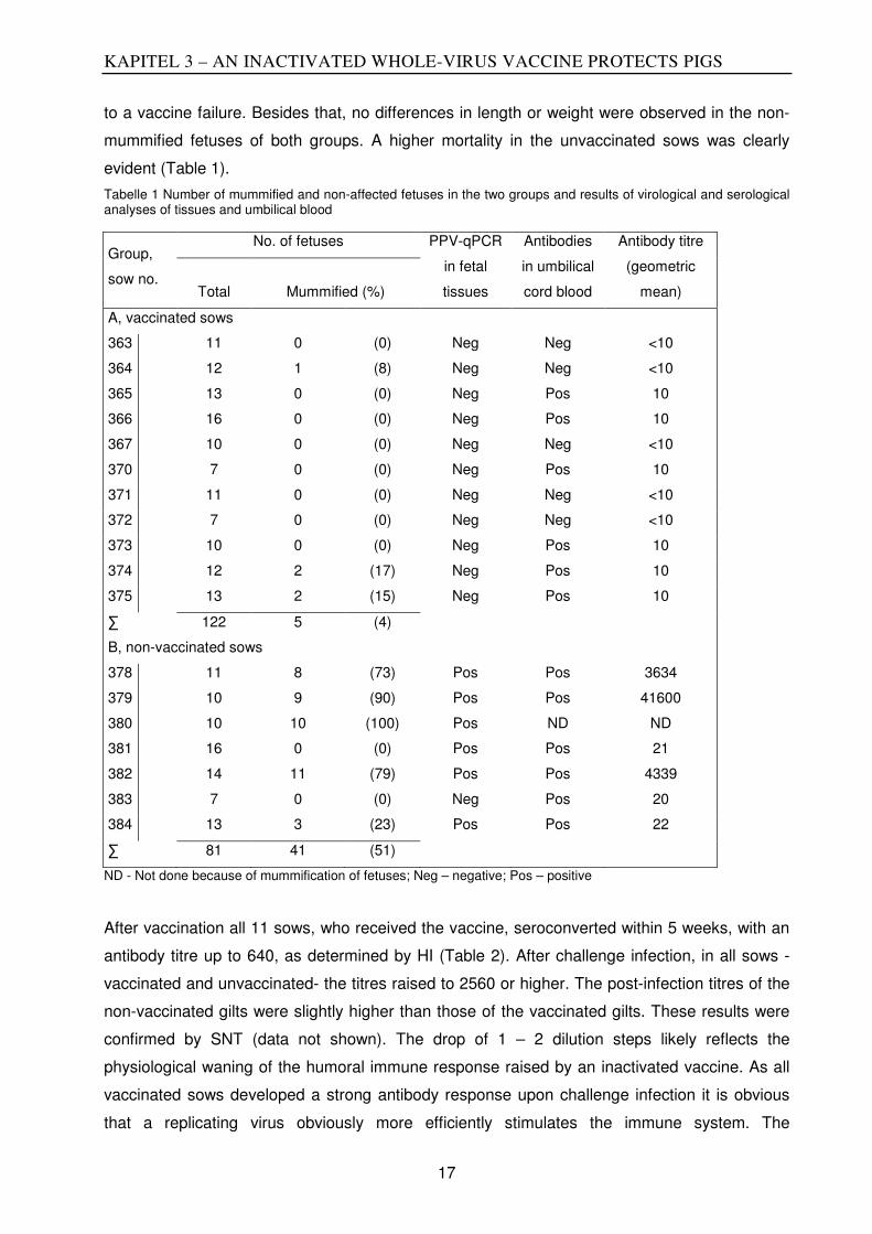
KAPITEL 3 – AN INACTIVATED WHOLE-VIRUS VACCINE PROTECTS PIGS
17
to a vaccine failure. Besides that, no differences in length or weight were observed in the non-
mummified fetuses of both groups. A higher mortality in the unvaccinated sows was clearly
evident (Table 1).
Tabelle 1 Number of mummified and non-affected fetuses in the two groups and results of virological and serological analyses of tissues and umbilical blood
Group,
sow no.
No. of fetuses PPV-qPCR
in fetal
tissues
Antibodies
in umbilical
cord blood
Antibody titre
(geometric
mean) Total Mummified (%)
A, vaccinated sows
363 11 0 (0) Neg Neg <10
364 12 1 (8) Neg Neg <10
365 13 0 (0) Neg Pos 10
366 16 0 (0) Neg Pos 10
367 10 0 (0) Neg Neg <10
370 7 0 (0) Neg Pos 10
371 11 0 (0) Neg Neg <10
372 7 0 (0) Neg Neg <10
373 10 0 (0) Neg Pos 10
374 12 2 (17) Neg Pos 10
375 13 2 (15) Neg Pos 10
∑ 122 5 (4)
B, non-vaccinated sows
378 11 8 (73) Pos Pos 3634
379 10 9 (90) Pos Pos 41600
380 10 10 (100) Pos ND ND
381 16 0 (0) Pos Pos 21
382 14 11 (79) Pos Pos 4339
383 7 0 (0) Neg Pos 20
384 13 3 (23) Pos Pos 22
∑ 81 41 (51)
ND - Not done because of mummification of fetuses; Neg – negative; Pos – positive
After vaccination all 11 sows, who received the vaccine, seroconverted within 5 weeks, with an
antibody titre up to 640, as determined by HI (Table 2). After challenge infection, in all sows -
vaccinated and unvaccinated- the titres raised to 2560 or higher. The post-infection titres of the
non-vaccinated gilts were slightly higher than those of the vaccinated gilts. These results were
confirmed by SNT (data not shown). The drop of 1 – 2 dilution steps likely reflects the
physiological waning of the humoral immune response raised by an inactivated vaccine. As all
vaccinated sows developed a strong antibody response upon challenge infection it is obvious
that a replicating virus obviously more efficiently stimulates the immune system. The

KAPITEL 3 – AN INACTIVATED WHOLE-VIRUS VACCINE PROTECTS PIGS
18
significance of the differences of the antibody titers of days 14 p.i., 21 p.i and 49 p.i. between
the vaccinated and non-vaccinated sows was calculated using the one-tailed Man Whitney-U
test. For all three time points the differences were found to be significant (p=0.0082, p=0.02559,
and 0.0004, respectively). It is hypothesized that the vaccine-induced antibody titers interfere
with the replication of the challenge virus and may hinder to some extend the induction of
antibodies. The drop of the antibody titers of the vaccinated sows from day -37 to the day of
challenge infection was surprising.
Tabelle 2. Antibody titre of the gilts after vaccination and challenge infection as determined by HI
Group,
sow no.
HI antibody titre at day:
-75
(1. vaccination)
-54
(2. vaccination) -37
0
(infection) 7 p.i. 1 4p.i. 21 p.i. 49 p.i.
A, vaccinated sows
363 <10 40 640 320 640 2560 2560 1280
364 <10 <10 160 160 160 1280 1280 640
365 <10 20 640 320 2560 5200 5200 2560
366 <10 40 320 160 1280 5200 2560 1280
367 <10 <10 160 <10 160 2560 1280 640
370 <10 <10 160 160 640 2560 1280 1280
371 <10 <10 160 80 1280 2560 1280 1280
372 <10 20 320 80 1280 5200 2560 1280
373 <10 10 320 80 640 10400 5200 1280
374 <10 <10 40 20 320 2560 1280 1280
375 <10 40 320 160 1280 2560 1280 1280
B, non-vaccinated sows
378 <10 <10 <10 <10 160 10400 5200 2560
379 <10 <10 <10 <10 80 10400 5200 5200
380 <10 <10 <10 <10 320 10400 5200 5200
381 <10 <10 <10 <10 80 10400 5200 5200
382 <10 <10 <10 <10 640 20800 5200 10400
383 <10 <10 <10 <10 80 2560 1280 2560
384 <10 <10 <10 <10 <10 5200 2560 5200
In both groups, fetuses with positive antibody titers in the umbilical blood were detected.
Curiously, low antibody titres were observed in vaccinated animals even in qPCR negative
fetuses. Since maternal antibody cannot cross the placental barrier (Mengeling et al., 2000), this
phenomenon most likely represents an euthanization artifact, resulting from a contamination
with maternal blood, instead of an active virus replication in immune-competent fetuses.

KAPITEL 3 – AN INACTIVATED WHOLE-VIRUS VACCINE PROTECTS PIGS
19
In contrast, in group A (vaccinated sows) only very low antibody titres up to 10 were observed in
13 of 122 fetuses, whereas antibody titres observed in 30 of 81 fetuses from the non-
vaccinated sows (group B) ranged from 10 up to 41600 (Table 1).
Viral DNA could not be detected by qPCR in lung or kidney of any of the 122 fetuses (0%) from
vaccinated sows (Table 3). However, in 55 of 81 (67.9%) fetuses from non-vaccinated sows
virus could be readily detected in the various tissues. The mean number of viral genomic copies
in those tissues was 7.99 x10⁹/µl. While only 41 of the 51 PPV-infected fetuses were
mummified, PPV was demonstrated in 14 apparently healthy fetuses, which did not show any
signs of disease.
After infection, virus was shed from animals of both groups, as determined by qPCR. High
shedding of virus could be detected until day 9, but some sows shed virus even up to 50 days
after infection. In the non-vaccinated sows virus DNA could be detected in 29.9% of all swabs,
whereas in the vaccinated sows only 4.1% were positive. The copy number of viral DNA differed
between the groups. Whereas in non-vaccinated sows viral genomic copies up to 7.79 x 10⁴/µl
(with a mean of 2.87 x 10³/µl) were detected, only a maximum number of genomic copies, range
to 2.831 x 10³/µl (with a mean of 3.04 x 10² /µl) was seen in vaccinated sows (Table 3).
Applying the two-tailed Mann Whitney U-test the combined values for nasal and fecal viral DNA
shedding was highly significant between the two groups (p<0,0001).
The weakly positive samples at day 2 and 4 post infection are most likely residual inoculum.
Considering the sensitivity of the PCR with 20 copies per reaction, only very small amounts of
DNA were detected. A contamination of the animals or the swabs taken from these animals
cannot be fully excluded in an animal experiment with pigs of more than 100 kg body mass and
housing in a group. In contrast, a contamination in the laboratory, can almost be excluded as all
appropriate negative controls for purification of the DNA and for the real-time PCR were always
negative. As a negative control water used used instead of the sample during DNA purification
and subsequent PCR, and furthermore a non-template control for the real time PCR was always
included.
All qPCR positive tested swabs were further investigated by cell culture for the presence of
infectious virus. After inoculation, a cytopathic effect could be observed in the first passage after
7 days in only four of these positives samples, all from three non-vaccinated sows (group B,
sow number 378, 380, 381).
The results of this study are very similar to the ones in a previous study (Jóźwik et al., 2009)
where an inactivated whole-virus vaccine was tested against a challenge infection with an
antigenically heterologous virus. It was then observed that vaccination prevented reproductive
disorders, but not infection and subsequent shedding of the heterologous challenge virus. The

KAPITEL 3 – AN INACTIVATED WHOLE-VIRUS VACCINE PROTECTS PIGS
20
data at that time were interpreted as an incomplete protection after heterologous challenge virus
infection.
Here, we were able to examine the protection after virus challenge with an antigenically
homologous virus, expecting protection against disease but also a complete prevention of virus
shedding after virus challenge. These results show that the experimental PPV-27a vaccine
prevented fetal death after homologous virus challenge with PPV-27a. However, as with the
heterologous challenge, all sows showed a substantial increase of antibody titres after infection,
suggesting that neither homologous nor heterologous vaccines could prevent virus infection.
Furthermore, shedding of genomic DNA could be detected in both vaccinated and non-
vaccinated sows after infection, although the number of sows that shed viral DNA and the
magnitude differed markedly between the groups (Table 3). Infectious virus was only recovered
from three non-vaccinated sows but not from any vaccinated sow. The sows that shed
infectious virus also shed high copy numbers of viral DNA.

KAPITEL 3 – AN INACTIVATED WHOLE-VIRUS VACCINE PROTECTS PIGS
21
Tabelle 3 Shedding of PPV from the day of challenge infection until day 49 p.i.
Group,
sow qPCR results at days post-challenge infection
Mean of DNA
in lung/ kidney
no. 0 2 4 7 9 11 14 16 18 21 49 of piglets
A, vaccinated sows
363 -/- -/- -/- -/- -/- -/- -/- -/- -/- -/- -/- -/-
364 -/- 74/- -/- -/- -/- -/- -/- -/- -/- -/- -/- -/-
365 -/- -/- -/- -/- -/- -/- -/69 -/- -/- -/- -/- -/-
366 -/- -/- -/- -/- -/- -/- -/- -/- -/- -/- -/- -/-
367 -/- -/2 -/- -/- -/- -/- -/- -/- -/- -/- -/- -/-
370 -/- 1/- -/- -/- 21/- -/- -/- -/- -/17 -/- -/- -/-
371 -/- 19/- -/- -/- -/- -/- -/- -/- -/- -/- -/- -/-
372 -/- -/- -/- -/- -/- -/- -/- -/- -/- -/- -/- -/-
373 -/- -/- -/- -/- -/- -/- -/- -/- -/- -/- -/36 -/-
374 -/- -/2 -/- -/- 2831/- -/- -/- -/- -/- -/- -/- -/-
375 -/- -/- -/- -/- -/- -/- -/- -/- -/- -/- -/- -/-
B, non-vaccinated sows
378 -/- -/- -/3 23155/97/+ 17995/274/+ 115/50 7/63 15/- 23/- -/- 35/- 3x10⁹/2x109
379 -/- -/- 13/- -/- 641/9 52/20 16/- -/- -/- -/- -/2 6x10⁹/1x109
380 -/- -/- -/- 186/809 77900/33/+ 49/20 228/7 -/- -/- 38/- 16/- 1x10⁹/6x109
381 -/- -/- -/- 1090/-/+ 222/11 2568/- -/- 1165/- -/- -/- -/3 335/1506
382 -/- -/- -/- 35/- 4696/25 214/- 21/- 6/- -/- -/- -/- 7x10¹⁰/7x109
383 -/- -/- -/- 36/41 -/8 -/- -/- -/- -/- -/26 11/- -/-
384 -/- -/- -/- -/- -/- -/- -/- -/- -/- -/- -/- 6/459
qPCR detection of PPV DNA from nasal/rectal swabs of sows and from fetal tissues. Numbers indicate the virus DNA copies/µl swab suspension and mean DNA copies/µg tissue found in lungs/kidneys of each litter. A dash indicates PPV DNA not detected (-/- i.e. DNA in nasal/rectal swab suspension not detected). Virus isolation was performed on all qPCR-positive swab samples. Growth of virus is indicated by +.

KAPITEL 3 – AN INACTIVATED WHOLE-VIRUS VACCINE PROTECTS PIGS
22
It is not clear whether the shedding of very low copy numbers of viral DNA is of
epidemiological importance, as infectious virus could not be recovered from any vaccinated
sow. Based on the challenge experiments described here and those reported previously
(Jóźwik et al., 2009) an inactivated vaccine does not induce a sterile immunity against a
challenge infection even against a homologous challenge virus and may not completely
prevent virus shedding.

KAPITEL 3 – AN INACTIVATED WHOLE-VIRUS VACCINE PROTECTS PIGS
23
References
Bergeron, J., Hébert, B. &Tijssen, P. (1996). Genome organization of the Kresse strain of
porcine parvovirus: identification of the allotropic determinant and comparison with those of
NADL-2 and field isolates. J Virol 70, 2508–2515.
Jòzwik, A., Manteufel, J., Selbitz, H. J. &Truyen, U. (2009). Vaccination against porcine
parvovirus protects against disease, but does not prevent infection and virus shedding after
challenge infection with a heterologous virus strain. J Gen Virol 90, 2437–2441.
King, A. (Hg.) (2012).Virus taxonomy. Classification and nomenclature of viruses, ninth
report of the International Committee on Taxonomy of Viruses. Amsterdam [u.a.]: Elsevier.
Mengeling, W. L., Lager, K. M. &Vorwald, A. C. (2000).The effect of porcine parvovirus and
porcine reproductive and respiratory syndrome virus on porcine reproductive performance.
Anim Reprod Sci 60-61, 199–210.
Nielsen, J.,Rønsholt, L.&Sørensen, K. J. (1991). Experimental in utero infection of pig
foetuses with porcine parvovirus (PPV). Vet. Microbiol.28, 1–11.
Streck, A. F.,Bonatto, S. L.,Homeier, T., Souza, C. K.,Gonçalves, K. R.,Gava, D., Canal, C.
W. &Truyen, U. (2011).High rate of viral evolution in the capsid protein of porcine parvovirus.
J. Gen. Virol. 92, 2628–2636.
Streck, A. F.,Homeier, T., Foerster, T., Fischer, S. &Truyen, U. (2013a). Analysis of porcine
parvoviruses in tonsils and hearts from healthy pigs reveals high prevalence and genetic
diversity in Germany. ArchVirol 158, 1173–1180.
Streck, A. F.,Homeier, T, Foerster, T&Truyen, U. (2013b). Population dynamics and in vitro
antibody pressure of porcine parvovirus indicate a decrease in variability. J. Gen. Virol. 94
2050–2055.
Streck, A. F., Hergemöller, F. Rüster, D, Speck, S & Truyen, U. (2015).A TaqMan qPCR for
quantitation of Ungulate protoparvovirus 1 validated in several matrices .
Truyen, U.& Streck, A. F. (2012).Porcine parvovirus. In Diseases of swine,pp. 447–455.
Edited by J. Zimmerman, Chichester, West Sussex, FL: Wiley-Blackwell,
Zeeuw, E. J. L., Leinecker, N., Herwig, V., Selbitz, H. J. &Truyen, U.(2007). Study of the
virulence and cross-neutralization capability ofrecent porcine parvovirus field isolates and
vaccine viruses inexperimentally infected pregnant gilts. J Gen Virol 88, 420–427.
Zimmermann, P., Ritzmann, M., Selbitz, H. J., Heinritzi, K. &Truyen, U. (2006). VP1
sequences of German porcine parvovirus isolates definetwo genetic lineages. J Gen Virol 87,
295–301.
Acknowledgements:
We thank Nadja Leinecker for expert technical assistance and Prof Martin Pfeffer for help with the statistical analyses.

KAPITEL 4 - POPULATION DYNAMICS OF PPV
24
KAPITEL 4 - POPULATION DYNAMICS AND IN VITRO ANTIBODY
PRESSURE OF PORCINE PARVOVIRUS (PPV) INDICATE A
DECREASE OF VARIABILITY
André Felipe Streck1,2, Timo Homeier1, Tessa Foerster1, Uwe Truyen1
Author’s affiliations:
1. Institute of Animal Hygiene and Veterinary Public Health, Faculty of Veterinary Medicine,
University of Leipzig, An den Tierkliniken 1, 04103, Leipzig, Germany.
2. CAPES Foundation, Ministry of Education of Brazil, Setor Bancário Norte, Quadra 2Bloco L,
Lote 06, 70040-020, Brasília, Brazil.
ABSTRACT
To estimate the impact of PPV vaccines on the emergence of new phenotypes, the population
dynamic of PPV was calculated, and an in vitro-model was designed to reproduce a possible
immune selection. A decrease in genetic diversity was observed in the presence of antibodies in
vitro or after vaccination. Since antibodies have reduced neutral selection, then the vaccine
failures and infections in non-vaccinated populations were most likely responsible for the
emergence of new PPV phenotypes.
Porcine parvovirus (PPV) infections are characterized by the reoccurrence of estrus, abortion
and the delivery of mummified and stillborn fetuses [summarized in TRUYEN and STRECK
(2012)]. The resulting economic losses for the pig industry led to the global use of inactivated
vaccines after the 1980s (TRUYEN and STRECK 2012).
In the last 15 years, several reports have been published about new PPV-phenotypes with
amino acid changes resulting in variations on the capsid surface (SOARES et al.2003;
ZIMMERMANN et al. 2006; STRECK et al. 2011). These amino acid substitutions may be
responsible for the dramatically different pathogenic properties of the virus (BERGERON et al.
1996). In addition, there is evidence that antibodies raised against most of the PPV vaccine
strains displayed a low heterologous neutralizing activity against these new phenotypes. This
led to the hypothesis that the vaccines may force a selective pressure on PPV field strains
resulting in “escape mutants”, which could have supported the emergence of new phenotypes
(ZEEUW et al. 2007).To address the question whether PPV vaccines have influence on the
emergence of new phenotypes, we analyzed the population dynamics of the virus in silico and
the antibody pressure was evaluated with an in vitro-model.
To test the hypothesis of an antigenic selection we designed an in vitro experiment in which
polyclonal serum generated from an experimentally NADL2-infected sow was used to neutralize

KAPITEL 4 - POPULATION DYNAMICS OF PPV
25
one homologous and one heterologous PPV virus isolates. The homologous strain used was
the NADL-2 (USA), a tissue culture-adapted vaccine virus(MENGELING. 1975) and the
heterologous strain used was the strain called “Challenge” (England), a virulent strain that
shows only a limited crossneutralization with the NADL-2 strain (ZEEUW et al. 2007). The
neutralization capacity of the polyclonal antibodies is 1:5120 for the NADL2 strain and 1:640 for
the Challenge strain(minimal serum dilution able to neutralize 50 TCID mL-1). Both strains
without antibodies were used as negative controls.
The first inoculations with Challenge and NADL-2 viruses were performed in PK-15cell-lines
(3x10⁵ cells/mL) with a minimal concentration of virus able to produce aTCID50 (in 96h) and
maintained in Dulbecco's Modified Eagle Medium (DMEM, Sigma–Aldrich, Germany)
supplemented with 5% (v/v) fetal bovine serum (FBS) at 37°C in 5%CO2. Additionally, different
dilutions of polyclonal serum were added to the inoculated cells (hemagglutination-inhibition titer
of 1:500 to 1:1.72 per 25 µL). After five days, the mixture with the minimal dilution of serum, but
still displaying signs of CPE was collected and frozen. A subsequent re-inoculation of cells with
different serum dilutions was performed with the collected viruses. After another five days, cells
with the minimal dilution of serum and still presenting signs of CPE were collected again. This
process was repeated for 21 passages. In the control, both viruses were inoculated in cells
without polyclonal serum, collected, frozen and further passed this way for 21 times. A
resistance was observed in the homologous NADL2 and heterologous Challengestrains
inoculated with the antibodies in the last two passages, and a cytopathiceffect was evident even
in the most concentrated antibody dilution (1:500). Thus, no further passages were performed.
The DNA amplification, sequencing, and assembly were performed as previously described
(STRECK et al. 2011). The sequences were deposited in GenBank under the accession
numbers (waiting for approval).
The VP gene sequence analysis revealed that the viruses grown without antibodies had more
substitutions than the viruses cultivated with antibodies after 21 passages (Table 3). That was
more evident in the homologous NADL2 strain without antibody pressure (with one synonymous
and six non-synonymous substitutions) compared to the NADL2 submitted to antibody pressure
(two synonymous substitutions). For the heterologous Challenge, the differences were blander:
three synonymous and three non-synonymous substitutions for the virus without the antibody
pressure and three non-synonymous and two non-synonymous substitutions for the virus
submitted to the antibody pressure.

KAPITEL 4- POPULATION DYNAMICS OF PPV
26
Tabelle 1 - Nucleotide (nuc.) and amino acid (aa.) substitution for the homologous NADL2 and heterologous Challenge strains after 21 passages with and without antibody (AB) pressure.
Nuc. position number 2551 2746 2943 3163 3242 3383 3522 3635 3768 3942 3958 4030 4115 4183 4474 4503
NADL2 original GAA AGA ACT GGA ATA AGT CTA TTA ATT GAT CAC AAG TCT CCT AAC AGA
NADL2 with AB AGT CAA
NADL2 without AB CTA ACT GGT CAA AAC AAA AAA
AA. position number 63A 128A 45B 118B 145B 192B 238B 276B 320B 378B 383B 407B 436B 458B 555B 565B
NADL2 original E R T G I S L L I D H K S P N R
NADL2 with AB S Q
NADL2 without AB T G Q N K K
Nuc. position number 2551 2746 2943 3163 3242 3383 3522 3635 3768 3942 3958 4030 4115 4183 4474 4503
Challenge original GAG AGA AGT GGG ATA AGT CTA TTA ATT GGT CAA AAG CCT CCC AAC AAA
Challenge with AB GAA ACT GGA TCT CCT
Challenge without AB GAA AGG CTA GGT CCA CCT AA. position number 63A 128A 45B 118B 145B 192B 238B 276B 320B 378B 383B 407B 436B 458B 555B 565B
Challenge original E R S G I S L L I G Q K P P N K
Challenge with AB T S
Challenge without Ab L G P AAmino acids numbers from VP1 protein BAmino acids numbers from VP2 protein
KAPITEL 4- POPULATION DYNAMICS OF PPV
27
Most of the changes were observed in positions known to be under a high selective pressure
(SHANGJIN et al. 2009). In the amino acid position 320 (isoleucine), a serine was inserted in
the NADL2 virus submitted to the antibody pressure and a threonine in the NADL2 virus grown
without the antibody pressure, indicating that this site is highly variable. Additionally, in site 436
of the Challenge virus grown with antibody pressure, a serine was inserted (presented also in
the original NADL2 strain). This site is located at the top of the 3-fold spike and is considered to
have a high binding potential to antibodies (SIMPSON et al. 2002). Since antibodies raised
against the NADL2 virus should now be able to strongly bind the Challenge virus with this
substitution, this change was not expected. Despite the antibody pressure, the P436S
substitution may have been selected because of replication advantages. A previous study with
different chimaeras between the virus Kresse and NADL2 revealed that genetic elements of
theNADL2 virus (including the site 436) are responsible for the replication efficiency in cell lines
(FERNANDES et al. 2011).
For the in silico analysis, all VP gene sequences deposited in the GenBank (up to May2012)
were retrieved from NCBI (http://www.ncbi.nlm.nih.gov/). Only viruses originally isolated from
swine were considered. The dataset consisted of 739 nucleotides (between nucleotide position
3701 and 4439) from 74 samples. The nucleotide and amino acid numbers used in this study
are based on the Kresse strain (GenBank accession number U44978).
The clock-like behavior of the dataset was visualized using the regression of the root to-tip
genetic distance inferred from the Maximum Likelihood tree against the sampling time in the
software Path-O-Gen v.1 (DRUMMOND et al. 2003). The populationdynamic was estimated
with a Bayesian Markov chain Monte Carlo (MCMC) method, using BEAST version 1.7.1
(DRUMMOND and RAMBAUT 2007). These analyses were run using the GTR DNA substitution
model with partitions into codon positions, performing 500 million generations through the
MCMC and subsampling each 10000generations. The population dynamic model used was the
Bayesian skyline coalescent (stepwise) model and the molecular clock model used was a
relaxed clock with an uncorrelated log-normal distribution of rates. Additionally, different models
testing the population size change through time were estimated (constant, lineal and stepwise)
using the GTR DNA substitution model with partitions into codon positions, performing10 million
generations through the MCMC and subsampling each 10 000 generations. The fitness of both
models was compared using the marginal likelihood, adopting the method of NEWTON AND
RAFTERY (1994), with modification (SUCHARD and REDELINGS 2006). The strength of the
statistical evidence was quantified using the Bayes factors (KASS and RAFTERY 1995).
The root-to-tip analysis was constructed to examine whether the samples exhibited adequate
temporal structure. Since the obtained value of R2 for the regression analysis was 0.9845, a
strong temporal fitness was confirmed for the dataset. Finally, the Bayesian skyline coalescent
model was used to estimate the epidemiological history and evolutionary dynamics of the PPV

KAPITEL 4 - POPULATION DYNAMICS OF PPV
28
over time. The comparison of the nullhypothesis (constant population size) with the hypothesis
in which population change is allowed indicated that the data fit better with the hypothesis that
allow variability (Table4). The resulting demographic inference is shown in Figure 10. The
effective population size of PPV underwent a period of relative steadiness until around 1982.
After 1982, a moderate but continuous decrease is evident until the recent strains. The
beginning of the population decrease matches with the period in which vaccination campaigns
were broadly introduced
Abbildung 1 -. Skyline plot of the PPV population dynamics. The solid black line represents the mean value of the skyline plots. The blue area represents the limits of 95% higher posterior probability density. Time is shown in years across the X bar and the effective population size is shown in the Y bar.

KAPITEL 4 - POPULATION DYNAMICS OF PPV
29
Tabelle 2- Comparison of the null-hypothesis (constant population size) with the hypothesis that allows population variability.
Model 1 Model 2 Model 3
Tree prior Constant Lineal Stepwise
Maximum likelihood -1770.3356 -1772.9462 -1762.8145
95% higher posterior probability density difference
49.00 43.66 43.56
Log 10 Bayes’ factor* 2.488 0.851 - *Values between 0.0-0.5 provide no evidence; 1.0-1.5 provide weak evidence; >1provide strong-to-decisive evidence [adapted from van BALLEGOOIJEN et al. (2009and KASS and RAFTERY (1995)].
A main explanation for this phenomenon could be the broad use of certain PPV vaccines in the
last 30 years. This can be stipulated, since a reduction in the geneticdiversity is assumed to be
strictly related to a more difficult or reduced virustransmission (HALLORAN and HOLMES 2009;
van BALLEGOOIJEN et al. 2009), as expected in a vaccinated population. Another important
factor that can influence the genetic diversity of the virus is the behavior of the host population
(HALLORAN and HOLMES 2009). In the last decades, the demographics of the virus infection
and its spreading potential with larger swine breeding/growth facilities possible changed.
Additionally, new sanitary measures and disinfectants were adopted in the 1980s, probably
affecting the viral population dynamics as well.
As several other factors could have influenced the PPV dynamic in the last decades, the in
vitro-model was used to show that antibody pressure is able to cause a higher or lower genetic
drift. In that model, we expected to observe a viral escape mechanism in viruses submitted to
the antibody pressure. However, for both strains (NADL2 and Challenge), we observed less
nucleotide and amino acids substitutions in the viruses when cultivated under antibody pressure
in comparison with the same viruses cultivated without this pressure. This was particularly
evident for the NADL2 strain submitted to the homologous antibody pressure. Regarding the
heterologous Challenge strain, the emergence of non-synonymous and synonymous
substitutions was quite similar for the virus cultivated with and without antibodies. As antibodies
raised against the NADL2 virus do not efficiently neutralize the Challenge virus (ZEEUW et al.
2007; JÓŹWIK et al 2009), the pressure implemented by the anti-NADL2-antibodies should be
milder compared to that raised against the homologous strain NADL2. Under a strong pressure,
most mutations may be deleterious, and the genetic diversity was lower for the NADL2 strain.
The selective forces that drive parvovirus evolution remain uncertain. Apparently, as in the in
vitro-model a lower genetic diversity was observed in the presence of antibodies neutral
selection seems to be more important for PPV than adaptive evolution.
In wild boar populations, phylogenetic analysis indicated that PPV is more diverse than in
domestic pigs (CADAR et al. 2012). The authors of that study observed that the amino acid

KAPITEL 4 - POPULATION DYNAMICS OF PPV
30
substitution rate per year was higher than that found in a study examining samples from
domestic pigs (STRECK et al. 2011). As possible explanations for the wild boar viruses’ higher
variability, the dissemination of the same virus in herds by mixing animals of different sources
and the extensive vaccination in domestic swine were hypothesized as factors that reduce PPV
variability in these populations. Taken into consideration with the findings in the in vitro and in
silico models, it can be assumed that vaccine failures and non-vaccinated populations (e.g. wild
boars) rather than escape mutations due to vaccine-pressure may have played a major role in
the emergence of new PPV-phenotypes.
In conclusion, the widely used vaccination programs had influenced the genetic diversity of
porcine parvoviruses. To further minimize the risk of the emergence of new phenotypes the use
of homologous vaccines along with a constant monitoring of the PPV appears advisable.
KAPITEL 4 - POPULATION DYNAMICS OF PPV
31
REFERENCES
Bergeron J, Hérbert B, Tijssen P. Genome organization of the Kresse strain of
porcineparvovirus: identification of the allotropic determinant and comparison with those
ofNADL-2 and field isolates. J Virol. 1996; 70:2508-2515.
Cadar D, Dán Á, Tombácz K, Lőrincz M, Kiss T, Becskei Z, Spînu M, Tuboly T,Cságola A.
Phylogeny and evolutionary genetics of porcine parvovirus in wild boars.Infect Genet Evol 2012;
12:1163-1171.
Drummond AJ, Pybus OG, Rambaut A. Inference of evolutionary rates from
molecularsequences. Adv Parasitol. 2003; 54:331-358.
Drummond AJ, Rambaut A. BEAST: Bayesian evolutionary analysis by sampling trees. BMC
Evol Biol. 2007; 7:214.
Fernandes S, Boisvert M, Tijssen P. Genetic elements in the VP region of porcine parvovirus
are critical to replication efficiency in cell culture. J Virol. 2011; 85:3025-3029.
Halloran ME, Holmes EC. Invited commentary: Evaluating vaccination programs using genetic
sequence data. Am J Epidemiol. 2009; 170:1464-1466.
Jóźwik A, Manteufel J, Selbitz HJ, Truyen U. Vaccination against porcine parvovirus protects
against disease, but does not prevent infection and virus shedding after challenge infection with
a heterologous virus strain. J Gen Virol. 2009; 90:2437-2441.
Kass RE, Raftery AE. 1995. Bayes factors. J Am Stat Assoc. 1995; 90(430):773-795.
Mengeling WL. Porcine parvovirus: frequency of naturally occurring transplacental infection and
viral contamination of fetal porcine kidney cell cultures. Am J Vet Res.1975; 36:41-44.
Newton MA, Raftery AE. 1994. Approximate Bayesian inference with the weighted likelihood
bootstrap. J R Stat Soc (B). 1994; 56:3-48.
Shangjin C, Cortey M, Segalés J. Phylogeny and evolution of the NS1 and VP1/VP2gene
sequences from porcine parvovirus. Virus Res. 2009; 140:209-215.
Simpson AA, Hébert B, Sullivan GM, Parrish CR, Zádori Z, Tijssen P, Rossmann M.The
structure of porcine parvovirus: comparison with related viruses. J Mol Biol. 2002;315:1189-
1198.
Soares RM, Cortez A, Heinemann MB, Sakamoto SM, Martins VG, Bacci M, DeCampos FM,
Richtzenhain LJ. Genetic variability of porcine parvovirus isolates revealed by analysis of partial
sequences of the structural coding gene VP2. J GenVirol. 2003; 84:1505-1515.
Streck AF, Bonatto SL, Homeier T, Souza CK, Gonçalves KR, Gava D, Canal CW, Truyen U.
High rate of viral evolution in the capsid protein of porcine parvovirus. J GenVirol. 2011;
92:2628-2636.
Suchard MA, Redelings BD. BAli-Phy: simultaneous Bayesian inference of alignmentand
phylogeny. Bioinformatics. 2006; 22(16):2047-2048.

KAPITEL 4 - POPULATION DYNAMICS OF PPV
32
Truyen U, Streck AF. Porcine parvovirus. In: Zimmerman J, Karriker L, Ramirez A, Schwartz K,
Stevenson G (ed). Diseases of Swine. 10th ed. Oxfort: John Wiley & SonsInc; 2012. p. 447-455.
van Ballegooijen WM, van Houdt R, Bruisten SM, Boot HJ, Coutinho RA, Wallinga J.Molecular
sequence data of hepatitis B virus and genetic diversity after vaccination. AmJ Epidemiol. 2009;
170:1455-1463.
Zeeuw EJL, Leinecker N, Herwig V, Selbitz HJ, Truyen U. Study of the virulence and cross-
neutralization capability of recent porcine parvovirus field isolates and vaccine viruses in
experimentally infected pregnant gilts. J Gen Virol. 2007; 88:420-427.
Zimmermann P, Ritzmann M, Selbitz HJ, Heinritzi K, Truyen U. VP1 sequences of German
porcine parvovirus isolates define two genetic lineages. J Gen Virol. 2006;87:295-301
ACKNOWLEDGMENTS
This research was supported by the Deutscher Akademischer Austausch Dienst(DAAD,
Germany) and Coordenação de Aperfeiçoamento Pessoal de Nível Superior(CAPES, Brazil).
We thank Dietlinde Woll, Linda Oettel and Martin Pfeffer for criticallyreading the manuscript.
KAPITEL5-HIGH PREVALENCE AND GENETIC DIVERSITY OF PPVS OBSERVED IN
GERMANY
33
KAPITEL 5 - ANALYSIS OF PORCINE PARVOVIRUSES IN TONSILS
AND HEARTS FROM HEALTHY PIGS REVEALEDHIGH PREVALENCE
AND GENETIC DIVERSITY IN GERMANY
André Felipe Streck1,2, Timo Homeier1, Tessa Foerster1, Stefan Fischer3, Uwe Truyen1
Author´s affiliations:
1. Institute of Animal Hygiene and Veterinary Public Health, Faculty of Veterinary Medicine,
University of Leipzig, An den Tierkliniken 1, 04103, Leipzig, Germany.
2. CAPES Foundation, Ministry of Education of Brazil, Setor Bancário Norte, Quadra 2, BlocoL,
Lote 06, 70040-020, Brasília, Brazil.
3. Veterinary and Food Monitoring Office, am Schlachthof 8a, 06667, Weißenfels, Germany.
ABSTRACT
In the present study, tonsils and hearts from 100 pigs were collected in a German
slaughterhouse in 2010 and tested for porcine parvoviruses (PPV, PPV2, PPV3 and
PPV4).PPV was observed in 60/100 hearts and 61/100 tonsils and, PPV2 in 55/100 hearts
and78/100 tonsils. PPV3 and PPV4 were found in 20/100 and 7/100 of the tonsils tested, but
not in the heart samples. Positive samples of PPV, PPV2 and PPV3 were analyzed by
nucleotide sequencing and their phylogenetic analysis revealed at least two distinct lineages for
each virus in the German samples. The high detection rate of the PPV, PPV2 and PPV3in
healthy animals and their genetic diversity highlights the importance of the continuous
monitoring of these viruses and their zoonotic potential.
Parvoviruses are small DNA viruses infecting a variety of animal hosts, in which they may cause
asymptomatic or sub-clinical infections as well as severe or even fatal disease. These viruses
belong to the Parvoviridae family, which is sub-classified into two subfamilies: Densovirinae and
Parvovirinae (TIJSSEN et al. 2001). The latter containing the most important genera for the
veterinary and human medicine in the whole family (Amdovirus, Bocavirus, Dependovirus,
Erythrovirus and Parvovirus).
Porcine parvovirus (PPV) is considered to be one of the major causes of reproductive failure in
swine. Clinical signs of PPV infections are characterized by the reoccurrence of estrus, abortion
and the delivery of mummified and stillborn fetuses [reviewed by TRUYEN and STRECK
(2012)] usually described with the acronym SMEDI (stillbirth, mummification, embryonic death
and infertility). It was already demonstrated that PPV has a high substitutionrate in the capsid
gene, similar to that in other parvoviruses and in RNA viruses (SHACKELTON and HOLMES
2006; SHACKELTON et al. 2005; STRECK et al. 2011a).Therefore, the emergence of new PPV

KAPITEL5-HIGH PREVALENCE AND GENETIC DIVERSITY OF PPVS OBSERVED IN
GERMANY
34
strains may interfere with the efficacy of the currently used vaccines, recommending the
continuous monitoring of the swine population for PPV.
In recent years, by means of molecular techniques, the DNA sequences of several novel
viruses have been identified. For parvoviruses affecting swine, the first of the newly identified
viruses was the virus H-1 in sera from pigs in Myanmar (HIJIKATA et al. 2001), originally
described as porcine parvovirus 2 (PPV2). In 2008, a virus closely related to the human
parvovirus 4 was found in slaughtered pigs in Hong Kong (LAU et al. 2008) and initially termed
as porcine hokovirus, here designated as porcine parvovirus 3 (PPV3). In 2010, another
parvovirus was found in lungs from a diseased pig co-infected with porcine circovirustype 2 and
designated porcine parvovirus 4 (PPV4) (CHEUNG et al. 2010). The impact of these viruses for
the pig industry remains unknown.
In order to monitor the occurrence of new strains of PPV and to study the distribution of novel
parvoviruses (PPV2, PPV3 and PPV4), hearts and tonsils of clinically healthy swine were
collected at a slaughterhouse and examined for these viruses.
MATERIAL AND METHODS
SAMPLES
Tonsils and hearts (apex) from 100 pigs (around six months old) were collected in a German
slaughterhouse in December 2010. The animals originated from thirteen farms (here termed A
to M) located in seven German Federal States (Hessen, Lower Saxony, Mecklenburg-
Vorpommern, Saxony, Saxony-Anhalt, Schleswig-Holstein and Thuringia). One gram of each
tissue was macerated using mortar and pestle and resuspended as a 20% (v/v) suspension in
phosphate-buffered saline solution (PBS, pH 7.2). Homogenates were cleared by centrifuging at
4000 g for 5 min and filtered (0.2 µm pore size). Clarified supernatants (200µL) were used for
total nucleic acid extraction using the QIAamp DNA Mini kit (Qiagen, Germany), according to
the manufacturer´s instructions.
DNA AMPLIFICATION AND SEQUENCE ANALYSIS OF THE VP-GENE AND FULL-
LENGTHGENOMES
Primers for the PPV2, PPV3 and PPV4 (Table 5) were designed using the Primer3
program(http://primer3.sourceforge.net/). The selected primers were locally aligned using the
BLAST
algorithm (http://blast.ncbi.nlm.nih.gov/Blast.cgi) against the GenBank content to avoid
homology with other viral sequences. The PCRs specific for PPV, PPV2, PPV3 and PPV4were
carried out in single reactions using ten nmol from each dNTP (Rapidozym, Germany), ten pmol
of the respective primer pair (Table 5), one unit of Taq polymerase (Rapidozym, Germany),
enzyme buffer (according to the manufacturer’s instruction) and 2 µL of DNA template in a total

KAPITEL5-HIGH PREVALENCE AND GENETIC DIVERSITY OF PPVS OBSERVED IN
GERMANY
35
volume of 25 µL completed with bi-distilled water. The amplification was performed with an
initial heating at 95°C for 2 minutes, followed by 35 cycles consisting of 30seconds at 95°C for
denaturation, 30 seconds at 55° C for annealing, 30 seconds at 72°C for extension, and a final
extension at 72°C for 5 minutes. Amplification products were submitted to electrophoreses in
1.5% agarose gels with 89 mM Tris-Base, 89 mM of boric acid, 2 m MEDTA Na2 (TBE, pH 8.0)
buffer and stained with 1,5 µL (per 100 mL of agarose gel) of MidoriGreen DNA Stain (Nippon
Genetics Europe, Germany).
Tabelle 3 - Primers for the detection of the PPV, PPV2, PPV3 and PPV4.
Primer Sequence Location
PPV_S GGGGGAGGGCTTGGTTAGAATCAC 2974-2997
PPV_AS ACCACACTCCCCATGCGTTAGC 3149-3170
PPV2_F AGATTCTTGCAGGCCGTAGA 909-928
PPV2_R CCAAGGGTCAGCACCTTTTA 1096-1115
PPV3_F GTGGCAGTGATATTGCATCG 992-1011
PPV3_R TGGCAGTCATTGAATGGAAA 1219-1238
PPV4_F ACAAGGTGGAGGAACGTTTG 3069-3088
PPV4_R TTCCATGAGGGAGAGGATTG 3289-3308
In order to determine the sensitivity of each PCR, a plasmid harboring the target sequence of
each PCR was generated. Briefly, PPV2, PPV3 and PPV4 positive samples were amplified as
described previously. The amplicons were purified with the NucleoSpin purification kit
(Macherey-Nagel, Germany) and cloned into the pDRIVE vector (Qiagen, Germany) using One
Shot® TOP10 Chemically Competent E. coli (Invitrogen, Germany). The transformed cells were
grown on LB (Luria Bertani, Carl Roth GmbH, Germany) plates supplemented with100 µg/ml
ampicillin and incubated overnight at 37°C. Positive colonies were subcultured in LB broth (Carl
Roth GmbH, Germany) with ampicillin and incubated at 37°C for 8 hours. Plasmids were
extracted using the PeqGOLD Plasmid Miniprep Kit I (Peqlab, Germany) and quantified by UV
spectroscopy. For PPV, a plasmid containing the whole virus (Kressestrain) was used
(FERNANDES et al. 2009). The copy number of each plasmid was calculated according the

KAPITEL5-HIGH PREVALENCE AND GENETIC DIVERSITY OF PPVS OBSERVED IN
GERMANY
36
equation described in WHELAN et al. (2003). Finally, successive10-fold dilutions of each
plasmid were performed and tested for the respective PCR reaction.
Randomly selected positive samples of PPV, PPV2 and PPV3 were further amplified using the
primer pairs listed in Supplementary Table 1. PCR was performed using the HotStarTaqMaster
Mix Kit (Qiagen, Germany) in a final volume of 25 µL. The PCR was performed underfollowing
conditions: one cycle at 94°C for 15 min followed by 40 cycles of 95°C for 1 min,55°C for 1 min
and 72°C for 1 min, with final elongation at 72°C for 5 min. PCR products were purified using
the NucleoSpin Gel and PCR Clean-up kit (Macherey-Nagel, Germany),sequenced using a
BigDye Terminator version 3.1 Cycle Sequencing kit (AppliedBiosystems, Germany) and
analyzed on an ABI3100 Genetic Analyzer (Applied Biosystems,Germany) at the Zentraler
Funktionsbereich DNA-Sequenzierung (University of Leipzig, Germany). Assembling of the
obtained sequences to a full-length sequence was performed using the SeqMan program of the
Lasergene software (DNASTAR). The nucleotide sequences of the complete VP1 gene of the
PPV and nearly full-length genomes of thePPV2 and PPV3 viruses were submitted to the NCBI
GenBank, and are available under the accession numbers KC296743- KC296752. The samples
were named according their sample number (1 to 100), followed by an H (sample from heart) or
a T (sample from tonsil) and the respective virus (PPV, PPV2 or PPV3).
PHYLOGENETIC ANALYSIS
Sequences deposited in GenBank (up to June 2012) were retrieved from the NCBI homepage
(http://www.ncbi.nlm.nih.gov/). In addition, the samples generated in this study were included.
Altogether, the PPV dataset consisted of 47 sequences of the VP1 gene. In this dataset only
sequences isolated after 2002 and standard sequences (143a, 225b, 27a, NADL2 and Kresse
strains) were used. The PPV2 and PPV3 datasets consist of 7 and 45 sequences of the putative
VP gene, respectively. The Open Reading Frames (ORFs) were found with Artemis software
(RUTHERFORD et al. 2000) and the alignment was performed with the MUSCLE software
(EDGAR. 2004).
The phylogeny was estimated with a Bayesian Markov Chain Monte Carlo (MCMC) method,
using BEAST version 1.7.2 (DRUMMOND and RAMBAUT 2007). These analyses were run
using the GTR substitution model with partitions into codon positions, performing 10 million
generations through the MCMC and sub-sampling each 1000 generations. The resulting data
were analyzed using Tracer software (http://tree.bio.ed.ac.uk/software/tracer/) after removing a
10% “burn in” for each data and compared with different priors. A maximum clade credibility tree
for each dataset was generated by the software Tree Annotator v.1.7.1 (BEAST package).
Phylogenetic trees were visualized with the software Fig Tree v.1.3.1 (http://tree.bio.ed.ac.uk/).
All nucleotide numbers used in the present study for the PPV refer to the Kresse strain
(GenBank accession number U44978). Amino acid numbers are according to the VP2protein of

KAPITEL5-HIGH PREVALENCE AND GENETIC DIVERSITY OF PPVS OBSERVED IN
GERMANY
37
the same strain. The nucleotide number for the novel parvoviruses refer to: PPV2strain YH14
(GU938301), PPV3 strain HK1 (EU200671) and PP4 strain JS0918-5598(GU978966).
CELL CULTURES
For virus isolation, the following cell lines were used: porcine kidney 15 (PK-15, CCL-33), swine
embryonic kidney (SPEV) and swine testicular epitheloid (STE, CRL 1746). PK-15 and SPEV
cells were propagated in Dulbecco's Modified Eagle Medium (DMEM, Sigma–Aldrich, Germany)
supplemented with 5% (v/v) Fetal Bovine Serum (FBS), grown and maintained at37°C in 5%
CO 2. STE cells were cultivated in DMEM supplemented with 10% (v/v) FBS and grown and
maintained at 37°C. For primary cultures, testicles, lungs and kidney from piglets (negative
tested for PPV, PPV2, PPV3 and PPV4) were washed in PBS (pH 7.2) and gingerly minced.
The enzymatic disaggregation was performed with Trypsin 0.25% (Sigma–Aldrich, Germany)
agitated using a magnetic stirrer at 37°C for 45 min. The cells were then clarified (centrifugation
at 500 g for 5 min), propagated in DMEM supplemented with 10%(v/v) FBS and grown and
maintained at 37°C in 5% CO 2. Finally, blood was obtained from 4months old pigs and
peripheral blood lymphocytes were separated from heparinized blood on Ficoll-Hypaque
(Sigma–Aldrich, Germany). After one wash with PBS (pH 7.2) and clarification (600 g per 5 min)
the cells were resuspended in Roswell Park Memorial Institute Medium (RPMI 1640, Sigma–
Aldrich, Germany). Non-adherent cells were collected and resuspended in RPMI 1640 medium
plus 20 µg/mL of concanaval in A (Lektin, Applichem, Germany).
All cell-lines and cell-cultures were inoculated with 100 µL of the inoculum (macerated tissues,
Supplementary Table 3), after five days of incubation the cells were frozen and thawed twice
and 500 µL of this mixture were used to inoculate new cells. The second cell passage was
incubated for 5 days and frozen-thawed twice again. DNA of the cell extract and medium
surface was extracted and submitted to the PCR.
STATISTICAL ANALYSIS
The data were statistically analyzed using the Chi-Square-test to evaluate differences in the
PCR results. All analyses were carried out using the statistical package IBM-SPSS Version20.
Values of P < 0.05 were considered statistically significant.

KAPITEL5-HIGH PREVALENCE AND GENETIC DIVERSITY OF PPVS OBSERVED IN
GERMANY
38
RESULTS
PCR
The sensitivities of the PCRs was determined to be 10, 50, 100 and 50 plasmid copies detected
per reaction for PPV, PPV2, PPV3 and PPV4, respectively. As shown in Table 6, amplification
products of the PPV could be observed in 60/100 hearts and 61/100 tonsils.PPV2 could be
identified in 55/100 hearts and 78/100 tonsils. None of the heart samples but20/100 tonsil
samples were positive for PPV3. Furthermore, none of the hearts was positive for PPV4
whereas in 7/100 a positive result was observed in tonsils. In total, 77 animals were positive
tested for PPV, 81 for PPV2, 20 for PPV3 and 7 for PPV4
Tabelle 4- Positive samples for the PPV, PPV2, PPV3 and PPV4 viruses according their respective herd (A-M) and federal state (HE: Hessen, NI: Lower Saxony, MV: Mecklenburg- Vorpommern, SN: Saxony, ST: Saxony-Anhalt, SH: Schleswig-Holstein and TH: Thuringia). Results displayed as (positive heart samples//positive tonsil samples)
Herd Fed. State PPV PPV2 PPV3 PPv4
A (n=10) TH 7//4 3//4 0//0 0//2
B (n=10) MV 8//10 8//9 0//0 0//0
C/D (n=10) SN 10//9 7//10 0//5 0//1
E/F (n=10) NI 8//6 5//8 0//4 0//0
G (n=10) ST 2//0 5//10 0//0 0//1
H (n=10) SH 6//9 5//5 0//2 0//0
I (n=10) HE 8//7 3//5 0//5 0//0
J/K (n=10) HE/NI 8//10 5//8 0//0 0//2
L (n=10) NI 1//6 5//9 0//4 0//1
M (n=10) SN 2//0 9//10 0//0 0//0
Total 60a//61a 55b//78b 0//20 0//7
Upper letters showed statistical association.
In the heart samples, PPV and PPV2 co-infection was observed in 32/100 samples. In the
tonsils, the greatest numbers of viral co-infection was observed for the PPV together withPPV2
(46/100), PPV together with PPV3 (15/100) and PPV2 together with PPV3 (15/100)(Table 7).
Co-infection with PPV, PPV2 and PPV3 was observed in 12/100 samples and2/100 samples
were positive tested for the four viruses. No contamination was observed in the negative
controls.

KAPITEL5-HIGH PREVALENCE AND GENETIC DIVERSITY OF PPVS OBSERVED IN
GERMANY
39
Tabelle 5 - Number of positive samples for each virus per tissue
Heart Tonsil
Virus PPV PPV2 PPV3 PPV4 PPV PPV2 PPV3 PPV4
PPV 60 61
PPV2 32 55 46 78
PPV3 0 0 0 15 15 20
PPV4 0 0 0 0 4 5 2 7
GENETIC CHARACTERIZATION
The four sequences of the PPV VP1 gene analyzed (6T-PPV, 37T-PPV, 55T-PPV and
80TPPV)show an identity of 98.4% - 100% with the sequences retrieved from the GenBank
(altogether 47 PPV sequences, nucleotide number 2287 - 2314 joined with 2388 - 4549).Among
the four sequences of this study the nucleotide identity ranged from 98.4% to 99.9%.The
synonymous substitution rate ranged from two to 21 (overall average of fourteen) and the non-
synonymous substitution rate from zero to fourteen (overall average of nine). ForPPV2, the
three sequences (6T-PPV2, 68T-PPV2 and 98T-PPV2) showed an identity of94.4% - 98.7%
with the sequences available (altogether seven PPV2 sequences, nucleotide number 240 -
5358).The identity between the three sequences was 94.7% - 98.4%. ForPPV3, the three
sequences (62T-PPV3, 70T-PPV3 and 85T-PPV3) had an identity of 95.3%- 99.7% with the
sequences available (altogether 34 PPV3 sequences, nucleotide number109 - 4180). Among
the three sequences, the identity was 97.4% - 99.2%.
At the protein level, the PPV sequences 6T-PPV and 55T-PPV displayed a higher number of
amino acid differences than 37T-PPV and 80T-PPV compared with standard strains
(NADL2and Kresse). A similar amino acid profile can also be found in the PPV strains 15425
(GenBank accession number JN400520) and 21620005_1h (GQ884035). On the other hand,
the sequences 37T-PPV and 80T-PPV were almost identical to the virulent 27a
strain(differences only at the sites A414S and E419Q) (Table 8).

KAPITEL5-HIGH PREVALENCE AND GENETIC DIVERSITY OF PPVS OBSERVED IN
GERMANY
40
Tabelle 6 - Amino acid substitution within PPV VP2 sequences.
VP sequence
Aminoacid VP2
NA
DL2
Kre
sse
27a
1542
5
6T
37T
55T
80T
20 T - - A A - A -
45 T S S S S S S S
82 R - - K K - K -
215 I T T T T T T T
226 Q - - E - - E -
228 Q - E - - E - E
233 S - - - T - T -
304 P - - T T - T -
320 I - - T T - T -
366 D - - N N - N -
378* D G G G G G G G
383* H Q Q Q Q Q Q Q
407 K - - N N - N -
414 A - S - S - S -
419 E - Q - Q - Q -
436* S P T P P T P T
565 R K K K K K K K
*Site considered responsible for the different tissue tropism (BERGERON et al. 1996).
PHYLOGENETIC ANALYSIS
In the phylogeny, groups with posterior probability ≥ 0.90 were designated clusters and groups
with low posterior probability (< 0.90) were designated clades. The Bayesian maximum clade
credibility tree generated for the complete VP1 gene of the PPV revealed three clusters [here
called A-PPV, B-PPV and D-PPV (posterior probability = 0.99, 0.97 and0.97)] and two different
clades [C-PPV and E-PPV (posterior probability < 0.90)]. The sequences 6T-PPV and 55T-PPV
were located in the cluster D-PPV and the sequences 37TPPVand 80T-PPV were located in the
cluster A-PPV. From the sequences (isolated after2002), the cluster A-PPV contains the higher
proportion of new sequences in the DNA databanks (Figure 11).
The tree generated for the PPV2 putative VP gene displayed two clusters [A-PPV2 and BPPV2
(posterior probability = 0.99 and 0.99]. The sequence 68T-PPV2 was located in the cluster A-
PPV2 and the sequences 6T-PPV2 and 98T-PPV2 were located in the cluster
BPPV2.Additionally, cluster A-PPV2 was formed with the sequence from Myanmar while cluster

KAPITEL5-HIGH PREVALENCE AND GENETIC DIVERSITY OF PPVS OBSERVED IN
GERMANY
41
B-PPV2 by sequences from China. Finally, the tree generated for the PPV3 putative VP gene
revealed two main clades [A-PPV3 and B-PPV3 (posterior probability < 0.90)]. The sequences
62T-PPV3 and 70T-PPV3 were located in the clade A-PPV3 and the sequence85T-PPV3 was
located in the clade B-PPV3. Clade A-PPV3 was formed mostly by sequences from China and
clade B-PPV3 included sequences from Germany, Romania and the United Kingdom.
PPV

KAPITEL5-HIGH PREVALENCE AND GENETIC DIVERSITY OF PPVS OBSERVED IN
GERMANY
42
PPV2

KAPITEL5-HIGH PREVALENCE AND GENETIC DIVERSITY OF PPVS OBSERVED IN
GERMANY
43
PPV3
Abbildung 2 - Phylogenetic tree for the PPV, PPV2 and PPV3 datasets. The scale axis indicates the substitutions per site. Posterior probabilities are indicated above the mainly branches. The GenBank accession number of each sequence is followed by the name (identification) and the location of isolation. The location is coded as: AUS (Austrian), CH (China), and EU [Europe (no precise location available)]. GE (Germany), MY (Myanmar), RO (Romania), SW (Switzerland), UK (United Kingdom) and USA (United States). The sequences from the present study are indicated with a black dot.
CELL CULTURE
After two passages in PK-15, SPEV and STE cell-lines and as well as in the primary
culture(kidneys, lungs, testes and macrophages) no virus specific CPE was observed and no
specific amplicons for PPV, PPV2, PPV3 or PPV4 could be generated in the PCR(supernatant
and cell debris).

KAPITEL5-HIGH PREVALENCE AND GENETIC DIVERSITY OF PPVS OBSERVED IN
GERMANY
44
STATISTICAL ANALYSIS
Statistical associations among hearts and tonsils samples were observed for PPV and PPV2,
individually. No statistical association was observed among different viruses (co-infection
between PPV, PPV2, PPV3 or PPV4).
DISCUSSION
The high PPV DNA prevalence in hearts and tonsils observed in healthy fattening pigs was
unexpected, since at that age these animals are usually separated from the sows (which can
shed highly contaminated material from abortions). The animals were not vaccinated against
PPV and, as in Germany no PPV live vaccines are licensed, the probability of vaccine DNA
detection is minute.
Samples of tonsils were chosen due to their predominant role in a PPV infection. Tonsils are
considered to be a primary replication site for PPV (TRUYEN and STRECK 2012). The high
PPV prevalence (> 50%) may indicate a function of secondary replication for this tissue as well.
As myocardium has been reported to support live-long presence of the human parvovirus B19
(KUETHE et al. 2009) and canine parvovirus (WALDVOGEL et al. 1991), heart samples were
also included in the present study. In humans, prevalence of low DNA level of parvovirus B19 in
the myocardium is a frequent finding in patients with heart disease and also in healthy patients
(LOTZE et al. 2010). In pigs, PPV has already been demonstrated by nested-PCR in hearts of
piglets presenting a non-suppurative myocarditis (BOLT et al. 1997), but its etiologic role is
unknown.
The prevalence of low level of parvoviral DNA in humans is hypothesized to be caused by DNA
of non replicating defective interfering particles or site specific integration in the chromosome
(SÖDERLUND-VENERMO et al. 2002). In the swine host, the presence of the PPV DNA in
serum with no correlation with the antibodies titers was already demonstrated(STRECK et al.
2011b), suggesting accordance with the hypothesis of DNA defective interfering particles, which
are not capable of any immune system stimulation. Further studies should be performed in
order to characterize the mechanism of persistence of PPV and parvovirus B19.
As the novel porcine parvoviruses are phylogenetically closely related to the novel human
parvoviruses, those porcine viruses have recently been investigated. For PPV2, a high
prevalence (20.7%) was already reported in pig lungs in USA in 2011 (XIAO et al. 2012).Here,
even higher detection rates were obtained from hearts (55%) and tonsils (78%),strongly indicate
that these tissues may be important for the viral infection. For PPV3, a high detection rate
(18.7%) was observed in lungs from growing-finishing pigs in the USA [26]. In Germany, this
virus was also detected in liver and serum (32.7%) from wild boars (ADLHOCH et al. 2010). For
PPV4 only low detection rates were observed, confirming other reports (HUANG et al. 2010).
Coincidently, in healthy humans a similar low to moderate DNA prevalence is observed for

KAPITEL5-HIGH PREVALENCE AND GENETIC DIVERSITY OF PPVS OBSERVED IN
GERMANY
45
Human parvovirus 4 (high nucleotide identity to the PPV3) and Human bocavirus (with a similar
genome structure to the PPV4) and a high prevalence is observed for parvovirus B19 as well
(CORCIOLI et al 2010; KUETHE et al 2011).
To examine the properties of the infection in heart and tonsils, several cell-lines and primary cell
cultures were inoculated with sixteen PCR-positive samples from PPV, PPV2, PPV3 andPPV4
(from tonsils and heart). PPV was firstly reported as a contaminant particle of primary cell
culture from kidney and testicle and replication of PPV in these cells was essential for the
further understanding of PPV biology {see SIEGL. (1976) for review}. Thus, it was expected that
the novel parvoviruses have similar properties. The lack of virus replication in these cells may
be due to the detection of non-infective particles of viral DNA or the primary cell cultures and the
cell lines lack some property needed for the replication. Further studies are ongoing to define
the tissue tropism of these viruses.
In the phylogenetic analysis, due to the high number of PPV sequences available from
GenBank, the tree generated for the complete VP1 gene was calculated with sequences
derived during the last ten years. In comparison with a previous phylogenetic study with PPV
samples collected until September 2010 (STRECK et al. 2011a) a new clade (E-PPV) was
formed with moderate posterior probability (0.70). To the standard strains 27a (sequence
located in cluster A-PPV) and 225b (sequence located in cluster D-PPV) the in vivo
pathogenicity was experimentally confirmed. Additionally, it was shown that antibodies raised
against the vaccine strains have a low neutralization activity against these strains (ZEEUW etal.
2007), but no information about cross-neutralization and virulence of strains from the cluster B-
PPV and clade E-PPV is available.
At the protein level, the sequences 37T-PPV and 80T-PPV were similar to the virulent strain27a
(AY684871). This strain was first detected in 2001 in Germany (ZIMMERMANN et al.2006) and
has since then been reported in several countries (STRECK et al. 2011a). The samples 6T-PPV
and 55T-PPV had higher similarity with the strain 15425, a new strain (STRECK et al. 2011a),
with unknown biological properties. A previous study with different PPV-single-mutants has
shown that the presence of amino acids 228 (E) and 436 (T) (as observed in the samples 37T-
PPV and 80T-PPV) as well as the amino acids 414 (S) and 419(Q) (as for samples 6T-PPV and
55T-PPV) increased the replication efficiency in vitro compared to the NADL2 original strain
(data not shown). The amino acids 226 (E) in the sample 55T-PPV as well as the amino acids
233 (T), 304 (T), 320 (T), 366 (N) and 407 (N) for the samples 6T-PPV and 55T-PPV are
located in the loops two, three and four of VP2protein (Protein Data Bank accession 1K3V), it
can be speculated that these substitutions may influence the immunogenicity or replication
efficiency as well. For all the new samples, the amino acids substitutions (I-215-T, D-378-G, H-
383-Q, S-436-P and R-565-K) that distinguish the non-virulent NADL2 strain (L23427) from the

KAPITEL5-HIGH PREVALENCE AND GENETIC DIVERSITY OF PPVS OBSERVED IN
GERMANY
46
virulent Kresse strain (U44978)were similar to the Kresse sequence. The only exception was
the site 436, at which the samples 37T-PPV and 80T-PPV have a serine (as in the strain 27a).
For the PPV2, there are few reports to enrich our phylogenetical analyses. It seems possible
that the virus is divided in two main clusters; however, more sequences are needed to come to
any meaningful conclusion. For PPV3, two distinct clades are evident. One is formed mostly by
samples from China and the other includes samples from Canada, Germany and Romania. In
Germany, five almost identical samples from PPV3 were already observed in wild boars
(ADLHOCH et al. 2010) and were similar to the sequence 85T -PPV3 described here. The
PPV2 and PPV3 sequences from this study were highly diverse, indicating that there is no
geographic predominance of a single lineage of PPV2 or PPV3 in Germany.
In conclusion, DNA of PPV as well as of the novel PPV2, PPV3 and PPV4 was present in
healthy animals (tonsils and hearts) and the phylogenetic analysis of the PPV, PPV2 andPPV3
demonstrated that these viruses have a high genetic diversity.

KAPITEL5-HIGH PREVALENCE AND GENETIC DIVERSITY OF PPVS OBSERVED IN
GERMANY
47
REFERENCES
Adlhoch C, Kaiser M, Ellerbrok H, Pauli G. High prevalence of porcine Hokovirus in German
wild boar populations. Virol J. 2010; 7:171.
Bergeron J, Hérbert B, Tijssen P. Genome organization of the Kresse strain of porcine
parvovirus: identification of the allotropic determinant and comparison with those ofNADL-2 and
field isolates. J Virol. 1996; 70:2508-2515.
Bolt DM, Häni H, Müller E, Waldvogel AS. Non-suppurative myocarditis in piglets associated
with porcine parvovirus infection. J Comp Pathol. 1997; 117:107-118.
Corcioli F, Zakrzewska K, Fanci R, De Giorgi V, Innocenti M, Rotellini M, Di Lollo S, Azzi A.
Human parvovirus PARV4 DNA in tissues from adult individuals: a comparison with human
parvovirus B19 (B19V). Virol J. 2010; 7:272.
Cheung AK, Wu G, Wang D, Bayles DO, Lager KM, Vincent AL. Identification and molecular
cloning of a novel porcine parvovirus. Arch Virol. 2010; 155:801-806.
Drummond AJ, Rambaut A. BEAST: Bayesian evolutionary analysis by sampling trees. BMC
Evol Biol. 2007; 7:214.
Edgar RC. MUSCLE: multiple sequence alignment with high accuracy and high throughput.
Nucleic Acids Res. 2004; 32:1792-1797.
Fernandes S, Tijssen P. Seamless cloning and domain swapping of synthetic and complex
DNA. Anal Biochem. 2009; 385:171-173.
Hijikata M, Abe K, Win KM, Shimizu YK, Keicho N, Yoshikura H. Identification of new parvovirus
DNA sequence in swine sera from Myanmar. Jpn J Infect. 2001; 54:244-245.
Huang L, Zhai SL, Cheung AK, Zhang HB, Long JX, Yuan SS. Detection of a novel porcine
parvovirus, PPV4, in Chinese swine herds. Virol J. 2010; 7:333.
Kuethe F, Lindner J, Matschke K, Wenzel JJ, Norja P, Ploetze K, Schaal S, Kamvissi
V,Bornstein SR, Schwanebeck U, Modrow S. Prevalence of parvovirus
B19 and humanbocavirus DNA in the heart of patients with no evidence of dilated
cardiomyopathy ormyocarditis. Clin Infect Dis. 2009; 49:1660-1666.
Lau SK, Woo PC, Tse H, Fu CT, Au WK, Chen XC, Tsoi HW, Tsang TH, Chan JS, TsangDN, Li
KS, Tse CW, Ng TK, Tsang OT, Zheng BJ, Tam S, Chan KH, Zhou B, Yuen KY. Identification of
novel porcine and bovine parvoviruses closely related to human parvovirus 4. J Gen Virol. 2008;
89:1840-1848.
Lotze U, Egerer R, Glück B, Zell R, Sigusch H, Erhardt C, Heim A, Kandolf R, Bock T, Wutzler
P, Figulla HR. Low level myocardial parvovirus B19 persistence is a frequent finding in patients
with heart disease but unrelated to ongoing myocardial injury. J MedVirol. 2010; 82:1449-1457.
Rutherford K, Parkhill J, Crook J, Horsnell T, Rice P, Rajandream MA, Barrell B. Artemis:
sequence visualization and annotation. Bioinformatics. 2000; 16:944-945.

KAPITEL5-HIGH PREVALENCE AND GENETIC DIVERSITY OF PPVS OBSERVED IN
GERMANY
48
Shackelton LA, Holmes EC. Phylogenetic evidence for the rapid evolution of human
B19erythrovirus. J Virol. 2006; 80:3666-3669.
Shackelton LA, Parrish CR, Truyen U, Holmes EC. High rate of viral evolution associated with
the emergence of carnivore parvovirus. Proc Natl Acad Sci USA. 2005; 102:379-384.
Siegl G. The Parvoviruses, in: Gard S, Hallauer C (eds) Virology Monographs, 15th ed. Wien,
New York: Springer-Verlag; 1976. p 47-52.
Söderlund-Venermo M, Hokynar K, Nieminen J, Rautakorpi H, Hedman K Persistence of human
parvovirus B19 in human tissues. Pathol Biol. 2002; 50:307-316.
Streck AF, Bonatto SL, Homeier T, Souza CK, Gonçalves KR, Gava D, Canal CW, Truyen U.
High rate of viral evolution in the capsid protein of porcine parvovirus. J GenVirol. 2011a;
92:2628-2636.
Streck AF, Gava D, Souza CK, Gonçalves KR, Bortolozzo FP, Wentz I, Canal CW. Presence of
porcine parvovirus in sera from pigs is independent of antibody titers. BerlMunch Tierarztl
Wochenschr. 2011b; 124:242-246.
Tijssen P, Agbandje-McKenna M, Almendral JM, Bergoin M, Flegel TW, Hedman K,
Kleinschmidt JA, Li D, Pintel D, Tattersall P. Parvoviridae In: King MQ, Adams MJ, Carstens E,
Lefkowitz EJ (eds) Virus Taxonomy: Classification and Nomenclature of Viruses: Ninth Report
of the International Committee on Taxonomy of Viruses. SanDiego: Elsevier; 2011.
Truyen U, Streck AF. Porcine parvovirus In: Zimmerman J, Karriker L, Ramirez A, Schwartz K,
Stevenson G (eds) Diseases of Swine, 10th Ed Oxfort: John Wiley & SonsInc; 2012. p 447-455.
Waldvogel AS, Hassam S, Weilenmann R, Tratschin JD, Siegl G, Hänichen T, Briner J,
Pospischil A. Retrospective study of myocardial canine parvovirus infection by in situ
hybridization. Zentralbl Veterinarmed B. 1991; 38:353-357.
Whelan JA, Russell NB, Whelan MA. A method for the absolute quantification of cDNA using
real-time PCR. J Immunol Methods. 2003; 278(1-2):261-269.
Xiao CT, Gerber PF, Giménez-Lirola LG, Halbur PG, Opriessnig T.Characterization of porcine
parvovirus type 2 (PPV2) which is highly prevalent in the USA. Vet Microbiol.2012; Epub ahead
of print
Xiao CT, Giménez-Lirola LG, Halbur PG, Opriessnig T. Increasing porcine PARV4 prevalence
with pig age in the US pig population. Vet Microbiol. 2012; Epub ahead of print
Zeeuw EJL, Leinecker N, Herwig V, Selbitz HJ, Truyen U. Study of the virulence and cross-
neutralization capability recent porcine parvovirus field isolates and vaccine viruses
experimentally infected pregnant gilts. J Gen Virol. 2007; 88:420-427.
Zimmermann P, Ritzmann M, Selbitz HJ, Heinritzi K, Truyen U. VP1 sequences of German
porcine parvovirus isolates define two genetic lineages. J Gen Virol. 2006; 87:295-301.

KAPITEL5-HIGH PREVALENCE AND GENETIC DIVERSITY OF PPVS OBSERVED IN
GERMANY
49
ACKNOWLEDGEMENTS
This research was supported by the Deutscher Akademischer Austausch Dienst
(DAAD,Germany) and Coordenação de Aperfeiçoamento Pessoal de Nível Superior
(CAPES,Brazil). We thank Dietlinde Woll and Martin Pfeffer for critically reading the manuscript.

KAPITEL 6 - DISKUSSION
50
KAPITEL 6 - DISKUSSION
Beim porzinen Parvovirus handelt es sich um ein weltweit verbreitetes Virus, das in allen
Bereichen der Schweinehaltung vorkommt und bei Sauen zu Reproduktionsstörungen führt. Um
die dadurch bedingten wirtschaftlichen Einbußen möglichst gering zu halten, werden seit 30
Jahren regelmäßig Impfungen der Zuchtsauen durchgeführt. Dabei kommen inaktivierte
Impfstoffe, die eine PPV-spezifische Antikörperbildung induzieren, zum Einsatz (TRUYEN und
STRECK 2012).
In den vergangen 10 Jahren wurden diverse neue PPV-Isolate identifiziert, deren genaue
klinische Auswirkungen auf die Schweinepopulation bisher noch ungeklärt sind. Anhand von
Sequenzanalysen wurden ausgetauschte Aminosäuren dicht an der Kapsidoberfläche entdeckt.
Diese könnten von wichtiger Bedeutung für die Zell-Virus Interaktion und die Immunität sein
(STRECK et al 2011). Da die Antikörper, die gegen die Impfstämme gebildet werden, eine
geringere Neutralisation der heterologen Feldstämme als der homologen Impfstämme
bewirkten, scheinen diese neuen Phänotypen im Vorteil zu sein (ZEEUW et al. 2007; JÓŹWIK
et al. 2009). Anhand von Infektionsversuchen wurde die Effektivität der aktuell gebräuchlichen
Impfstoffe untersucht. Beispielsweise wurde in der Arbeit von JÓZWIK et al (2009)
exemplarisch die Wirksamkeit eines kommerziell erhältlichen und eines experimentellen
Impfstoffes, an dem zu ihnen heterologen hochvirulenten PPV-Isolat 27a, in tragenden Sauen
getestet. Dabei wurde gezeigt, dass die getesteten Impfstoffe durchaus in der Lage sind, einen
zufriedenstellenden Schutz der Feten vor einer klinischen Manifestation des Virus-Isolates zu
bewirken. Dennoch kam es zu einer Infektion der Sauen mit Virusreplikation und Ausscheidung
über Kot und Nasensekret.
Das Ziel der ersten Arbeit dieser Dissertation mit dem Titel „An inactivated whole-virus porcine
parvovirus vaccine protects pigs against disease but does not prevent virus shedding even after
homologous virus challenge“ war es, zu überprüfen, ob sich dieses hochvirulente Isolat (PPV-
27a) dazu eignet, einen inaktivierten Impfstoff zu entwickelt. Dieser sollte bei homologer
Infektion nicht nur einen zuverlässigen Schutz der Feten bewirken, sondern auch die Infektion
der Sauen und damit die Virusreplikation und Virusausscheidung verhindern. Auf diese Weise
soll er zu einer vollständigen Elimination des Virus beitragen. Hierfür wurden zunächst 18
spezifisch pathogenfreie, primipare Jungsauen in zwei Gruppen unterteilt. Eine Gruppe erhielt
den experimentellen Impfstoff, die zweite ein Placebo. Alle Jungsauen wurden am 40. Tag ihrer
Trächtigkeit infiziert, wobei ihnen das zum Impfstamm homologe PPV Isolat 27a appliziert
wurde. Am 90. Trächtigkeitstag erfolgten die Euthanasie der Sauen und die Entnahme der
Feten. Die Untersuchung der Feten sowie der während des Versuches gesammelten Proben
erwiesen sich erstaunlicherweise als konform zu den von Józwick et al. 2009 ermittelten

KAPITEL 6 - DISKUSSION
51
Ergebnissen. Es kam in der aktuellen Studie zur Ausscheidung von Virus DNA in der
ungeimpften wie auch in der geimpften Gruppe. Dies belegt, dass der Einsatz von homologen
Impfstoffen eine Infektion nicht verhindert. Die Ausscheidung von Virus DNA über Kot und
Nasensekret bei den ungeimpften, homolog infizierten Tieren (PPV-Isolat 27a infiziert) war
deutlich höher als die der geimpften, homolog infizierten Tiere (PP-Isolat 27a infiziert). Daraus
lässt sich schließen, dass der neue Impfstoff ein gutes Mittel darstellt, um die
Virusausscheidung innerhalb einer Herde zu reduzieren. Außerdem konnte in den fetalen
Organen der geimpften Sauen, im Gegensatz zu denen der ungeimpften Sauen, kein Virus
nachgewiesen werden, womit ein Schutz der Feten aufgrund des Impfstoffes belegt ist.
Zusammenfassend ist zu sagen: Der experimentelle Impfstoff auf Basis des hochvirulenten
PPV Isolates 27a verhindert die klinische Manifestation des Virus innerhalb der Feten und
reduziert die Virusausscheidung der Sauen, unterbindet sie jedoch nicht vollständig. Eine
effektivere Methode zur Eliminierung des Virus aus den Schweinepopulationen wird erst
möglich sein, sobald die genaue Pathogenese des Virus, mit all ihren virulenten genetischen
Eigenschaften, den Ausweichmechanismen und der vollständigen Immunreaktion des Wirtes
verstanden wird. Ein möglicher Ansatz könnte auch die Entwicklung von modifizierten
Lebendimpfstoffen, wie sie im Umgang mit den verwandten caninen und felinen Parvoviren
Verwendung finden, sein. Diese bewirken eine länger anhaltende Immunantwort, die einen
Schutz bis zu mehreren Jahren gewährleistet. Vielleicht könnten diese oder Vektorimpfstoffe,
welche eine zelluläre Immunantwort induzieren, einen Fortschritt in der Bekämpfung der
porzinen Parvoviren bewirken.
Seit gezeigt wurde, dass mittels der Antikörper die Neutralisation der neuen PPV Phänotypen
gegenüber denen der Impfstämme reduziert ist, wird diskutiert, ob die Impfungen das Virus
unter einen Selektionsdruck setzen und so die Bildung neuer Phänotypen, in einer Art „escape“
Reaktion, ermöglichen (ZEEUW et al. 2007). Allerdings konnte bisher nicht geklärt werden, ob
die Hauptunterschiede im VP1 Genom der einzelnen Isolate eine Entwicklung der letzten 10-30
Jahre (also dem Impfzeitraum) sind oder es sich um einen kontinuierlichen Verlauf handelt. Mit
Hilfe sogenannter phylogenetischer Trees in Verbindung mit einer molekularen Uhr sollte diese
Frage geklärt werden.
Ihr wurde in der zweiten Veröffentlichung mit dem Tilel “Population dynamics and in vitro
antibody pressure of porzine parvovirus indicates decrease of variability” nachgegangen.
Anders als erwartet, gelang es mit einem in silico Model einen leichten aber kontinuierlichen
Abfall der genetischen Vielfallt ab 1985 nachzuweisen. Etwa zum gleichen Zeitpunkt wurde mit
den regelmäßigen Impfungen der Sauen begonnen. Diese Feststellung steht im Widerspruch
zur ursprünglichen Hypothese, wird aber durch das hier herangezogene in vitro Modell
unterstützt. Hierzu wurden mit spezifischen Antikörpern (gegen das PPV Isolat-NADL2)

KAPITEL 6 - DISKUSSION
52
versehene PK15 Zellen mit entweder zum Impfisolat homologen oder heterologen PPV Isolaten
infiziert und über 21 Passagen vermehrt. Zum Vergleich wurden in 21 Zellpassagen (ohne
Antikörperzusatz) dieselben Virusisolate vermehrt. Erstaunlicherweise fanden sich in den
Kulturen mit Antikörpern weniger Mutationen, als in denen ohne Antikörperzusatz. Dies war
besonders deutlich für das NADL2 Isolat. Der Unterschied zwischen der Probe des Isolats mit
Antikörpern und der ohne Antikörper betrug vier ausgetauschte Aminosäuren. Für das zum
Antikörper heterologe Isolat Challenge konnte in der Kultur ohne Antikörperzusatz nur ein
Aminosäureaustausch mehr gefunden werden, als in der Kultur mit Antikörpern. Der
Unterschied zwischen den beiden Isolaten könnte dadurch bedingt sein, dass der
Antikörperdruck auf das NADL2 Isolat höher ist, da Antikörper gegen diesen Stamm gebildet
wurden.
Wenn man bedenkt, dass Impfungen zu erschwerten und reduzierten Virusübertragungen
führen (van BALLEGOOIJEN et al. 2009), erscheint es logisch, dass es nur wenigen
Mutationen gelingt, sich bei den erschwerten Gegebenheiten zu etablieren. Die
Populationsdynamik könnte jedoch auch leicht von anderen Prozessen, wie zum Beispiel dem
Aufteilen von Schweinen in Kategorien oder Desinfektionsmaßnahmen in Betrieben beeinflusst
worden sein. Außerdem ist zu bedenken, dass der Probenumfang dieser Studie größenmäßig
anderen viralen Datensammlungen entspricht, allerdings die Herkunftsorte nicht zufällig verteilt
sind. Innerhalb der wildlebenden Schweinepopulation wurde jedoch eine stärker ausgeprägte
genetische Vielfalt (mit pro Jahr mehr Aminosäuresubstitutionen) dokumentiert, als bei den
domestizierten Hausschweinen (CADAR et al. 2012). Eventuell könnte dies bedingt sein durch
eine höhere Variabilität der Viren, eine bessere Durchmischung der Populationen, Infektionen
mit mehreren Isolaten oder das Ausbleiben von Impfungen.
Zusammengefasst: Es wurde erwartet, mit Hilfe des in vitro und des in silico Models einen
Anstieg der genetischen Vielfalt, im Rahmen eines „viralen escape“, zu dokumentieren.
Gefunden wurde jedoch das Gegenteil. Die Virus-Isolate waren bei Anwesenheit von
Antikörpern genetisch stabiler, anstelle des erwarteten Anstieges wurde mit Beginn der
Impfroutine ein Abfall der PPV-Vielfallt dokumentiert. Demzufolge scheint es wahrscheinlich,
dass die Impfungen zu einem selektiven Druck führen, der es nur wenigen Mutationen gestattet,
sich zu etablieren, wodurch die genetische Vielfalt reduziert wird. Demnach ist die natürliche
Evolution für die Entstehung neuer Phänotypen wahrscheinlicher, als eine durch Antikörper
provozierte Evolution.
Analysiert man die neuen Phänotypen, stellt man fest, sie unterscheiden sich vor allem
hinsichtlich der Aminosäurezusammensetzung ihrer Kapsidoberfläche. Viele durch sie
verursachte Infektionen verlaufen asymptomatisch und wurden erst in den letzten Jahren mit
Hilfe neu entwickelter Technologien entdeckt (SIJUN et al. 2011, ALTSCHUL et al. 1990). Es

KAPITEL 6 - DISKUSSION
53
zeigte sich, dass durch herkömmliche Vakzine gebildete Antikörper eine niedrigere
Neutralisation dieser Parvoviren bewirken (ZEEUW et al. 2007, JÓŹWIK et al. 2009).
Shackelton et al. zeigten 2005 am Beispiel von CPV2, welches innerhalb weniger Jahre durch
genetische Mutation aus dem FPL-V hervorgegangen war, dass Parvoviren unter bestimmten
Voraussetzungen in der Lage sind, in kürzester Zeit andere Arten zu infizieren.
Die Gefahr, die aus diesem Potential hervorgeht, verdeutlicht die Notwendigkeit einer genauen
Studie der aktuell auftretenden Parvoviren.
Daher befasst sich die dritte Veröffentlichung dieser Arbeit mit dem Titel “Analysis of porzine
parvoviruses in tonsils and hearts from healthy pigs revealed high prevalence and genetic
diversity in Germany” mit der Analyse von in 100 Tonsillen und Herzen deutscher
Schlachtschweine exprimierter Parvoviren. Getestet wurde auf PPV und die neuen Parvoviren,
das porzine parvovirus 2 (PPV2), porzine parvovirus 3 (PPV3, oder porzine hokovirus) und
porzine parvovirus 4 (PPV4). Besonders häufig wurden dabei die Typen PPV und PPV2 in den
Herzen und Tonsillen sowie PPV3 in den Tonsillen gefunden.
Die Wahl fiel auf die genannten Organe, da Tonsillen für die Replikation von PPV ein
bevorzugtes Gewebe zu sein scheinen und bereits 1997 in der Untersuchung von Bolt et al.
PPV in Herzen nachgewiesen werden konnte (TRUYEN und STRECK 2012). Warum das Virus
ausgerechnet eine Affinität zu diesen Geweben aufweist, ist für PPV2, PPV3 und PPV4 immer
noch unbekannt. Da Schweineherzen jedoch zum Verzehr in den Verkehr gebracht werden,
besteht mitunter die Gefahr einer Zoonose. Diese wird noch unterstützt durch die Untersuchung
von Hallauer et al. (1971). Hier konnte gezeigt werden, dass PPV auch in der Lage ist, sich in
humanen Zelllinien zu vermehren.
2001 wurde in Deutschland erstmals der virulente Stamm 27a isoliert (ZIMMERMANN et al.
2006). Dieser Phänotyp weist in der Analyse der Aminosäuren nahezu identische Sequenzen
zu 37T-PPV und 80T-PPV auf. Unter Ausbildung deutlicher Krankheitssymptome gelang es 27a
sich über mehrere Länder auszubreiten. Die ihm ähnlichen Isolate konnten jedoch in
ungeimpften und klinisch gesunden Schweinen nachgewiesen werden.
Die phylogenetische Analyse von PPV wurde durchgeführt an Sequenzen, die während der
letzten 10 Jahre gesammelt wurden. Dabei galten die Isolate 143a, 225b, 27a NADL2 und
Kresse als Standard.
Die Sequenzen 6T-PPV und 55T-PPV konnten dem Cluster D-PPV und die dem Isolat 27a
ähnlichen Sequenzen (37T-PPV und 80T-PPV) dem Cluster A-PPV zugeordnet werden.
Außerdem entstand innerhalb von zwei Jahren eine neue Clade (E-PPV). Leider sind über die
kreuzneutralisierenden und virulenten Eigenschaften der Beispiele dieser Clade, als auch des
Clusters B keine Informationen verfügbar. Um die veterinärmedizinische Relevanz dieser

KAPITEL 6 - DISKUSSION
54
Proben zu verstehen, sind daher weitere Studien nötig. PPV ist der phylogenetischen Analyse
nach in zwei Cluster unterteilt. Um jedoch eine sinnvolle Schlussfolgerung ziehen zu können,
sind mehr Sequenzen und Berichte nötig. Auch PPV3 besteht aus zwei Clades. Geographische
Zusammenhänge können dahingehend beobachtet werden, als das eine Clade fast
ausschließlich von Proben aus China, die andere von Proben aus Kanada, Deutschland und
Rumänien gebildet wird. In der Wildschweinpopulation von Deutschland konnten bereits fünf
nahezu homologe Proben von PPV3 isoliert werden. Diese sind annähernd identisch mit der in
dieser Studie beschriebenen Sequenz (ADLHOCH et al. 2010).
Es gilt: Für PPV, PPV2 und PPV3 konnte die phylogenetische Analyse zeigen, dass dieses
Virus über eine hohe genetische Diversität verfügt und keine geographische Vorherrschaft
innerhalb Deutschlands besteht.

KAPITEL 7 - SCLUSSFOLGERUNG
55
KAPITEL 7 - SCHLUSSFOLGERUNG
Ein neuer, inaktivierter Impfstoff, auf der Basis des hochvirulenten und verbreiteten PPV-
Isolates 27a, verhindert die klinische Ausprägung sowie die Virusreplikation in Feten, reduziert
die Virusausscheidung durch die Sau, kann aber eine Infektion nicht verhindern.
Mittels in vitro und in silico Modellen konnte gezeigt werden, dass die Anwesenheit von
Antikörpern, eventuell durch Unterdrückung der natürlichen Selektion, zur Verringerung der
genetischen PPV Vielfallt führt. Daher scheint es wahrscheinlich, dass ungeimpfte Herden und
eventuelles Impfversagen verantwortlich für das Auftreten neuer PPV Phänotypen sind.
PPV DNA konnte in hohem Ausmaß in Herz- und Tonsillen-Proben von klinisch gesunden
Schweinen nachgewiesen werden. Wobei PPV3 ausschließlich in Tonsillen, PPV und PPV2
dagegen in Tonsillen und Herzen isoliert wurden.
Die Phylogenetische Untersuchung zeigt, dass die Viren von PPV, PPV2 und PPV3 genetisch
vielfältig sind und implizieren, dass kein einzelner Phänotyp in Deutschland dominant ist.

KAPITEL 8 - ZUSAMMENFASSUNG
56
KAPITEL 8 - ZUSAMMENFASSUNG
Verfasser: Tessa Foerster
Titel: Die genetische Varianz des Porzinen Parvovirus und die Wirksamkeit einer neuen
experimentellen Vakzine
Institut: Institut für Tierhygiene und Öffentliches Veterinärwesen
der Veterinärmedizinischen Fakultät, Universität Leipzig
Eingereicht im Mai 2016
59 Seiten, 5 Abbildungen, 9 Tabellen, 103 Literaturangaben
Schlüsselwörter: porzines Parvovirus, Schweine, PCR, SNT, HI
Einleitung
Die Infektion mit dem porzinen Parvovirus (PPV) ist weltweit in allen Arten von
Schweinebetrieben endemisch und verursacht große wirtschaftliche Verluste aufgrund von
Fruchtbarkeitsstörungen, wie Unfruchtbarkeit, verzögerter Östrusbeginn, Aborten und
mumifizierten Ferkeln. In den letzten 10 Jahren konnten neue porzine Parvoviren isolierte
werden, deren klinische Bedeutung noch ungeklärt ist.
Ziele der Untersuchung
Da diese oder andere neue PPV-Typen die Effektivität der derzeit verwendeten Impfstoffe
reduzieren könnten, wurde in der vorliegenden Dissertation anhand von drei Studien die
genetische Variabilität von PPV, die Entstehung neuer PPV-Typen, die Prävalenz der
neuartigen Parvoviren sowie die Wirksamkeit eines neuen Impfstoffes gegen PPV untersucht.
Material und Methoden
Die erste Arbeit befasst sich mit der Effektivität eines neuen Impfstoffes auf Basis des
hochvirulenten und weit verbreiteten PPV Isolates 27a. Es wurden 18 spezifisch pathogen freie
primipare Jungensauen in zwei Gruppen unterteilt. Eine Gruppe erhielt den neuen Impfstoff, die
andere ein Placebo. Anschließend wurden die trächtigen Tiere mit einem zum Impfstamm
homologen PPV-Isolat infiziert. Am 90. Trächtigkeitstag wurden die Feten entwickelt und mittels
Hämagglutinationshemmtest (HI), Serumneutralisationstest (SNT) und
Polymerasekettenreaktion (PCR) auf das Vorhandensein von Antikörpern und Viren untersucht.
Die zweite Studie analysierte die in öffentlichen Datenbanken (Gen Bank) vorhandenen
vollständigen PPV-Proteingene sowie Teilsequenzen und bestimmte so die Populationsdynamik
der verschiedenen PPV-Isolate bei Schweinen. Hierzu wurde die Bayesian-Methode mit einem
Bayesian skyline coalescent Model und außerdem eine in-vitro Untersuchung von 21
aufeinanderfolgenden Zellkultur-Passagen durchgeführt. Für das in vitro Modell wurden ein

KAPITEL 8 - ZUSAMMENFASSUNG
57
virulenter Feldstamm (PPV-Isolat Challenge) und ein Impfstamm (PPV-Isolat NADL2) auf mit
polyklonalen Antikörpern gegen NADL2 versehenden PK15 Zellen vermehrt, wobei eine
Negativkontrolle ohne Antikörper-Zusatz mitgeführt wurde. Herzen und Tonsillen von 100
Schweinen wurden 2010 auf einem deutschen Schlachthof für die dritte Studie gesammelt und
auf das Vorhandensein von PPV, porzinem Parvovirus 2 (PPV2), porzinem Parvovirus 3 (PPV3)
und porzinem Parvovirus 4 (PPV4) mittels PCR untersucht und alle positiven Proben
anschließend sequenziert.
Ergebnisse
Die Feten der geimpften Sauen verfügten über einen Schutz gegen die Infektion, dennoch kam
es bei den Sauen zur Infektion mit Virusausscheidung. Mit der Bayesian Analyse konnte die
Abnahme der Populationsdiversität im Laufe der Zeit und die Dominanz einzelner PPV-Stämme
gezeigt werden und es wurde festgestellt, dass beide Viren ohne den Zusatz von Antikörpern
deutlich mehr Mutationen entwickelten, als die Vergleichspassagen mit Antikörpern. 60 Herzen
und 61 Tonsillen wiesen PPV DNA, 55 Herzen und 78 Tonsillen PPV2 DNA, 20 Tonsillen PPV3
DNA und sieben Tonsillen DNA von PPV4 auf. Dabei wurden für PPV, PPV2 und PPV3 je
mindestens zwei Cluster oder Clades entdeckt.
Schlussfolgerungen
PPV entwickelt sich kontinuierlich weiter, auch wenn die Verwendung von PPV-Impfstoffen in
den letzten 30 Jahren zur Abnahme der genetischen Diversität des Virus geführt hat. Da PPV,
PPV2 und PPV3 eine hohe Prävalenz besitzen und auch neu entwickelte Impfstoffe noch kein
Garant für die Eliminierung des Virus sind, empfiehlt sich die strickte Überwachung der
aktuellen PPV Situation.
KAPITEL 9 - SUMMERY
58
KAPITEL 9 - SUMMARY
Author: Tessa Foerster
Title: The genetic variance of Porcine Parvovirus and the effectiveness of a new experimental
vaccine Institute: Institute of Animal Hygiene and Veterinary Public Health
Faculty of Veterinary Medicine, University of Leipzig
Submitted in May 2016
59 Pages, 5 Figures, 9 Tables, 103 References
Keywords: Porcine parvovirus, pig, PCR, SNT, HI
Introduction
The infection with the porcine parvo virus (PPV) is worldwide, in all kinds of pig farms, endemic
and causes great economic losses due to reproductive disorders like infertility, delayed estrus,
abortions and mummified piglets. In the last 10 years, new porcine parvovirus with still unclear
clinical significance could be isolated.
Aim of the study
As these and other new PPV types could reduce the effectiveness of the currently used
vaccines in the present work, the genetic variability of PPV, the emergence of new types of
PPV, the prevalence of novel parvoviruses as well as the efficacy of a new vaccine against PPV
was examined by three studies.
Material and Methods
The first paper deals with the effectiveness of a new vaccine on the basis of the highly virulent
predominant PPV isolate 27a. 18 specific pathogen–free, primiparous gilts were divided into two
groups. One group received the new vaccine, one a placebo. Subsequently, the pregnant
animals were infected with a to the vaccine homologous PPV isolate. On the 90th day of
pregnancy, the fetus has been developed and tested by hemagglutination inhibition test (HI),
serum neutralization test (SNT) and polymerase chain reaction (PCR) for the presence of
antigen and virus. The second study analyzed the on public databases (GenBank) existing full
PPV protein gene and partial sequences and determined the population dynamics of different
PPV isolates in pigs. For this purpose, the Bayesian method using a Bayesian skyline
coalescent model and also an in vitro study of 21 passages consecutive cell culture were
carried out. For the in vitro model a virulent field-(PPV isolate Challenge) and a vaccine-strain
(PPV isolate NADL2) were increased, on PK15 cells supplemented with polyclonal antibodies
against NADL2, a negative control without antibody addition was carried with.

KAPITEL 9 - SUMMERY
59
Heart and tonsils of100 pigs were collected in 2010 on a German slaughterhouse for the third
study and were tested by PCR for the presence of PPV, porcine parvovirus 2 (PPV2), porcine
parvovirus 3 (PPV3) and porcine parvovirus 4 (PPV4) All positive samples were subsequently
sequenced.
Results
The fetuses of vaccinated sows possessed a protection against infection but in the sows it came
to an infection and virus shedding. With the Bayesian analysis, the decrease in the population
diversity over time and the prevailing position of individual PPV strains could be shown and it
was found that both viruses without the addition of antibodies developed significantly more
mutations than the comparative passages with antibodies. 60 hearts and 61tonsils exhibited
PPV DNA, 55 hearts and 78 tonsils PPV2 DNA, 20 tonsils PPV3 and seven tonsils PPV4. In this
process for each of PPV, PPV2 and PPV3 at least two cluster of clades were discovered.
Conclusions
PPV is continuously evolving, even if the use of vaccines against PPV in the last 30 years has a
decrease of the genetic diversity of the virus resulted. Since PPV, PPV2 and PPV3 have a high
prevalence and also newly developed vaccines are no guarantee for the elimination of virus,
knitted monitoring of the current situation PPV is recommended.
ABBILDUNGSVERZEICHNIS
60
LITERATURVERZEICHNIS
Adlhoch C, Kaiser M, Ellerbrok H, Pauli G. High prevalence of porcine Hokovirus in German
wild boar populations. Virol J. 2010; 7:171.
Altschul SF, Gish W, Miller W, Myers EW, Lipman DJ. Basic local alignment search tool.J Mol
Biol. 1990; 215(3):403-10.
Andrews CH. Generic names of viuses of vertebrates. Virology. 1970; 40:1070-1
Bachmann PA. Vorkommen und Verbreitung von Picodna-(Parvo-)Virus beim Schwein. Zbl Vet
Med. 1969; 16:341–45.
Bachmann PA, Sheffy BE, Vaughan JT. Experimental in utero infection of fetal pigs with a
porzine parvovirus.Infect Immun. 1975; 12(3):455-60.
Bachmann PA. Parvovirus beim Schwein. Zbl Vet Med. 1970; 17:192–4.
Bergeron J, Hérbert B, Tijssen P. Genome organization of the Kresse strain of porcine
parvovirus: identification of the allotropic determinant and comparison with those of NADL-2 and
field isolates. J Virol.1996; 70:2508-15.
Bergeron J, Menezes J, Tijssen P. Genomic organization and mapping of transcription anf
translation products of the NADL-2 strain of porzine parvovirus. Virology. 1993; 197(1):86-98.
Berns K. Parvovirus Replication. Microbiol Rev. 1990; 54(3):316-29.
Bolt DM, Häni H, Müller E, Waldvogel AS. Non-suppurative myocarditis in piglets associated
with porcine parvovirus infection. J Comp Path. 1997, 117(2):107-18.
Boisvert M, Fernandes S, Tijssen P. Multiple pathways invovled in porcine parvovirus cellular
entry and trafficking toward the nucleus. J Virol.2010; 84:7782-92.
Brown T T Jr, Paul PS, Mengeling WL. Response of conventionally raised weanling pigs to
experimental infection with a virulent strain of porzine parvovirus. Am J Vet Res. 1980;
41(8):1221-24.
Burger D, Gorham JR, Ott RL. Protection of cats against feline panleukopenia following mink
virus enteritis vaccination.Small Anim Clin.1963; 3:611-4.

ABBILDUNGSVERZEICHNIS
61
Cadar D, Dán Á, Tombácz K, Lőrincz M, Kiss T, Becskei Z, Spînu M, Tuboly T, Cságola A.
Phylogeny and evolutionary genetics of porcine parvovirus in wild boars. Infect Genet Evol.
2012; 12:1163-71.
Cartwright SF, Huck RA. Virus Isolated in Association with Herd Infertility, Abortions and
Stillbirths in Pigs. Vet Rec. 1967; 81:196-7.
Cartwright SF, Lucas M, Huck RA. A small hemagglutinating porzine DNA virus. I. Isolation and
properties. J comp Path.1969; 79:371-7.
Cartwright SF, Lucas M, Huck RA. A small hemagglutinating porzine DNA virus.II.Biological and
serological studies.J Comp Pathol.1971; 81:145-55.
Chapman MS, Rossmann MG. Comparison of surface properties of picornaviruses: strategies
for hiding the receptor site from immune surveillance. Virology. 1993a; 195(2):745-56.
Chapman MS, Rossmann MG.Structure, sequence and function ceorrelations among
parvoviruses. Virology. 1993b; 194(2):491-508.
Chen HY, Li XK, Cui BA, Wei Zy, Li XS, Wang YB, Zhao L, Wang ZY.A TaqMan-based real-
time polymerase Chain reaction for the detection of porcine parvovirus.J Virol Methods. 2009;
156:84-8.
Cheung AK, Wu G, Wang D, Bayles DO, Lager KM, Vincent AL. Identification and molecular
cloning of a novel porcine parvovirus. Arch Virol.2010; 155:801-6.
Cutlip RC, Mengeling WL. Experimentally induced infection of neonatal swine with porzine
parvovirus. Am J Vet Res. 1975; 36(08):1179-82.
Dea S, Elazhary MA, Marineau GP, Vaillancourt J. Parvovirus-like particles associated with
diarrheain unweaned piglets. Can J Comp Med. 1985; 53(1):343-5.
Dias AS, Gerber PF, Araújo AS, Auler PA, Gallinari GC, Lobato ZI. Lack of antibody protetction
against Porcine circovirus 2 and Porcine parvovirus in naturally infected dams and their
offspring. Res Vet Sci. 2012; 94(2):341-5.
Farr GA, Zang L, Tattersall P. Parvoviral virions deploy a capsid-tethered lipolytic enzyme to
breach the endosomal membrane during cell entry. Prod Nat Acad Sci USA. 1005; 102:17148-
53.
Forman AJ, Lenghaus C, Hogg GG, Hale CJ. Association of a parvovirus with an outbreak of
foetal death and mummification in pigs.Aust Vet J. 1977; 53:326-9.

ABBILDUNGSVERZEICHNIS
62
Fujisaki Y, Morimoto T, Sugimori T, Suziki H.Experimental infection of pigs with porzine
parvovirus. Natl Ins Anim Health. 1975; 22:36–7.
Girod A, Wobus CE, Zádori Z, Tied M, Leike K, Tijssen P, Kleinschmidt JA, Hallek M. The VP1
capsid protein of adeno-associated virus type 2 is carrying a phospholipase A2 domain required
for virus infectivity. J Gen Virol. 2002; 83:973-8.
Hallauer C, Kronauer G. Nachweis von Gelbfiebervirus-Haemagglutinin in menschlichen
Explanaten . Arch Gesamte Virusforsch. 1960; 10:267-86.
Hallauer C, Kronauer G, Siegl G. Parvoviruses as contaminants of permanent cell lines. 1.
Isolations from 1960-1970. Arch Gesamte Virusforscchung. 1971; 35:80-90.
Hampton EG. Viral antigen in rat embryo in culture infected with the H-I virus isolated from
transplantable human tumours: cytochemical studies. Cancer Res. 1964; 24:1534-43.
Hao X, Lu Z, Sun P, Fu Y, Cao Y, Li P,Bai X, Bao H, Cie B, Chen Y, Li D, Liu Z. Phylogenetic
analxsis of porcine parvoviruses from swine samples in China. Viro J. 2011; 8:320.
Harbinson CE. Chiorini JA, Parrish C. The parvovirus capsid odyssey: from the cell surface to
the nucleus. Trends in Microbiol. 2008; 16:208-14.
Hijisaka M, Abe K, Win KM, Shimizu YK, Keicho N, Yoshikura H. Identification of new
parvovirus DNA sequence in swine sera from Myanmar. Ipn Infect Dis. 2001; 54:244-5.
Jenkins CE. An enzyme-linked immunosorbent assay for detection of porzine parvovirus in fetal
tissues. J Virol Methods. 1992; 39(1-2):179-84.
Johnson RH, Collings DF. Eperimental infection of piglets and pregnant gilts with a parvovirus.
Vet Rec. 1969; 85(16):446–7.
Johnson RH, Collings DF. Transplacental infection of piglets with a porzine parvovirus. Res Vet
Sci. 1971; 12(6):570-2.
Johnson RH, Donaldson-Wood CR, Joo HS, Allender U. Observations on the epidemiology of
porzine parvovirus. Aust Vet J. 1976; 52(2):80–4.
Johnson RH, Collings DF. Eperimental infection of piglets and pregnant gilts with a parvovirus.
Vet Rec. 1969; 85(16):446–7.
Johnson RH.Isolation of swine parvovirus in Queensland.Aust Vet J. 1973; 49:157–9.

ABBILDUNGSVERZEICHNIS
63
Joo HS, Donaldson-Wood CR, Johnson RH.Observations on the pathogenesis of porzine
parvovirus infection.Arch Virol. 1976; 51(1-2):123-9.
Joo HS, Donaldson-Wood CR, Johnson RH.Rapid diagnostic techniques for the detection of
porzine parvovirus in mummified fetuses.Aust Vet J. 1976; 52:51-2.
Joo HS, Donaldson-Wood CR, Johnson RH.A standardised haemagglutination inhibition test for
porzine parvovirus antibody.Arch Virol.1976; 52:422-4.
Joo HS, Donaldson-Wood CR, Johnson RH. Pathogenesis of porzine parvovirus infection:
pathology and immunofluorescence in the fetus.J Comp Pathol.1976; 87:383-91.
Joo HS, Johnson RH. Serological response in pigs vaccinated with inactivated porzine
parvovirus. Aust Vet J. 1977; 53:550-2.
Józwik A, Manteufel J, Selbitz HJ, Truyen U. Vaccination against porcine parvovirus protects
against disease but does not prevent infection and virus shedding of a heterologous challenge
virus. J Gen Virol. 2009; 90:2437-41.
Kilham L, Olivier LJ. A latent virus of rats isolated in tissue culture. Virology.1959; 7:428-37.
Ladekjaer-Mikkelsen AS, Nielsen J. A longitudinal study of cell-mediated immunity in pigs
infected with porcine parvovirus. Viral Immunol. 2002; 15:373-84.
Lau SK, Woo PC, Tse H, Fu CT, Au WK, Chen XC, Tsoi HW, Tsang TH, Chan JS, Tsang DN, Li
KS, Tse CW, Ng TK, Tsang OT, Zheng BJ, Tam S, Chan KH, Zhou B, Yuen KY. Identification of
novel porcine and bovine parvoviruses closely related to human parvovirus 4. J Gen Virol. 2008;
89:1840-8.
Lawson JR. Infectios infertility of swine.Reports of the Meeting of the Expert Panel of Livestock
Infertility; Rome, Italy; 1961. p-67.
Lenghaus C, Forman AJ, Hale CJ. Experimental infection of 35, 50 and 60 day old pig foetuses
with porzine parvovirus.Aus Vet J. 1978; 54:418-22.
Madsen ES, Madsen KG, Nielsen J, Jensen MH, Lei JC, Have P. Detection of antibodies
against porzine parvovirus nonstructural protein NS 1 may distinguish between vaccinated and
infected pigs. Vet Microbiol. 1997; 54:1-16.
Mayor DH, Melnick JC.Small deoxyribonucleic acid containing viruses (picodnavirus).Nature.
1966; 210:331-2.

ABBILDUNGSVERZEICHNIS
64
Mayr A, Mahnel H. Züchtung von Schweinepestvirus in Schweinenieren-Kulturen mit
cytopathogenem Effekt. Zbl Bakt I Abt Orig. 1964; 195:157–66.
Mayr A, Bachmann PA, Sheffy BE, Siegl G. Characterization of a small porzine DNA virus.Arch
ges Virusforsch. 1968; 25(1):38-51.
Mengeling WL, Cutlip RC. Pathogenesis of in utero infection: experimental infection of five-
week-old porzine fetuses with porzine parvovirus. Am J Vet Res. 1975; 36(08):1173-7.
Mengeling WL, Cutlip RC. Reproductive disease experimentally induced by exposing
pregnantgilts to porzine parvovirus. Am J Vet Res. 1976; 37(12):1393-1400.
Mengeling WL. Prevalence of porzine parvovirus-induced reproductive failure: An abattoir study.
J Am Vet Med Assoc. 1978; 172:1291-4.
Mengeling WL. Prenatal infection following maternal exposure toporzine parvovirus on either
the seventh of fourteenth day of gestation. Can J Comp Med. 1979; 43(1):106-9.
Mengeling WL. Porcine parvovirus: frequency of naturally occurring transplacental infection and
viral contamination of fetal porcine kidney cell cultures. Am J Vet Res. 1975; 36:41-4.
Mengeling WL, Paul PS, Lager KM. Virus-induced maternal reproductive failure of swine. J Am
Vet Med Assoc. 1993; 203(9):1268-72.
Mengeling WL. Porzine Parvovirus: Properties and prevalence of a strain isolated in the United
States. Am J Vet Res. 1972; 33:2239-48.
Miao LF, Zhang CF, Chen CM, Cui SJ. Real-time PCR to detect and analyze virulent PPV loads
in artificially challenged sows and their fetuses. Vet Microbiol. 2009; 138:145-9.
Molitor TW, Joo HS. Clinical features of porzine-parvovirus-related disease and its diagnosis. In
Handbook of Parvoviruses, Boca Raton: CRC Press 1990; vol II. (Tjissen P., ed.): 135-50.
Molitor TW, Joo HS, Collett MS. Porzine Parvovirus DNA: Characterization of the genomic and
replicative form DNA of two virus isolates. Virology. 1983; 137:241-54.
Morimoto T, Kugori H, Miura Y, Sugimori T, Fujisaki Y. Isolation of Japanese encephalitis virus
and a hemagglutinating DNA Virus from the brain in swine fetuses naturally infected with
porzine parvovirus. Natl Inst Anim Health. Q(Tokyo). 1972; 12:137-44.
Morimoto T, Fujisaki Y, Ito Y, Tanaka Y. Biological and physiochemical properties of porzine
parvovirus recovered from stillborn piglets. Nat Ins Anim Health Quart. (Tokyo) 197; 12:137-44.

ABBILDUNGSVERZEICHNIS
65
Narita M, Inui S, Kawakami Y, Kitamura K, Maeda A. Histopathological changes of the brain in
swine fetuses naturally infected with porzine parvovirus-induced reproductive disease in pigs.
Natl Inst Anim Health Q(Tokyo). 1975; 15:24-8.
Nielsen J, Rønsholt L, Sørensen KJ. Experimental in utero infection of pig foetuses with porzine
parvovirus (PPV).Vet Microbiol. 1991; 28(1):1-11.
Oravainen J, Heinonen M, Tast A, Virolainen Jv, Peltoniemi OAT. High Porcine parvovirus
antibodies in sow herd: prevalence and associated factors. Reprod Domest Anim. 2005; 40:57-
61.
Paul PS, Mengeling WL, Brown TT. Replication of porzine parvovirus in peripheral blood
lymphocytes, monocytes, and peritoneal macrophages.Infect Immun. 1979; 25(3):1003-7.
Paul PS, Mengeling WL, Brown TT. Effect of vaccinal and passive immunity on experimental
infection of pigs with porzine parvovirus. Am J Vet Res. 1980; 41(9):1368-71.
QinqL, Lv J. Li H. Tan Y. Hao H. Chen Z. Zhao J. Chen H. The recombinant nonstructural
polyprotein NS1 of porcine parvovirus (PPV) as diagnostic antigen in ELISA to differentiated
from vaccinated pigs. Vet Res Commun. 2006; 30:175-90.
Rasebech NO.A review of the causes of reproductive failure in swines. Brit Vet J. 1969.
Redman DR, Bohl EH, Ferguson LC.Porzine parvovirus: natural and experimental infections of
the porzine fetus and prevalence in mature swine. Infect Immun. 1974; 10(4):718-23.
Rodeffer HE, Leman AD, Dunne HW, Cropper M, Sprecher DJ. Reproductive failure in swine
associated with maternal seroconversion for porzine parvovirus. J Am Vet Med Assoc.1975;
166(10):991-2.
Rose JA, Berns KI Hoggan MD, Koczot FJ. Evidence for a single-stranded adenovirus
associatedvirus genome: formation of a DMA density hybrid on release of viral DNA. Proc Nat.
Acad Sci USA. 1969; 64:863-9.
Ruckerbauer G M, Dulac G C, Boulanger P. Demonstration of parvovirus in Canadian swine
and antigenic relationships with isolates from other countries. Can J Comp Med. 1978; 42:278-
85.
Rudek Z, Kwiatkowska L. The Possibillity of detection getal lymphocytes in the maternal blood
of the domestic pig.Sus scrofa.Cytogenet Cell Genet. 1983; 36:580-3.

ABBILDUNGSVERZEICHNIS
66
Shackelton LA, Parrish CR, Truyen U, Holmes EC. High rate of viral evolution associated with
the emergence of carnivore parvovirus. Proc Natl Acad Sci USA. 2005; 102:379-84.
Shangjin C, Cortey M, Segalés J. Phylogeny and evolution of the NS1 and VP1/VP2 gene
sequences from porcine parvovirus. Virus Res. 2009; 140:209-15.
Siegl G. The Parvoviruses, in: Gard S, Hallauer C (eds) Virology Monographs, 15th ed. Wien,
New York: Springer-Verlag.; 1976. p 47-52.
Simpson AA, Hébert B, Sullivan GM, Parrish CR, Zádori Z, Tijssen P, Rossmann M. The
structure of porcine parvovirus: comparison with related viruses. J Mol Biol. 2002; 315:1189-98.
Soares RM, Durigon EL, Bersano JG, Richtzenhain LJ. Detection of porcine parvovirus DNA by
the polymerase chain reaction assay using primers to the highly conserved nonstructural protein
gene, NS-1. J Virol Methods. 1999; 78(1-2):191-8.
Soares RM, Cortez A, Heinemann MB, Sakamoto SM, Martins VG, Bacci M, De Campos FM,
Richtzenhain LJ. Genetic variability of porcine parvovirus isolates revealed by analysis of partial
sequences of the structural coding gene VP2.J Gen Virol. 2003; 84:1505-15.
Sørensen KJ, Askaa J, Dalsgaard K. Assay for antibody in pig fetuses infected with porzine
parvovirus. Acta Vet Scand. 1980; 21(3):312-7.
Streck AF, Bonatto SL, Homeier T, Souza CK, Gonçalves KR, Gava D, Canal CW, Truyen U.
High rate of viral evolution in the capsid protein of porcine parvovirus.J Gen Virol. 2011a;
92:2628-36.
Steitz TA. Structural studies of protein-nucleic acid interaction: the sources of sequences-
specific binding. Q Rev Biophys. 1990; 23:2005-80.
Szelei J. Zadori Z, Tijssen P. Porcine parvovirus. In: Kerr JR, Cotmore SF, Bloom ME, Linden
RM, Parrish CR, Eds. Parvoviruses. 1. edn. London: Jodder Arnold Pulblication; 2006. p. 435-
46.
Tattersall P. Replication of the parvovirus MVM. I. Dependence of virus multiplication and
plaque formation on cell growth. J Virol. 1972; 10:586-90.
Thompson GR, Prozesky L. Porcine parvovirus infection. In: Coetzer JAW. Thomson GR, Tustin
RC, Eds. Infectious Diseases of Livestock. South Afrrica: Oxford University Press. 1994. p. 888-
94.

ABBILDUNGSVERZEICHNIS
67
Tijssen P, Agbandje-McKenna M, Almendral JM, Bergoin M, Flegel TW, Hedman K,
Kleinschmidt JA, Li D, Pintel D, Tattersall P. Parvoviridae In: King MQ, Adams MJ, Carstens E,
Lefkowitz EJ (eds) Virus Taxonomy: Classification and Nomenclature of Viruses: Ninth Report
of the International Committee on Taxonomy of Viruses. San Diego: Elsevier; 2011.
Truyen U, Streck AF. Porcine parvovirus In: Zimmerman J, Karriker L, Ramirez A, Schwartz K,
Stevenson G (eds) Diseases of Swine, 10th Ed Oxfort: John Wiley & Sons Inc; 2012. p 447-55.
Tsao J, Chapman MS, Wu H, Agbandje M, Keller W, Rossmann MG.Structure deteernination of
monoclinic canine parvovirus. Acta Crystallogr B. 1992, 48:75-88.
van Ballegooijen WM, van Houdt R, Bruisten SM, Boot HJ, Coutinho RA, Wallinga J. Molecular
sequence data of hepatitis B virus and genetic diversity after vaccination. Am J Epidemiol.
2009; 170:1455-63.
Van Leengoed LA, Vos J, Gruys E, Rondhuis P, Brand A. Porzine Parvovirus infection: review
and diagnosis in a sow herd with reproductive failure. Vet Q. 1983; 5(3):131-41.
Vendeville A. Ravallec M, Jousset FX. Devisse M, Mutuel D, López-Ferber M. Fournier P,
Dupressoir T, Ogliastro M. Densovirus infectios pathway requires clathrin-mediated endocytosis
followed by trafficking to the nucleus. J Virol. 2009; 83:4678-89.
Vihinen-Ranta M, Wang D, Weicher WS, Parrish CR. The VP1 N-terminal sequence of canine
parvovirus affects nuclear transport of capsids and efficient cell infection .J Virol. 2002;
76(4):1884-91.
Wilhelm S, Zimmermann P, Selbitz HJ, Tuyen U. Real-time PCR protocol forthe detection of
porcine parvovirus in field samples.J Virol Methods. 2006; 134(1-2):257-60.
Yasuhara H, Matsui O, Hirahara T, Ohgitani T, Tanaka M, Kodama K, Nakai M, Sasaki N.
Characterization of a parvovirus isolated from the diarrheic feces of a pig. Nippon Juigaku
Zasshi.1989; 51(2):337-44.
Zeeuw EJL, Leinecker N, Herwig V, Selbitz HJ, Truyen U. Study of the virulence and cross-
neutralization capability recent porcine parvovirus field isolates and vaccine viruses
experimentally infected pregnant gilts. J Gen Virol. 2007; 88:420-27.
Zimmermann P, Ritzmann M, Selbitz HJ, Heinritzi K, Truyen U. VP1 sequences of German
porcine parvovirus isolates define two genetic lineages. J Gen Virol. 2006; 87:295-301.
Zimmermann P, Untersuchungen der genetischen Varianz aktueller Isolate des porzinen
Parvovius (Dissertation med.vet.) LMU München: Tierärztliche Fakultät; 2003.

ABBILDUNGSVERZEICHNIS
68
ABBILDUNGSVERZEICHNIS
Abbildung 1- Darstellung der Genomorganisation des PPV. Die Lage von Startsignal (P4, P40),
Leserahmen (ORF 1-3), Nichtstrukturprotein (NS1-NS3) und Strukturprotein (VP1, VP2) ist
dargestellt. Die Farbe zeigt, welcher ORF das Protein translatiert. (Zimmermann, 2003) 5
Abbildung 2 – Kapsid eines porzinen Parvovirus. schematisch dargestellt (Swiss Institute of
Bioinformatics) ........................................................................................................................... 6
Abbildung 3 – Konsequenzen einer PPV- Infektion zu unterschiedlichen
Trächtigkeitszeitpunkten ............................................................................................................ 9
Abbildung 4 – Wurf am 90. Trächtigkeitstag einer infizierten Sau. Ferkel zeigen unterschiedliche
Ausprägungen der Infektion. Die Position der Ferkel entspricht ihrer Lage im Uterus, wobei die
ovarial gelegenen Ferkel unter abgebildet sind. (Zeeuw et al 2007) ......................................... 10
Abbildung 5 -. Skyline plot of the PPV population dynamics. The solid black line represents the
mean value of the skyline plots. The blue area represents the limits of 95% higher posterior
probability density. Time is shown in years across the X bar and the effectivie population size is
shown in the Y bar. ................................................................................................................... 28
Abbildung 6 - Phylogenetic tree for the PPV, PPV2 and PPV3 datasets. The scale axis indicates
the substitutions per site. Posterior probabilities are indicated above the mainly branches. The
GenBank accession number of each sequence is followed by the name (identification) and the
location of isolation. The location is coded as: AUS (Austrian), CH (China), and EU [Europe (no
precise location available)]. GE (Germany), MY (Myanmar), RO (Romania), SW (Switzerland),
UK (United Kingdom) and USA (United States). The sequences from the present study are
indicated with a black dot. ........................................................................................................ 43

TABELLENVERZEICHNIS
69
TABELLENVERZEICHNIS
Tabelle 1 Number of mummified and non affected fetuses in the two groups and results of
virological and serological analyses of tissues and umbilical blood .......................................... 17
Tabelle 2. Antibody titre of the gilts after vaccination and challenge infection as determined by
HI 18
Tabelle 3 Shedding of PPV from the day of challenge infection until day 49 pi 21
Tabelle 4 - Nucleotide (nuc.) and amino acid (aa.) substitution for the homologous NADL2 and
heterologous Challenge strains after 21 passages with and without antibody (AB) pressure. ... 26
Tabelle 5- Comparison of the null-hypothesis (constant population size) with the hypothesis that
allows population variability. ..................................................................................................... 29
Tabelle 6 - Primers for the detection of the PPV, PPV2, PPV3 and PPV4. ............................... 35
Tabelle 7- Positive samples for the PPV, PPV2, PPV3 and PPV4 viruses according their
respective herd (A-M) and federal state (HE: Hessen, NI: Lower Saxony, MV: Mecklenburg-
Vorpommern, SN: Saxony, ST: Saxony-Anhalt, SH: Schleswig-Holstein and TH: Thuringia).
Results displayed as (positive heart samples//positive tonsil samples) ..................................... 38
Tabelle 8 - Number of positive samples for each virus per tissue ............................................. 39
Tabelle 9 - Amino acid substitution within PPV VP2 sequences. .............................................. 40

DANKSAGUNG
70
DANKSAGUNG
An dieser Stelle möchte ich all denen danken, die mich während der Arbeit an dieser
Dissertation in jeder erdenklichen Form unterstütz haben.
Ein besonderer Dank hierbei gilt:
• Herrn Prof. Dr. Uwe Truyen für die Ermöglichung sowie wissenschaftliche Betreuung
dieser Promotionsarbeit
• Herrn Dr. Thomas Lindner für die Unterstützung bei der Probennahme sowie für die gute
Zusammenarbeit
• Frau Dr. Stephanie Speck für die wissenschaftliche Unterstützung
• dem gesamten Team „Tierhygiene“ und hier vor allem Dr. André Felipe Streck, Dr.
Anneliese Balling, Dr. Carolin Karnath, Dana Rüster, Nadja Leinecker, Monika
Schneider, Marc Ullrich und Prof. Dr. Martin Pfeffer für ihre Freundschaft, Hilfe in allen
Lebenslagen und aufbauenden Worte, wann immer die Motivation zu sinken drohte
• der Firma IDT für die Finanzierung und gute Zusammenarbeit
• den Mitarbeitern der Ambulatorischen und Geburtshilflichen Tierklinik sowie Frau
Rauwolf vom Lehr- und Versuchsgut Oberholz für die Hilfe bei der Besamung der Sauen
Außerdem danke ich:
• Dirk Krämer, ohne den ich diese Dissertation nie begonnen hätte
• meinen Eltern und Großeltern für die finanzielle Unterstützung während der Zeit des
Studium und darüber hinaus
• und Patrick Seiler für den emotionalen Beistand in allen Höhen und Tiefen